Reduced Activity and mRNA Level of the Na/Ca Exchanger in Pressure‑Overload Hypertrophy in Rat Hearts
Shingo OHSAWA,Takehiko IZUMI,Ryukou ANZAWA,and Shingo SEKI
Division of Cardiology, Department of Internal Medicine, The Jikei University School of Medicine
ABSTRACT
Little is known about the relationship between the function and gene expression of Na/
Ca ‑exchanger(NCX)in cardiac hypertrophy. Few experiments using cardiac hypertro- phy models in the same disease stage have examined the correlations between NCX functions and changes in the levels of gene expr ession. In this study,we used whole hearts with moderate‑to‑severe hypertrophy from Gol dblatt renovascular hypertensive rats and investigated the dynamics and physiological act ivity of the NCX by observing intracellular Ca handling in the perfused hearts loaded wi th fluorescent Ca indicator,fura‑2. The correlation with genetic changes in the hyper trophied heart at the same disease stage was then examined. The following three results wer e obtained:(1)decreases in the velocities of intracellular Ca influx and efflux induced by transient depletion of extracellular Na ; (2)a decrease in NCX activity assessed by transient depletion of extracellular Na with ryanodine treatment;and(3)a decrease in mes senger RNA expression of NCX in hyper- trophic hearts.Impaired Ca handling might be partially related to genetic changes of the NCX in cardiac hypertrophy. (Jikeikai Med J 2006;53:111‑20)
Key words:Ca handling,cardiac hypertrophy,Goldblatt rat,Na/Ca ‑exchanger,messenger RNA
INTRODUCTION
Progressive cardiac hypertrophy causes diastolic dysfunction,which is often observed in heart failure.
Impaired muscular relaxation is a characteristic of hypertrophic myocardium. I n patients with car- diomyopathy,the myocardium contracts and relaxes more slowly than does nor mal myocardium . Because calcium ions(Ca )in the cytoplasm play a central role in the proces s of excitation‑contraction coupling,which is an impor tant element in contraction and relaxation,prolonged cyt oplasmic Ca attenua- tion during the Ca transient period is believed to lead to muscular relaxation i mpairement . Experi-
ments using cardiac myocytes isolated from hypertro- phic hearts have shown prolonged attenuation of the intracellular Ca transient . Thes e experiments have shown that changes i n Ca dynamics related to the prolonged relaxation of the myocardium in the hypertrophic heart.Na/Ca ‑exchanger( NCX)and sarcoplasmic reticulum (SR)ar e the main regulatory mechanisms of intracellul ar Ca . In the hypertro- phic heart,Ca uptake by the SR is slowed and may be related to inadequat e myocyte relaxation or intracellular Ca overload.
Gene targeting analysis has been used recently to elucidate the mechanisms of functional changes in the ionic transportation s ystem. For example,2
Received for publication,May 23,2006 大澤 真悟,和泉 武彦,安澤 龍宏,関 晋吾
Mailing address:Shingo SEKI,Division of Cardiology,Department of Internal Medicine,Aoto Hospital,The Jikei University School of Medicine,6‑41‑2,Aoto,Katsushika‑ku,Tokyo 125‑8506,Japan.
E‑mail:ms3114@jikei.ac.jp
111
isoforms of SR Ca ‑ATPase(SERCA2)have been analyzed genetically,and s ome researchers have re- ported lower levels of this messenger(m)RNA in pressure‑overloaded hyper trophied hearts . How- ever,other studies have suggested that the expression of these genes either i ncreases or does not change . Some studies have suggested that genetic changes are related to the degree of hypertrophy in the heart. Specifically,SERCA2a mRNA expression increases in the mildly hyper trophic heart and decreases in the severely hyper trophic heart.
Another mechanism of functional change is Ca release channels (ryanodine receptors)in the SR.
Some experiments in aortic stenosis model rats or cardiomyopathy model hams ters have shown de- creased expression of mRNA in the hypertrophic heart. In human,the mRNA expr ession of ryanodine receptors is suppressed in i schemic cardiomyopathy,
but does not change in idiopathic cardiomyopathy . One report states that severe heart failure decreases the density of Ca release channel s,and,as a compen- satory mechanism,Ca release channels become more sensitive .
Little is known about the functions and gene expression of NCX in car diac hypertrophy,although many studies have shown t hat the expression of NCX is up‑regulated and compens ates for decreased expression of SERCA2a i n heart failure. Further-
more,few experiments using models of cardiac hyper- trophy in the same stage of the disease have examined the correlations between t he exchanger functions and changes in the levels of gene expression. Studies in a rat model of cardiac hyper trophy showed no signifi- cant correlation between exchanger mRNA levels and exchanger activity.
Therefore,we investigated the dynamics and physiological activity of t he NCX by observing intracellular Ca handling in the hearts of Goldblatt rats with renovascular hyper tension. The isolated hearts were perfused and l oaded with a Ca ‑sensitive fluorescent indicator,and t he correlation with genetic changes in cardiac hypertr ophy at the same stage was examined.
MATERIALS AND METHODS
1. Animals
Ten 4‑week‑ol d Sprague‑Dawley rats weighing 120 to 130 g were divided i nto 2 groups of 5 rats each.
One group,the cardiac hypertrophy group,comprised Goldblatt model rats with r enovascular hypertension.
The other group was the sham‑operated control group. All rats in both gr oups were anesthetized by intraperitoneal injections of 30 mg/kg sodium pentobarbital. Incisions wer e made through the skin in the left dorsal area to expos e the left renal artery.
The left renal arteries of the rats in the cardiac hypertrophy group were cons tricted with 0.35‑mm‑
diameter silver clips. The sham‑operated control rats underwent the same pr ocedure without constric- tion. After each operation,blood pressure in the caudal artery was periodical ly measured with the tail cuff method over time. Si x weeks after the opera-
tion,the heart was removed for a series of experi- ments. Care of the animals was based on the Animal Handling Policies of The Ji kei University School of Medicine.
2. Measuring cardiac function
The heart was r emoved under anesthesia with intraperitoneal injection of 30 mg/kg sodium pentobarbital. The heart was then retrogradely per-
fused through the aorta with a constant flow of 14 ml/ min using a peristaltic pump. Tyrode solution buffer- ed with N‑2‑hydroxy‑ethylpiperazine‑N′‑2‑eth- anesulfonic acid(HEPES)(140 mM NaCl,6 mM KCl, 1 mM MgCl,2 mM CaCl,10 mM glucose,and 10 mM HEPES,pH 7.4)was used as the perfusate and was maintained at 37°C in an aer obic condition by bubbling with 100% O gas. A cat heter was inserted into the left ventricle via the left atrium. Left ventricular pressure was measured wi th a polygraph system (Nihon Kohden, Tokyo). The heart rate was monitored with an electrode attached to the surface of the heart by surface tension.
3. Intracellular Ca measurement
The intracellul ar Ca concentration was mea- sured by loading the heart with fluorescent dye,fura‑
2‑acetoxymethyl ester(fura‑2,Dojindo Laboratories, Kumamoto). The details of this procedure have been previously described . Fi rst,fura‑2 was dissolved in dimethyl sulfoxide,aft er which 25 ml of Tyrode solution was added to produce a loading solution with a final fura‑2 concentration of 4μM. The heart was initially perfused for 15 minut es with standard Tyrode solution and then was perf used with loading solution, which was recirculated for 30 minutes to load the myocardium with the fluor escent dye. After loading, the heart was perfused again with standard Tyrode solution for 20 minutes to was h away extracellular fluorescent dye. The fluor escence was measured with optic fibers,a common pr obe,and an ion anal-
yzer(CAF110 and CA200DP,Japan Spectroscopic Co., Japan). Ultraviolet lights from a xenon lamp excited the left ventricular myocyt es through the fiber when the probe was attached t o the surface of the left ventricle. Another fiber was used to collect the fluo- rescence from the ventricle. Intracellular Ca was measured by the ratio of the fura‑2 fluorescence inten- sity of 500 nm excited at 340 nm and 380 nm of UV light. Figure 1 shows the original recordings of 500‑
nm fluorescence intensity excited at 340 nm,380 nm, the ratio,and the left ventricular pressure. Fluores- cence intensity was not affected by motion artifacts produced by the beating hear t. All variables were
monitored simultaneously.
4. Ca influx and efflux during Na ‑free perfusion After being perfused wi th standard Tyrode solu- tion,the heart was perfused for 20 seconds with a Na‑free Tris solution(Na :0 mM)in which NaCl was replaced by Tris aminomet hane(Sigma‑Aldrich, St.Louis,MO,USA). The solution was then changed again to standard Tyrode s olution.
The depletion of extracellular Na produces a
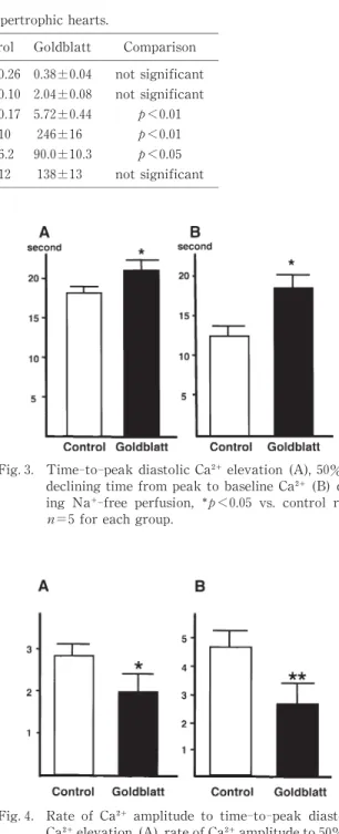
Fig.1. Original traces in a spontaneously beating heart during standard perfusion. Upper ,fura‑2 fluores- cence excited at 340 nm and 380 nm ;middle,fluo- rescence ratio of 340 nm and 380 nm ;lower,left ventricular pressure.
Fig.2. Original traces of changes in intracellular Ca and left ventricular pressure in sham‑operated control (upper)and Goldblatt(lower)rats in constricted scale. When the heart was perfused with a Na‑free solution for 20 seconds,Goldblatt rats showed a slower Ca increase and Ca decrease than did control rats.