Gastric cancer progression may involve a shift in HLA-E profile from an intact heterodimer to
β 2microglobulin-free monomer
HLA-E during progression of gastric cancer
Tumor cell-surface HLA-E binds to CTL/NKT inhibitory-receptors to escape cytotoxicity.
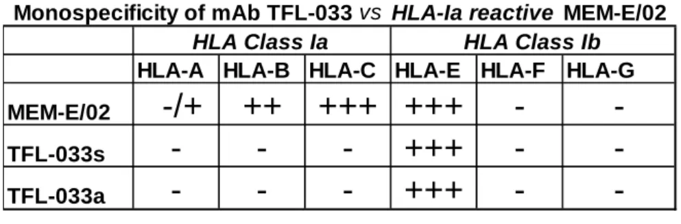
HLA-E-expression includes intact-heterodimer and β2m-free-monomers. The mAb TFL-033 is HLA-E monospecific; MEM-E/02 reacts to both HLA-E and HLA-Ia β2m-free-monomers.
Only TFL-033 stained normal-mucosa cytoplasm; Incidence of cell-surface staining with TFL-033 is higher for diffused carcinoma, early stages (I/II) and non-or undifferentiated tumors; the incidence of MEM-E/02 staining is higher for adenocarcinoma and metastatic lesions.
β2m-free-monomers increased with tumor-progression and in the presence of IFN
γ.(74)
Toshiyuki Sasaki
1,2, Mepur H. Ravindranath
1, Paul I Terasaki
1, Maria Cecilia Freitas
1, Satoru Kawakita
1, Vadim Jucaud
11
Terasaki Foundation Laboratory, Los Angeles, CA 90064
2
Department of Surgery, Jikei University School of Medicine, Tokyo, Japan
Abtstract (words 248) Text: (words 4279), Table: 1, Figures: 5, References: 38
Key words: Gastric cancer, gastric mucosa, carcinoma, adenocarcinoma, tumor progression, HLA-E, anti-HLA-E antibodies, β2microglobulin, heterodimer, monomer
Abbreviations: HLA: human leukocyte antigen;
β2m:
β2-microglobulin; mAb: monoclonal antibody; CTL: cytotoxic lymphocytes; NKT natural killer T cells; IFNγ: interferon-gamma;
TMA:Tissue Microarrays; MFI: mean fluorescent intensity;
TNFα: tumor necrosis factor-alpha;Corresponding author:
Mepur H. Ravindranath, Research Scientist, Terasaki Foundation Laboratory (TFL), 11570 W.
Olympic Blvd. Los Angeles, CA 90064, Phone: 310-479-6101 ext. 103; Fax: 310-445-3381, email: [email protected]
東京慈恵会医 科大学
電子署名者 : 東京慈恵会医科大学 DN : cn=東京慈恵会医科大学, o, ou, [email protected], c=JP 日付 : 2019.01.21 13:08:29 +09'00'
Abstract
Phenotypic expression of HLA-E on the surface of tumor lesions include intact heterodimer (HLA-E heavy chain and β2-microglobulin (β2m) and β2m-free monomer Anti- HLA-E monoclonal antibodies (mAbs), MEM-E/02 or 3D12 bind to the peptide sequences, in
β2-microglobulin-free HLA-E, which is common and shared with HLA-Ia monomers. A newlydeveloped monospecific anti-HLA-E mAb (TFL-033) recognizes HLA-E-restricted peptide sequences on α1 and α2 helices, away from β2-m-site. Tumor progression may involve shedding of β2-m from HLA-E or overexpression of β2m-free monomers. There is a need to identify and distinguish the different phenotypic expression of HLA-E, particularly the intact heterodimer from the β2m-free monomer on the surface of tumor lesions. Due to the unique peptide binding affinities of the mAbs, it is hypothesized that TFL-033 and MEM-E/02 may distinguish the phenotypic expressions of cell surface HLA-E during stages of tumor
progression. Only TFL-033 stained diffusely the cytoplasm of normal mucosa. The incidence and intensity of TFL-033 staining of the cell-surface in early stages, poorly or undifferentiated and non-nodal lesions, and in diffuse carcinoma is greater than that of MEM-E/02. Whereas, MEM-E/02 stained terminal stages, adenocarcinoma and lymph node metastatic lesions
intensely, either due to increased expression of β2m-free HLA-E with tumor progression or due to expression of HLA-Ia molecules. This study evaluates the relative diagnostic potential of HLA-E-monospecific TFL-033 and the HLA-Ia-reactive MEM-E/02 for determining the specific distribution and immunodiagnosis of different phenotypic expression HLA-E in tumor lesions, and the structural and functional alterations undergone by HLA-E during tumor
progression. (Words: 244)
Introduction
Structural and functional alterations of human leukocyte antigens (HLA) are implicated in tumor immune escape (1). The escape mechanism involves loss or downregulation of
classical HLA class I antigens (HLA-A,-B,-C) and/or aberrant overexpression of non-classical HLA class I antigens (HLA-E, -G). Consequently, these changes in HLA classes lead to decreased recognition or destruction of tumor cells by immune cytotoxic effectors, mainly cytotoxic lymphocytes (CTL) and natural killer T (NKT) cells (1).
On the surface of both normal and cancer cells, HLA class I molecules are expressed in at least two distinct phenotypic forms: as heterodimers associated with β2-microglobulin (β2m), recognized by the monoclonal antibody (mAb) W6/32 (1); and as β2m-free HLA class I
α−
chains or heavy chains, recognized by mAbs like LA-45 (2, 3), HC10 (4), L31 and M38 (5).
Phenotypic expression of HLA-E also include an intact heterodimer (with β2m) and in β2m-free
α-chain monomer. Since HLA-E expressed on tumor cells has been shown to protect tumor cellsfrom cytotoxic killing CD8+ T cells (6-8) and to serve as a major ligand for inhibitory receptors CD94/NKG2A on CD8+ CTL and NK T cells (NKT) (9-12), precise characterization of the two forms of phenotypic expressions of HLA-E would enable pathologists to distinguish stages of tumorigenesis and oncologist to evaluate the functional implications of phenotypic alterations to develop suitable immunotherapeutic strategies. Furthermore, the proinflammatory cytokine, interferon-gamma (IFNγ), upregulates HLA-E on the surface of tumor cells (13, 14). Such upregulation may result in the loss of the β2m followed by shedding of heavy chains of HLA-E in the tumor microenvironment (1, 8, 15). Therefore, anti-HLA-E mAbs that can distinguish intact from β2m-free HLA-E would be valuable diagnostic tools for defining the structural and functional alterations associated with HLA-E during tumorigenesis and tumor progression.
The presence of HLA-E has been identified using one of the two commercially available
mAbs, MEM-E/02 or 3D12, in breast cancer (16, 17), ovarian and cervical cancer (8, 16, 18, 19),
melanoma (13, 14, 16,19), glioblastoma (20-24) laryngeal carcinoma (25), colorectal carcinoma
(26-28) renal cell carcinoma (29, 30) , lymphoma (31) and Hodgkin (32). However both MEM-
E/02 and 3D12 lack specificity for HLA-E, since they also react with several alleles of HLA
class Ia molecules (HLA-A, -B and -Cw) (for details see, 33, 34). It was documented (33, 34)
that the reactivity of MEM-E/02 and 3D12 to HLA-E as well as HLA class Ia molecules is
inhibited by peptides (
137DTAAQI
142,
115QFAYDGKDY
123,
126LNEDLRSWTA
135) that are
cryptic and masked by β2m in intact heterodimer of HLA-E (for structural figures of crypticity,
see 37). It was established clearly, that MEM-E/02 and 3D12 recognize β2m-free HLA-E and HLA class Ia alleles (33, 34). In our earlier work (see Table 2 in 34), we have listed amino acid sequences specific (or restricted) to HLA-E, and thus not found in other class I HLA alleles.
These peptides are located on the surface of α1 and α2 helices, not masked by β2m. We have developed several mouse mAbs using heavy chain of HLA-E. One of these mAbs (TFL-033) recognizes the HLA-E-specific peptide sequences, and is considered monospecific for HLA-E
1. A mAb that recognizes specifically β2m-associated intact heterodimer of HLA-E (TFL-033) together with MEM-E/02, that recognizes β2m-free monomer of HLA-E would be valuable in the characterization of alternations undergone by the phenotype of HLA-E during tumorgenesis and tumor progression.
Since TFL-033 is a novel mAb, the immune reactivity is characterized by peptide inhibition, documenting its unique specificity and selectivity. The reliability and the relevance of the specificity is further assessed after exposing the cells to proinflammatory cytokine IFNγ which is known to upregulate the expression of both forms of HLA-E. We hypothesize that both mAbs, TFL-033 and MEM-E/02, may serve to distinguish intact heterodimer of HLA-E from
β2m-free monomer of HLA-E, because identifying the two distinct forms of HLA-E sheds light on the functional implications during malignant transformation and tumor progression.
Furthermore, in order to evaluate the diagnostic potential of the transformation undergone by tumor-associated HLA-E molecule per se, the expression and distribution of intact heterodimer and β2m-free monomer of HLA-E in normal gastric mucosa is compared with primary and metastatic phases of tumor progression of the gastric cancer, known to express HLA-E (35, 36).
Materials and Methods mAbs
The HLA-E-specific mAb TFL-033 was produced following the guidelines in the report
generated by the National Research Council’s Committee on Methods of Producing Monoclonal Antibodies
20. The recombinant polypeptides of HLA-E
R107(heavy-chain only; 10 mg/ml in MES buffer) were obtained from the Immune Monitoring Lab, Fred Hutchinson Cancer Research Center (University of Washington, Seattle, WA). Each antigen was immunized in two different
1 The monospecific monoclonal anti-HLA-E mAbc TFL-033c was developed by Dr. Mepur H. Ravindranath and Prof.
Dr. Paul I. Terasaki in 2011/12c and is under patent consideration.
mice. Fifty µg of the antigen (β2m-free heavy chain) in 100 µl of PBS (pH 7.4) were mixed with 100 µl of TiterMax® Gold adjuvant (CytRx Corp., San Diego, CA) before injection into the footpad and intraperitoneum of the mice. Three immunizations were given at about 12-day intervals. The clones were cultured in a medium containing RPMI 1640 w/L-glutamine and sodium bicarbonate (Sigma-Aldrich, St. Louis, MO, cat. no. R8758), 15% fetal calf serum 0.29mg/ml L-glutamine/Pen-Strep (Gemini-Bio, MedSupply Partners, Atlanta, GA, cat. no. 400- 110), and 1mM sodium pyruvate (Sigma, cat. no. S8636). Several clones were also grown using Hybridoma Fusion and Cloning Supplement (HFCS) (Roche Applied Science, Indianapolis, IN, cat. no. 11363735001). Isotypes of the mAbs were characterized, and no IgM Abs were detected.
The TFL-033 mAbs purified from HLA-E hybridoma supernatant (TFL-033s) and those purified from ascites of hybridoma immunized in BALB/c mice (TFL-033a) were used. Both mAbs were used for immunohistochemistry, and TFL-033a was also used for flow cytometry. The mAb MEM-E/02 (Thermo, NJ, cat. no. MA1-19309), which binds the heavy chain polypeptide of HLA-E, was used for both immunohistochemistry and flow cytometry.
Tissue Microarrays (TMA)
Three TMA (US Biomax, Inc., Rockville, MD) were carefully selected for
immunohistochemistry, based on the detailed information provided by the supplier. The cores of all TMA were 1.5 mm in diameter and 5 µm thick. In the TMA (BN01013a) of normal gastric mucosa (n = 68), four cores were excluded because there was no normal construction of
stomach mucosa or not enough to read. TMA (ST8015) of primary gastric cancer includes 30
adenocarcinoma (row A1-10, B1-9, C1, F4-10,G1-3) 40 diffuse carcinoma (B10, C2-10, entire
row of D & E, F1-3, G4-10) and 10 normal gastric mucosa (entire row H) with information on
pathological diagnosis, TNM grading, and cancer grade. The array includes well differentiated
(row A1-3), moderately differentiated (rows A4-10, entire B, C1), poorly differentiated (row
C2-10 entire D, E & F, G1-6), and undifferentiated G7-10). The array also includes stages I to
IV as follows: Stage I: A1, 4, 10, C2, D6, 7, G7, 8, 9 (n=9), Stage II (n = 38) Stage III: A2,7,
B5-7, 9, D1, 4, 8, E2, 5, 8, 10, F1,2,7, 9, 10, G3, 5, 6 (n = 21). Stage IV: E9, G4 (n =2);), TMA
(ST8013) of metastatic gastric cancer includes 5 peritoneal metastasis(entire row A), 3 Liver
metastasis (row B1-3), 5 Ovarian metastasis (row B4-5, C1-3), 27 Lymph node metastasis (row
C4-5, entire rows of D,E,F,G & H).
Cell lines
Four gastric cancer cell lines were obtained in 2012 from the American Type Culture Collection (Rockville, MD): AGS gastric adenocarcinoma, primary (ATCC Cat. No:CRL-1739), SNU-1 gastric carcinoma, peritoneal metastasis (CRC-5971), KATO-III gastric carcinoma, liver metastasis (HTB-103) and NCI-N87 gastric carcinoma, lymph node metastasis (CRL-5822) (derived from primary lesion, peritoneal metastasis, lymph node metastasis and liver metastasis).
ATCC certificate/authentication details of these cell lines are available on line with product information sheet at www.atcc.org/
and order in voice (# SOE69838) is appended as supplemental files. All cell lines were supplemented with RPMI 1640 and 15% fetal bovine serum, and cultured in 5% CO
2and 95%
air at 37°.
Immunoassay to characterize the specificity of mAbs
To determine the monospecificity of the mAbs for HLA-E, multiplex Luminex
®-based immunoassay (One Lambda, Inc., Canoga Park, CA) was used, as described elsewhere (33, 34, 37). The purified culture supernatants and ascites of mAb TFL-033 and of MEM-E/02 from commercial sources were serially diluted with PBS, pH 7.2, and added to the wells containing the microbeads coated with HLA-E heavy chain and HLA class Ia alleles. Using dual-laser flow cytometry (Luminex xMAP
®multiplex technology), single antigen assays were carried out for data acquisition and analysis of anti-HLA-Ia and anti-HLA-E Abs (33, 34, 37). The
LABScreen
®Single Antigen (One Lambda) assay consists of a panel of color-coded
microspheres (single antigen beads), coated with HLA antigens to identify antibody specificities.
The single recombinant HLA-Ia antigens in LS1A04-lot 005 were used for screening the mAbs.
This lot contains 31 HLA-A, 50 HLA-B and 16 HLA-Cw allelic molecules. The beads supplied by the manufacturer may have two categories of proteins attached to the beads: HLA heavy- chain polypeptide only or heavy-chain polypeptides in association with β2microglobulin. For coating the microbeads with HLA-E, the recombinant HLA-E, HLA-F and HLA-G folded heavy chains (10 mg/ml in MES buffer) were obtained from the Immune Monitoring Lab, Fred
Hutchinson Cancer Research Center. The recombinant HLA-E, HLA-F and HLA-G heavy
chains are individually attached by a process of simple chemical coupling to 5.6 µm polystyrene
microspheres, which are internally dyed by One Lambda with infrared fluorophores. The HLA-
Ia microbeads have built-in control beads: positive beads, coated with human IgG and negative
beads, coated with serum albumin (Human or bovine). For HLA-E, HLA-F and HLA-G, the
control beads (both positive and negative) were added separately. Data generated with Luminex Multiplex Flow Cytometry (LABScan 100) were analyzed using the same computer software and protocols reported earlier. For each analysis, at least 100 beads were counted. Mean and standard deviation of MFI for each allele were recorded. All the data were stored and archived at the Terasaki Foundation Laboratory; basic statistical analyses were made with Excel software.
Peptide inhibition assays
Peptide inhibition assays were performed as described earlier for mAbs MEM-E/02 and 3D12 (33, 34, 37). In contrast to the peptides used for those mAbs, two different HLA-E
restricted peptides,
65RSARDTA
71,
143SEQKSNDASE
152were used for inhibiting the binding of mAb TFL-033 to HLA-E. The peptides were synthesized by GenScript Corporation
(Piscataway, NJ), purified in a reversed-phase 4.6 x 150 mm HPLC column (VP-ODS, Shimadzu, Columbia, MD)—with pump A containing 0.065% trifluoroacetic acid in 100%
water, and pump B containing 0.05% trifluoroacetic acid in 100% acetylnitrile—at a flow rate of 1.0 ml/min and measured at 214 nm (detector A, Ch1). Various concentrations of peptides (serially diluted from the initial concentration of 100 µg to 100 µl) were added to the mAb TFL- 033 (7 µl) and further diluted by adding 14 µl of PBS-BSA pH 7.0 (final dilution 1/1200) then exposed to 2
µl of beads. PE-conjugated anti-mouse IgG was used for the LabScreen assay, and each experiment was done in triplicate.
Immunohistochemistry
Immunohistochemistry was performed using TMA described above. Slides were
dewaxed, rehydrated and heated in a water bath at 99°C for 20 minutes in sodium citrate buffer (pH 6.0), followed by cooling at room temperature for antigen retrieval. Endogenous peroxidase activity was inhibited with dual endogenous enzyme block (Dako, Carpenteria, CA). Sections were blocked with 10% normal goat serum. Primary Abs MEM-E/02, TFL-033a and TFL-033s were diluted, respectively, 1/100, 1/10 and 1/10 in 3% normal goat serum, and added to each section. Primary Abs were incubated for 60 minutes at room temperature. The secondary Ab, a biotinylated goat anti-mouse IgG (Vector Laboratories, Inc., Burlingame, CA), diluted 1/200, was incubated for 30 minutes at room temperature. Sections were incubated with
immunoperoxidase (ABC Kit, Vector), washed, and developed with diaminobenzidine (DAB kit,
Vector). Slides were counterstained with hematoxylin. Negative control was prepared by
omission of the primary Ab. Positivity of TFL-033 was defined as being positive for both TFL- 033a and TFL-033s.
Immunofluorescence analysis
Each cell line was incubated 72 hours under the conditions described above with or without three dilutions (200, 400, 800 IU/ml) of IFNγ (R&D Systems®, Minneapolis, MN, cat.
no. EA5412041), (50, 100, 200 IU/ml) of TNFα, and (400, 800, 1600 IU/ml) of IL-6. For detection of HLA-E on cell surfaces, cells were stained with MEM-E/02, TFL-033a, CEA and CA19-9 mAb for 60 minutes at 4°C. After washing, cells were incubated for 30 minutes with the secondary FITC-labeled mAb and analyzed on a FACScan™ flow cytometer using CellQuest™ software (BD Biosciences, San Jose, CA). Positive signal for HLA-E expression was calculated as being the ratio of positive signal with the specific mAb minus the ratio of positive signal with the negative control.
Results
Monospecificity of mAb TFL-033 and inhibition of HLA-E binding with HLA-E-specific peptide sequences.
Table 1A compares mAb MEM-E/02 that binds not only with HLA-E but also with several HLA-Ia alleles with the purified culture supernatants (s) and purified ascities (a) of mAb TFL-033 that binds and recognizes specifically HLA-E. The mAb TFL-033 did not react with HLA-A, -B or -Cw nor with HLA-F or HLA-G at any of the dilutions tested. Importantly, TFL- 033 was inhibited by two HLA-E-restricted peptides,
65RSARDTA
71, and
143
SEQKSNDASE
152found on α1 and α2 helices, at a site away from the location of β2-m (Table 1B).
. The peptide
143SEQKSNDASE
152showed stronger inhibition of TFL-033 binding to the HLA-E, on the the microbeads (Table 1C). Table 1D shows the amino acids in
65
RSARDTA
71that are recognized by the CD94 receptor present on the surface of CD8+
cytotoxic T cells and NKT cells, which are involved in binding to HLA-E. Similarly, one of the
amino acids in the peptide sequence
143SEQKSNDASE
152is recognized by NKG2a receptor on
the surface of the CD8+ T cells, thus pointing out that the mAb TFL-033 recognizes the peptide
epitopes recognized by CD94 and NKG2a receptors of CD8+ T cells.
Evaluation of immunostaining of normal gastric mucosa and gastric cancer HLA-E positivity to MEM-E/02 and TFL-033 can be evaluated as follows:
1. positive to both TFL-033 and MEM-E/02 may indicate reactivity with intact as well as β2m-free HLA-E;
2. TFL-033-positive and MEM-E/02-negative confirms the presence of intact HLA-E;
3. TFL-033-negative and MEM-E/02-positive may indicate the presence of β2m- free heavy chains of HLA-class I molecules; or the β2m-free heavy chains of HLA-E monomer may denatured in such a way that TFL-033 cannot recognize its specific epitopes.
Negative controls were completely negative in all cores (Figure 1.I A-C). Table 2 and Figure 6 summarize the immunostaining by the three mAbs.
Differential staining of normal tissues by mAbs MEM-E/02 and TFL-033.
Normal stomach mucosa consists of mucous neck cells (goblet cells), acid producing parietal cells, pepsinogen-secreting chief cells and the muscular layer of mucosa (Figure 1.I D-I).
Among these cellular components of the cytoplasm of the normal stomach mucosa, the chief cells and the parietal cells stained intensely by TFL-033a (purified from ascities) (arrows in Figures 1.IE & H) and TFL-033s (purified from culture supernatant) (arrows in Figures 1.IF &
I); the cytoplasm of the chief cells had strong intensity (arrows) in each core (Figure 1.IH & I).
Mucous neck cells and muscular layer stained feebly or failed to stain in each core. Positive staining was consistently observed in the cell cytoplasm. Intensity of staining by TFL-033a was higher than by TFL-033s in all cores. Staining of the cell surface could not be distinguished.
MEM-E/02 failed to stain normal gastric mucosa (Figures 1.ID & G). In the Table 2 the
immunostaining analyses of an array (BN01013a, n =68) of normal stomach mucosal tissue
samples are summarized in a single row. MEM-E/02 stained weakly only 2 of 68 (3.1%)
samples in the cytoplasm region. With both MEM-E/02 and TFL-033, 11 of 68 (17.4%) tissue
samples stained in the cytoplasmic regions of the tissues. MEM-E/02 failed to stain 55 of 68
(88.9%) tissue samples, whereas TFL-033a stained intensely 50 of 68 (79.3%) tissue samples in
the cytoplasmic regions. More samples were stained by TFL-033s (89.7%) (Table 2). These
normal gastric mucosa tissue samples were obtained from the cancer patients and whether the
normal stomach mucosa includes any inflamed tissue could not be confirmed by microscope in all cores.
Differences in the staining patterns of an array of gastric cancer tissues
Primary gastric cancer tissues (ST 8015)Gastric cancer TMA consist of adenocarcinoma (n=30) and diffuse carcinoma (n=40) (Figure 1.II A-F). Table 2 summarizes the immunostaining of the different tumor stages examined, which include T1, T2 (n=15) and T3, T4 (n=55), and the primary with negative (n=41) and positive (n=29) nodal involvement. In addition, 47 tissues at the early stages (I & II) and 23 tissues at the advanced stages (III & IV) were examined; they included 21 well
differentiated types (grades 1 & 2) and 47 poorly differentiated types (grades 3 & 4). With mAb MEM-E/02, 21 of the 70 primary gastric cancers stained. In comparison, TFL-033a and TFL- 033s stained, respectively, 26 and 27 primary cancer tissues (Table 2).
In adenocarcinoma, immunostaining was more intense on the cell surface and also in the cytoplasmic region of the gland. Immunostaining was more intense with MEM-E/02 (Figure 1- II A and D). There was no difference in the basic staining pattern (cell surface more intense than the cytoplasm between MEM-E/02 and TFL-033a/s. MEM-E/02 stained 12 of 30 tissue samples (40%) and the incidence is higher than that of TFL-033 (27-30%)
In diffuse carcinoma, positive staining was also prominent on the cell surface and in the cytoplasm of the cancer cells (Figure 1-II E and F). TFL-033 stained 18 of 40 tissue samples (45%), a higher incidence of staining than that of MEM-E/02 (23%) (Table 2). The primary, when the node was negative for metastasis, had significantly more positive staining with TFL- 033 than when node-positive. The staining intensity was greater in early stages than in advanced stages.
Metastatic region of gastric cancer (ST 8013):
TMA of metastatic gastric cancer included 5 peritoneal, 3 liver, 5 ovarian and 27 lymph node metastases. With the mAb MEM-E/02, 21 of the 40 metastatic carcinomas stained
intensely (53%), higher than that of TFL-033 (38-40%) (Table 2).
Lymph node metastasis stained with higher intensity than other metastatic lesions. The
staining was observed in both the cell surface and the cytoplasm. In peritoneal metastasis, with
all the mAbs, positive staining was observed mainly in cytoplasm (Figure 2-IA-C). Staining of
liver metastasis was much stronger by MEM-E/02 (Figure 2-ID) than by TFL-033a and TFL- 033s (Figure 2-IE & 5F). In contrast, TFL-033a and TFL-033s stained both ovarian (Figures 5G-I) and lymph node metastasis (Figures 5J-L) more intensely, the staining intensity greater than by MEM-E/02. With lymph node metastasis, the incidence of staining by TFL-033 was about 40%, whereas the incidence of staining by MEM-E/02 is much higher (55.6%) (Table 2).
Effect of proinflammatory cytokines on the cell surface expression of HLA-E by gastric cancer cell lines
To assess the role of proinflammatory cytokines on surface expression of HLA-E, HLA- E expression in four human gastric cancer cell lines was examined after exposing the cell lines to the three kinds of cytokines and by immunostaining with MEM-E/02 and TFL-033. The flow cytometric profile of IFNγ-treatment of different gastric cancer cell lines (AGS, primary
adenocarcinoma cell line; SNU-1, peritoneal metatstatic cell lines; KATO III, lymph node metastatic cell lines; and NCI-N87, liver metastatic cell lines) by MEM-E/02 and TFL-033a reveal that different cell lines respond differently to IFNγ. MEM-E/02 stains HLA-E post- IFNγ in SNU-1, KATO-III and NCI-N87 cell lines, whereas TFL-033 stains KATO-III and NCI-N87. The positive signals of KATO-III and NCI-N87 were amplified by using IFNγ, dose- dependently, up to 20% (Figure 3) but were not amplified using either TNFα (6%) or IL-6 (0.6%).
Discussion
A number of investigators have studied HLA-Ia expression in normal gastric mucosa
(38-41). Using mAb PA2.6 (HLA-E reactivity not known), the “heavy chains of HLA-Ia” was
found to be “only weekly detectable” (38). Using mAb W6/32, Garrido and co-investigators
(39-41) have also studied the expression of HLA-Ia alleles on normal gastric mucosa to be
feebly reactive. W6/32 staining was more intense in actively dividing gastric cancer cells and
the tumors tend to lose the expression. Consequently, 10% of gastric carcinomas did not show
W6/32 reactivity on the surface and the majority of gastric cancers reacted weakly to W6/32. A
similar decrease in the expression of HLA class Ia molecules in gastric cancer was reported
subsequently (42). While examining HLA-Ia expression in breast cancer tissues, Torigoe et al
(43) relied upon paraffin sections for observing significant decrease in the expression of HLA-Ia
antigens. However, these investigators are not aware at that time that the mAbs W6/32 or
HCA2, or HC10 are reactive to HLA-E heavy chains (44, 45). Since mAb W6/32 reacts to the
heavy chains of both HLA-E and HLA-Ia molecules similar to MEM-E/02, the observations can be compared with that of MEM-E/02 or even 3D12, but cannot be related to the staining of HLA-E-monospecific mAb TFL-003.
The enhanced reactivity of the mAb MEM-E/02 in gastric cancer tissues obtained from late stages, adenocarcinoma and lymph node metastatic lesions suggests increased expression of
β2m-free HLA-E with tumor progression (Figure 4B). This inference is based upon our earlierobservations (33. 34, 37) that the binding site (peptide epitope or sequence) of MEM-E/02 is located at the site at which β2-microglobulin is present (see Figure 4 in 37). These peptide sequences are considered cryptic, since they are masked by β2-microglobulin (see also Figure 4B). MEM-E/02 or 3D12 or any other mAb cannot assess to this site when HLA-E is intact and expressed as an heteromer with β2-microglobulin. Furthermore, our earlier data (33, 34, 37) show how we have identified these cryptic peptides as the binding sites for the MEM-E/02 using the cryptic peptides (e.g.,
115QFAYDGKDY
123) (see Figures, 2 and 4B in 33) for inhibiting the binding MEM-E/02 to HLA-E. Most importantly, this peptide sequence
(
115QFAYDGKDY
123) shared by the monomers of both HLA-E and other HLA class Ia alleles (33,34,37), not only inhibited the binding of MEM-E/02 to HLA-E but also to HLA-B and HLA-Cw alleles In the intact HLA-E or HLA-E heteromer, these shared peptide sequences are masked by the presence of β2m. However, upon dissociation and shedding of β2m, they are exposed in the heavy chain of HLA-E (33, 37). Since these peptide sequences are hidden by
β2m in intact HLA-E, MEM-E/02, may not be useful for monitoring the expression of intactHLA-E—although immunostaining with MEM-E/02 may be useful to monitor β2m-free HLA-E.
It is obvious that MEM-E/02 may recognize monomers of both HLA-E and HLA-Ia molecules on the cells.
However, since the peptide sequences recognized by MEM-E/02 are not only restricted to HLA-E but are also present on several HLA-Ia molecules (33, 34), MEM-E/02 positivity should be verified by staining with TFL-033. As indicated in Figure 2C, MEM-E/02 positivity in conjunction with TFL-033 negative staining does not indicate the presence of HLA-E. Our results show that MEM-E/02, in contrast to TFL-033, rarely stains normal gastric mucosa but does stain gastric cancer tissues.
Our observations showed that TFL-033 recognizes the two HLA-E-restricted peptides—
65
RSARDTA
71and
143SEQKSNDASE
152—located on α1 and α2 helices of intact HLA-E. It is
important to note that these two HLA-E-restricted peptides may also occur in the β2m-free
heavy chains of HLA-E. The cytoplasm of normal gastric mucosa was stained by both TFL-033
and TFL-033s but not by MEM-E/02, confirming the presence and distribution of intact HLA-E in normal mucosa, mostly in chief and parietal cells. Note, also, that the intensity of TFL-033 staining of gastric cancer cells is considerably less that when it stains normal gastric mucosa, indicative of the alteration undergone by HLA-E when normal gastric mucosa transforms into cancer. The alteration of cancer cells during malignant transformation and tumor progression is confirmed by positive and intense immunostaining with MEM-E/02. The flow cytometric profile of IFNγ-activated metastatic cell lines showed higher intensity of immunostaining by both TFL-033 and MEM-E/02, indicative of overexpression of β2m-free HLA-E, which has the propensity to shed in the tumor microenvironment and circulation (13).
TFL-033a and TFL-033s showed no cross-reactivity with other HLA class I alleles—
HLA-A, -E, -Cw, -F or -G (Figure 1). Although TFL-033a, purified from ascites, had a stronger positive signal than TFL-033s, the staining pattern was almost identical between the two mAbs (82.6% identical in cancer cell and 96.8 % identical in normal stomach cell). The incidence and percentage of staining of gastric adenocarcinoma by MEM-E/02 was notably higher than that by TFL-033, whereas the reverse was true for diffused carcinoma, suggesting that β2m-free heavy chains may be more prevalent in adenocarcinoma than in diffuse carcinoma. Diffuse carcinoma, including signet-ring cell and mucinous carcinoma, showed greater positivity with TFL-033 than did adenocarcinoma.
Tumor progression, as evidenced by the infiltration and invasion of the primary tumor to
lymph nodes and other organs (T1 to T4), is indicated by differences in the incidence of staining
of both MEM-E/02 and TFL-003. A careful comparison of the immunostaining of the two
different antibodies presented in Table 2 and summarized in Figure 4A reveal changes
undergone by the HLA-E phenotypes (intact HLA-E or HLA-E heterodimer and β2m-free
heavy chains of HLA-E or HLA-E monomer). First of all, the incidence of staining of TFL-033
is greater for T1 and T2 than for T3 and T4, Stages I and II than for stage III and IV, as well as
for undifferentiated (grade 4) and poorly differentiated (grade 3) (Figure 4A) than for grade 1
and grade 2 tumor tissues suggesting the higher prevalence of intact HLA-E heteromer than the
β2m-free monomers. Secondly, the reverse is true for mAb MEM-E/02 indicative of progressiveincrease in the expression of β2m-free heavy chains when primary tumor infiltrates serosa and
organs, lymph nodes, when the stage progresses to T3 and T4 and when moderately or well
differentiated tumors (Figure 4A). These two salient findings with the two mAbs suggest that
the prevalence of intact HLA-E heteromers in early stages of tumorigenesis and possible
emergence of β2m-free heavy chains due to loss or shedding of β2m at later stages of tumor
progression (Figure 4B). It is important to note that α1 and α2 helices of either intact HLA-E or the heavy chain only can contribute to the inactivation of cytotoxic CD8+ T cells and NKT cells by binding to CD94 and NKG2a receptors on the cytotoxic cells. The relationship between different phenotypes of HLA-E overexpression and cancer progression has been reported frequently (13-32). HLA-E molecules have often been implicated in tumor progression because of their capacity for binding the inhibitory receptor CD94/NKG2A expressed by CTL and NKT.
It was proposed that HLA-E overexpression favors tumor cell escape from CTL and NKT immunosurveillance (6-12).
Proinflammatory cytokines may activate the expression of HLA-E (13, 14). The positive signals of metastastic cell lines KATO-III and NCI-N87 were amplified with IFNγ treatment in a dose-dependent manner. Since both MEM-E/02 and TFL-033 recognizes the IFNγ-mediated HLA-E overexpression in KATO-III and NCI-N87, it is inferred that these cell lines overexpress HLA-E without β2-microglobulin. Specific staining of SNU-1 by MEM-E/02 but not by TFL- 033 points out that the staining may not be due to HLA-E but could be due to HLA-Ia alleles.
These results support the previous findings (13, 14) to show that IFNγ induces anti-tumor responses by HLA-E upregulation. It is important to note that immunostaining of the live metastatic cell lines by MEM-E/02 is more intense that that by TFL-033. Not only this
observation confirms our observations made on paraffin sections (presented in Table 2) but also point out that IFNγ may contribute to the overexpression of β2m-free HLA-E. The high
intensity of the live cell staining with MEM-E/02 may not only confirm upregulation of β2m- free HLA-E, but also upregulation of β2m-free HLA-Ia alleles, since this particular mAb binds to the peptide sequence shared by both HLA-E and HLA-Ia molecules. Possibly, strategies to regulate secretion of IFNγ in the tumor microenvironment may be useful to control tumor progression.
Thus our results confirm the presence of different phenotypes of HLA-E, namely the presence of intact heterodimer (with β2m) and β2m-free α-chain monomer. HLA class-I
antigens after presentation of the peptides undergo structural changes as illustrated in Figure 4B.
After the loss of peptide, β2m does not remain associated with the heavy chain. Loss of β2m exposes the cryptic epitopes hidden by β2m. The heavy chain monomer may undergo
denaturation on the cell surface or even after shedding into the surrounding microenvironment.
We envisage that the cryptic epitopes as well as the altered monomers are recognized by B cells
for antibody production. Figure 4B succinctly summarizes the diagnostic potential of the HLA-
E monospecific mAb TFL-033 compared to MEME/02, which is also reacts with HLA class Ia alleles, like another commercial anti-HLA-E mAb 3D12. The figure documents the different sites at which mAbs TFL-033 and MEM-E/02 may bind, based on our peptide inhibition studies carried out in this study, as well as in an earlier study (33). As noted in this and other
publications (34, 37, 46), MEM-E/02 binding site is shared by both HLA-E and several HLA-Ia alleles. Most importantly mAb TFL-033 does not recognize this region but only the sequences in the α1 & α2 helices, a site at which CD8+ T cell and NKT receptors. CD94 (binds to amino acid at positions 66, 68 and 73, see Table 1D) and NKG2a (binds to amino acid at position 151).
The binding sites recognized by TFL-033 occur in intact HLA-E with or without β2- microglobulin as well as in denatured heavy chains as illustrated in the figure.
In conclusion, this is the first study to compare expression of heterodimers of HLA-E and β2m-free HLA-E in normal gastric mucosa and gastric cancer. Any histological or histo- immunochemical documentation of HLA-E either on normal cells or inflamed cells or on cancer cells requires such a monospecific mAb. TFL-033 is the only reliable mAb for this purpose since all the known commercial anti-HLA-E mAbs—such as MEM-E/02, MEM-E/06, MEM-E/07 and 3D12—cross-react with HLA class Ia molecules(33.34), and hence a reasonable conclusion of the presence of HLA-E on tissues cannot be derived. Literature cited below will reveal that all the publications on HLA-E have used these commercial mAbs without evaluating their specific affinity for HLA-E. Therefore, using TFL-033 in conjunction with such commercial mAbs is necessary to confirm the distribution and presence of the two distinct forms of HLA-E. Using just MEM-E/02 or 3D12 may not provide a true picture of the expression of HLA-E in human cancer; what’s more, those Abs may not reveal the intact HLA-E but only β2m-free HLA-E.
Although the results of our study emphasize the need to use TFL-033, an HLA-Ia non-reactive, monospecific mAb, for evaluating the relative distribution of the phenotypic expressions of HLA-E (i.e., with or without β2m
), using both HLA class-Ia reactive (MEM-E/02) or non- reactive (TFL-033) mAbs is useful for evaluating the specific distribution and diagnosis of intact HLA-E in the cell surface of human cancers, and for evaluating the structural and functional alterations undergone by HLA-E during tumorigenesis, as illustrated in Figure 4B.
ACKNOWLEDGEMENTS
The entire project is supported by grants from Terasaki Family Foundation. Thanks are due to Mr. Tho Pham for his untiring help in raising TFL-033 in the initial stages of
development. We are greatly indebted to Dr. Mein Hardy, pathologist in the Department of
Pathology at Marina Del Rey Hospital, Los Angeles, CA, for critically verifying through the slides of immunostaining of normal gastric mucosa tissue and cancer tissues and concurring with the interpretation of the slides.
AUTHORS’ CONTRIBUTIONS
Professor. Paul I. Terasaki, Director of TFL, suggested the project, supervised the entire study, participated in conceptual discussion and editing and provided support by periodic monitoring.
Toshiyuki Sasaki, visiting surgical gastroenterologist, undertook the entire investigation, designed the testing of the hypothesis, carried out immunostaining of normal and cancer tissues and flow cytometric analyses using cell lines (Figure 5), photographed the critical observations, assembled the photographs in Figure 3 and 4, analyzed the data (Table 1) and wrote the first draft of the paper.
Mepur H. Ravindranath, research scientist at TFL, assisted in formulating and testing the hypothesis, carried out experiments presented, assisted in consolidating the figures and writing the manuscript.
Maria Cecilia Freitas, visiting nephrologist at TFL with expertise in histopathology, provided the immunoassay protocols, monitored and assisted in developing the assay, evaluated the histopathological data.
Vadim Jucaud and Satoru Kawakita, Research Associates at TFL, actively involved in performing the assays, participated in the critical discussion of the data and significantly contributed to the revised written version of the manuscript.
CONFLICT OF INTEREST DISCLOSURE
None of the authors have any conflicts of interest.
References
1. Algarra I, Garcia-Lora A, Cabrera T, Ruiz-Cabello F, Garrido F. The selection of tumor variants with altered expression of classical and nonclassical MHC class I molecules:
implications for tumor immune escape. Cancer Immunol Immunother 2004;53(10): 904–
910.
2. Schnable E, Stockinger H., Majdic O, Gaugitisch H, Lindley IJ, Maurer D, Hajek- Rosenmayr A, Knapp W. Activated human T lymphocytes express MHC class I heavy chains not associated with β2-microglobulin. J Exp Med 1990;171:1431-38.
3. Madrigal AJ, Belich MP, Benjamin RJ, Little AM, Hildebrand WH, Mann DL Parham P.
Molecular definition of a polymorphic antigen (LA45) of free HLA-A and -B heavy chains found on the surfaces of activated B and T cells. J Exp Med 1991;174:1085-93.
4. Stam NJ, Spits H. Ploegh HL. Monoclonal antibodies raised against denatured HLA-B locus heavy chains permit biochemical characterization of certain HLA-C locus products.
J Immunol 1986;137:2299-2307.
5. Grassi F, Meneveri R, Gullberg M, Lopalco L, Rossi GB, Lanza P, De Santis C, Brattsand G, Butto S, Ginelli E, Beretta A, Siccardi AG. Human immunodeficiency virus type 1 gpl20 mimics a hidden monomorphic epitope borne by class I major histo- compatibility complex heavy chains. J Exp Med 1991;174:53-8.
6. Nguyen S, Beziat V, Dhedin N, Kuentz M, Vernant JP, Debre P, Vieillard V. HLA-E upregulation on IFN-gamma-activated AML blasts impairs CD94/NKG2A-dependent NK cytolysis after haplo-mismatched hematopoietic SCT. Bone Marrow Transplant 2009;43(9): 693-9.
7. Berezhnoi AE, Chernisheva AD, Zakeeva IR, Danilova AB, Danilov AO, Moiseenko VM, Geraghty D, Gnuchev NV, Georgiev GP, Kibardin AV, Larin SS. HLA-E molecule induction on the surface of tumor cells protects them from cytotoxic lymphocytes. Vopr Onkol 2009;55(2): 224-9.
8. Gooden M, Lampen M, Jordanova ES, Leffers N, Trimbos JB, van der Burg SH, Nijman H, van Hall T. HLA-E expression by gynecological cancers restrains tumor-infiltrating CD8(+) T lymphocytes. Proc Natl Acad Sci U S A 2011;108(26): 10656-61.
9. Lo Monaco E, Tremante E, Cerboni C, Melucci E, Sibilio L, Zingoni A, Nicotra MR, Natali PG, Giacomini P. Human leukocyte antigen E contributes to protect tumor cells from lysis by natural killer cells. Neoplasia 2011;13(9): 822-30.
10. Iwaszko M, Bogunia-Kubik K. Clinical significance of the HLA-E and CD94/NKG2 interaction. Arch Immunol Ther Exp (Warsz) 2011;59(5):353-67.
11. Lee N, Llano M, Carretero M, Ishitani A, Navarro F, López-Botet M, Geraghty DE.
HLA-E is a major ligand for the natural killer inhibitory receptor CD94/NKG2A. Proc Natl Acad Sci U S A 1998;95(9): 5199-204.
12. Kaiser BK, Pizarro JC, Kerns J, Strong RK. Structural basis for NKG2A/CD94 recognition of HLA-E. Proc Natl Acad Sci U S A 2008;105(18): 6696-701.
13. Derre L, Corvaisier M, Charreau B, Moreau A, Godefroy E, Moreau-Aubry A, Jotereau F, Gervois N. Expression and release of HLA-E by melanoma cells and melanocytes:
potential impact on the response of cytotoxic effector cells. J Immunol 2006;177(5):
3100-7.
14. Coupel S, Moreau A, Hamidou M Horejsi V, Soulillou JP, Charreau B. Expression and release of soluble HLA-E is an immunoregulatory feature of endothelial cell activation.
Blood 2007;109(7): 2806-14.
15. Allard M, Oger R, Vignard V, Percier JM, Fregni G, Périer A, Caignard A, Charreau B, Bernardeau K, Khammari A, Dréno B, Gervois N. Serum soluble HLA-E in melanoma:
a new potential immune-related marker in cancer. PLoS One 2011;6(6): e21118.
16. Sensi M, Pietra G, Molla A, Nicolini G, Vegetti C, Bersani I, Millo E, Weiss E, Moretta L, Mingari MC, Anichini A. Peptides with dual binding specificity for HLA-A2 and HLA-E are encoded by alternatively spliced isoforms of the antioxidant enzyme peroxiredoxin 5. Int Immunol 2009;21(3):257-68.
17. de Kruijf EM, Sajet A, van Nes JG, Natanov R, Putter H, Smit VT, Liefers GJ, van den Elsen PJ, van de Velde CJ, Kuppen PJ. HLA-E and HLA-G expression in classical HLA class I-negative tumors is of prognostic value for clinical outcome of early breast cancer patients. J Immunol 2010;185(12):7452-9.
18. Goncalves MA, Le Discorde M, Simões RT, Rabreau M, Soares EG, Donadi EA,
Carosella ED. Classical and non-classical HLA molecules and p16(INK4a) expression in precursors lesions and invasive cervical cancer. Eur J Obstet Gynecol Reprod Biol 2008;141(1):70-4.
19. Marin R, Ruiz-Cabello F, Pedrinaci S, Méndez R, Jiménez P, Geraghty DE et al.
Analysis of HLA-E expression in human tumors. Immunogenetics 2003;54(11):767-75.
20. Wischhusen J, Friese MA, Mittelbronn M, Meyermann R, Weller M. HLA-E protects glioma cells from NKG2D-mediated immune responses in vitro: implications for immune escape in vivo. J Neuropathol Exp Neurol 2005;64(6):523-8.
21. Mittelbronn M, Simon P, Löffler C, Capper D, Bunz B, Harter P, Schlaszus H, Schleich A, Tabatabai G, Goeppert B, Meyermann R, Weller M, et al. Elevated HLA-E levels in human glioblastomas but not in grade I to III astrocytomas correlate with infiltrating CD8+ cells. J Neuroimmunol 2007;189(1-2):50-8.
22. Levy EM, Sycz G, Arriaga JM, Barrio MM, von Euw EM, Morales SB, González M, Mordoh J, Bianchini M. Cetuximab-mediated cellular cytotoxicity is inhibited by HLA- E membrane expression in colon cancer cells. Innate Immun 2009;15(2):91-100.
23. Kren L, Muckova K, Lzicarova E, Sova M, Vybihal V, Svoboda T, Fadrus P, Smrcka M, Slaby O, Lakomy R, Vanhara P, Krenova Z, et al. Production of immune-modulatory nonclassical molecules HLA-G and HLA-E by tumor infiltrating ameboid
microglia/macrophages in glioblastomas: a role in innate immunity? J Neuroimmunol 2010;220(1-2): 131-5.
24. Kren L, Slaby O, Muckova K, Lzicarova E, Sova M, Vybihal V, Svoboda T, Fadrus P, Lakomy R, Vanhara P, Krenova Z, Sterba J, et al. Expression of immune-modulatory molecules HLA-G and HLA-E by tumor cells in glioblastomas: an unexpected prognostic significance? Neuropathology 2011;31(2): 129-34.
25. Silva TG, Crispim JC, Miranda FA, Hassumi MK, de Mello JM, Simões RT, Souto F, Soares EG, Donadi EA, Soares CP. Expression of the nonclassical HLA-G and HLA-E molecules in laryngeal lesions as biomarkers of tumor invasiveness. Histol Histopathol 2011;26(12): 1487-97.
26. Levy EM, Bianchini M, Von Euw EM, Barrio MM, Bravo AI, Furman D, Domenichini E, Macagno C, Pinsky V, Zucchini C, Valvassori L, Mordoh J. Human leukocyte antigen-E protein is overexpressed in primary human colorectal cancer. Int J Oncol 2008;32(3): 633-41.
27. Benevolo M, Mottolese M, Tremante E, Rollo F, Diodoro MG, Ercolani C, Sperduti I, Lo Monaco E, Cosimelli M, Giacomini P. High expression of HLA-E in colorectal carcinoma is associated with a favorable prognosis. J Transl Med 2011;9:184-9.
28. Bossard C, Bézieau S, Matysiak-Budnik T, Volteau C, Laboisse CL, Jotereau F, Mosnier
JF. HLA-E/beta2 microglobulin overexpression in colorectal cancer is associated with
recruitment of inhibitory immune cells and tumor progression. Int J Cancer 2012;
131(4): 855-63.
29. Hanak L, Slaby O, Lauerova L, Kren L, Nenutil R, Michalek J. Expression pattern of HLA class I antigens in renal cell carcinoma and primary cell line cultures:
methodological implications for immunotherapy. Med Sci Monit 2009;15(12): CR638-43.
30. Kren L, Valkovsky I, Dolezel J, Capak I, Pacik D, Poprach A, Lakomy R, Redova M, Fabian P, Krenova Z, Slaby O. HLA-G and HLA-E specific mRNAs connote opposite prognostic significance in renal cell carcinoma. Diagn Pathol 2012;7:58.
31. Ge SJ, Duan LN, Luo Y, Suo TL, Lu CR, Tang J. Expression of NKG2D and NKG2A with their ligands MHC-I A/B and HLA-E in acute leukemia patients and its significance.
Zhongguo Shi Yan Xue Ye Xue Za Zhi 2011;19(2):312-6.
32. Kren L, Fabian P, Slaby O, Janikova A, Soucek O, Sterba J, Krenova Z, Michalek J, Kral Z. Multifunctional immune-modulatory protein HLA-E identified in classical Hodgkin lymphoma: possible implications. Pathol Res Pract 2012;208(1):45-9.
33. Ravindranath MH, Taniguchi M, Chen CW, Ozawa M, Kaneku H, El-Awar N, Cai J, Terasaki PI. HLA-E monoclonal antibodies recognize shared peptide sequences on classical HLA class Ia:relevance to human natural HLA antibodies. Mol Immunol 2010;47(5):1121-31.
34. Ravindranath MH, Pham T, El-Awar N, Kaneku H, Terasaki PI. Anti-HLA-E mAb 3D12 mimics MEM-E/02 in binding to HLA-B and HLA-C alleles: Web-tools validate the immunogenic epitopes of HLA-E recognized by the antibodies. Mol Immunol 2011;48(4): 423-30.
35. Dutta N, Majumder D, Gupta A, Mazumder DN, Banerjee S. Analysis of human lymphocyte antigen class I expression in gastric cancer by reverse transcriptase- polymerase chain reaction. Hum Immunol 2005;66(2):164-9.
36. Dutta N, Gupta A, Mazumder DN, Banerjee S. Down-regulation of locus-specific human lymphocyte antigen class I expression in Epstein-Barr virus-associated gastric cancer:
implication for viral-induced immune evasion. Cancer 2006;106(8): 1685-93.
37. Ravindranath MH, Kaneku H, El-Awar N, Morales-Buenrostro LE, Terasaki PI.
Antibodies to HLA-E in nonalloimmunized males: pattern of HLA-Ia reactivity of anti- HLA-E-positive sera. J Immunol 2010;185(3): 1935-48.
38. Daar AS, Fuggle SV, Fabre JW, Ting A, Morris PJ. The detailed distribution of HLA-A, B, C antigens in normal human organs. Transplantation 1984; 38(3):287-92.
39. Ruiz-Cabello F, Lopez Nevot MA, Gutierrez J, Oliva MR, Romero C, Ferron A, Esteban F, Huelin C, Piris MA, Rivas C, et al. Phenotypic expression of histocompatibility antigens in human primary tumours and metastases. Clin Exp Metastasis 1989; 7(2):213- 26.
40. Ferron A, Perez-Ayala M, Concha A, Cabrera T, Redondo M, Oliva MR, Ruiz-Cabello F, Garrido F. MHC class I and II antigens on gastric carcinomas and autoogous Mucosa.
J Immunogenet 1989; 16(4-5):413-23.
41. López-Nevot MA, Esteban F, Ferrón A, Gutiérrez J, Oliva MR, Romero C, Huelin C, Ruiz-Cabello F, Garrido F. HLA class I gene expression on human primary tumours and autologous metastases: demonstration of selective losses of HLA antigens on colorectal, gastric and laryngeal carcinomas. Br J Cancer 1989; 59(2):221-6.
42. Ishigami S., Natsugoe S, Nakajo A, Arigami T, Kitazono M, Okumura H, Matsumoto M, Uchikado Y, Setoyama T, Sasaki K, Aikou T. HLA-class I expression in gastric cancer.
J Surg Oncol 2008;97(7): 605-8.
43. Torigoe T, Asanuma H, Nakazawa E, Tamura Y, Hirohashi Y, Yamamoto E, Kanaseki T,
Hasegawa T, Sato N. Establishment of a monoclonal anti-pan HLA class I antibody
suitable for immunostaining of formalin-fixed tissue: unusually high frequency of down- regulation in breast cancer tissues. Pathol Int. 2012; 62(5):303-8.
44. Braud V, Jones EY, McMichael A. The human major histocompatibility complex class Ib molecule HLA-E binds signal sequence-derived peptides with primary anchor residues at positions 2 and 9. Eur J Immunol. 1997; 27(5):1164-9.
45. Seitz C, Uchanska-Ziegler B, Zank A, Ziegler A. The monoclonal antibody HCA2 recognises a broadly shared epitope on selected classical as well as several non-classical HLA class I molecules. Mol Immunol. 1998; 35(13):819-27.
46.
Ravindranath MH, Terasaki PI, Kawakita S. Naturally occurring anti-HLA-E autoantibodies:Evidences of HLA-Ia reactivity of anti-HLA-E antibodies. In:
Autoantibodies
Eds.Shoenfeld, Y., Gershwin, ME., Meroni, PI,
2014, Chapter 35, 1-9.Elsevier B.V.
http://dx.doi.org/10.1016/B978-0-444-56378-1.00035-6.
Table 1. HLA-E and HLA-Ia reactivity of TFL-033a, TFL-033s and MEM-E/02. (A) The reactivity of TFL-033 and MEM-E/02 to HLA-Ia/-E. (A) E-restricted (65RSARDTA71 and
143SEQKSNDASE152) recognized by TFL-033 and shared Peptide sequences
(115QFAYDGKDY123) recognized by MEM-E/02. For details of the peptides recognized by MEM-E/02 see Refs. 33 and 34. (C). Inhibition of Protein-G purified culture supernatants of TFL-033 with two HLA-E-restricted peptides at 0.27 µg/14 µl/well concentration. (D) Peptide recognized by mAb TFL-033 is also the binding of site of CD94, the CD8+ T cell receptor.
A
HLA-A HLA-B HLA-C HLA-E HLA-F HLA-G
MEM-E/02
(-/+ (++) +++ +++ - -
TFL-033s
- - - +++ - -
TFL-033a
- - - +++ - -
Monospecificity of mAb TFL-033 vs HLA-Ia reactive MEM-E/02 HLA Class Ib HLA Class Ia
B
HLA-A HLA-B HLA-C HLA-F HLA-G
65RSARDTA71 HLA-E restricted TFL-033 0 0 0 0 0
143SEQKSNDASE152 HLA-E restricted TFL-033 0 0 0 0 0
115QFAYDGKDY123d Polyspecific MEM-E/02 1 104 75 0 0
HLA Class Ia HLA Class Ib Sequence Specificity Affinity
Recognition sequences of TFL-033 and MEM-E/02 in HLA Class I
C
E- specific Peptides MFI without peptide MFI with peptide P-value
65RSARDTA71 3759 ± 118 3371 ± 172 0.03
143SEQKSNDASE152 3958 ± 118 3155 ± 117 <0.003
*: antibody concentration (0.7 µg.14 µl/well):
** peptide concentration (0.27 µg.14 µl/well)
Inhibition of TFL-033* by HLA-E-specific peptides**
D
65 66 67 68 69 70 71 72
R S A R D T A Q
The peptide sequence that inhibits binding of TFL-033 to HLA-E on solid matrix is located in the region of the epitope recognized by of CD8+ T cell receptor CD94 on
cell surface HLA-E (The shared amino acids are indicated in Bold and in shaded boxes ) TFL-033 inhibitory peptide sequence (α1)
P a g e | 22
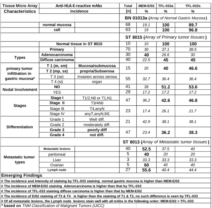
Table 2. Summary of the results of immunostaining of normal gastric tissue and primary and metastatic gastric cancers during different stages of tumor progression with mAbs MEM-E/02 and TFL-033a (purified from ascites) and TFL-033s (purified from culture supernatant).
Tissue Micro Array Total MEM-E/02 TFL-033a TFL-033s
Characteristics [n] % % %
normal mucosa 68 19.1 100 89.7
cell 63 19 100 96.8
10 10 100 100
Primary 70 30 37.1 38.5
Adenocarcinoma 30 40 26.6 30
Diffuse carcinoma 40 22.5 45 45
T 1 (m, sm) Mucosa/submucosa T 2 (mp, ss) propria/Subserosa
T 3 (se) Invasion across serosa
T 4 (si) organ
NO 41 39 51.2 53.6
YES 29 17.2 17.2 17.2
Stage I T1/2,N0 or T1,N1
Stage II T3/4N0
Stage III
T2,N3 or T3,N2/3 or T4,anyN
Stage IV anyT,anyN,M1
Grade 1 Well diff.
Grade 2 moderately diff.
Grade 3 poorly diff
Grade 4 not diff.
Metastatic lesions 40 52.5 37.5 40
peritoneal 5 40 20 20
Liver 3 33.3 33.3 33.3
Ovarian 5 60 40 40
Lymph node 27 55.6 40.4 44.4
* based on TNM Classification of Malignant Tumors (UICC)
ST 8015 (Array of Primary tumor tissues ) BN 01013a (Array of Normal Gastric Mucosa )
# The incidence and Intensity of staining by TFL-033 staining normal gastric mucosa is higher than MEM-E/02
# The incidence of MEM-E/02 staining Adenocarcinoma is higher than that by TFL-033
# The incidence of TFL-033 staining diffuse carcinoma is higher than that by MEM-E/02
# The incidence of E/02 staining of T3 & T4 is higher than the staining of T1 & T2, no such difference is seen by TFL-033
# Of all metastatic lesions, the Lymph node lesions stain well with all mAbs in the following order: MEM-E/02 > TFL-033
Emerging Findings
36.4
21.7
38.1 38.1
23.4 36.2 38.3
47 21
ST 8013 (Array of Metastatic tumor tissues )
15 20 40 46.6
55 32.7 36.4
46.8 23 17.4 26.1
47 36.2 42.6
42.9 Anti-HLA-E-reactive mAbs
Incidence
Metastatic tumor types Stages Nodal Involvement
primary tumor infiltration in gastric mucosa*
Types
Differentiation
Normal tissue in ST 8015
P a g e | 23
LEGEND FOR FIGURES
Figure 1. Immunostaining of the Tissue Microarray (TMA) of normal gastric mucosal and primary gastric cancer tissues with mAbs MEM-E/02 and TFL-033s & TFL-033a.
I.
Normal mucosal tissues. A: Normal Mucosa; B: Adenocarcinoma; C: Diffuse Carcinoma, Controls stained without primary mAbs: D (x40) & G (x200); MEM-E/02: E (x40) & H (x200):TFL-033a: (E: x40; H: x200); TFL-033s: F(x40) & I (x200). Red arrows indicate intense staining of chief cells by TFL-033.
II.
Primary cancer tissues. Adenocarcinoma: (A) MEM- E/02, (B) TFL-033a, and (C) TFL-033s, x200. (A) Red arrows indicate cytoplasmic staining (B& C) of glandular epithelia and the intense staining of the cell surface of the glandular epithelia.
Diffuse Carcinoma stained intensely with MEM-E/02 (D) compared to TFL-033a (E) or TFL- 033s (F),x400.
III
. Normal mucosa and different stages of the primary adenocarcinoma, diffuse carcinoma.: positive for both TFL-033 and MEM-E/02, : positive by TFL-033 only, : positive by MEM-E/02 only, : negative for both mAbs. (A) Staining of normal mucosa with TFL-033 and MEM-E/02. (B) Primary adenocarcinoma and diffuse carcinoma at different stages of
tumorigenesis by TFL-033 and MEM-E/02.
Figure 2. Immunostaining of array of metastatic gastric cancer tissues. Arrows indicate the staining intensity on the same core of tissues.
I.
Metastatic tissues. Peritoneal metastasis (A) MEM-E/02, (B) TFL-033a, and (C) TFL-033s, x200. Liver metastasis (D) MEM-E/02, (E) TFL- 033a, and (F) TFL-033s, x200. Staining by TFL-033 was performed on a different core.Ovarian metastasis (G) MEM-E/02, (H) TFL-033a, and (I) TFL-033s, x200. Arrows: staining on the cell surface. Lymph node metastasis (J) MEM-E/02, (K) TFL-033a, and (L) TFL-033s, x200. Arrows: the staining on the cell surface as well as cytoplasm.
II.
A. Positivity of mAbs.The meaning of circles , interpretation of the findings and a detailed summary of the incidence of staining with different mAbs used are indicated for Array of normal stomach mucosa
(BN010123a), primary gastric cancer tissues (ST8015) and metastatic gastric cancer tissues (ST8013). B. Staining of an array of metastatic tissues. The details regarding open, closed and semi circles are presented in Figure 1.
Figure 3. Flow cytometric profile of staining of different gastric cancer cell lines: AGS (primary adenocarcinoma cell line), SNU-1 (peritoneal metatstatic cell lines), KATO III (lymph node metastatic cell lines) and NCI-N87 (liver metastatic cell lines) by MEM-E/02 and TFL-033a after exposing the cell lines to IFNγ (at varying concentrations, see x-axis) for 72 hours.
Different cell lines respond differently to the treatment of IFNγ. MEM-E/02 identifies the upregulation of HLA-E post-IFNγ by SNU-1, KATO-III and NCI-N87 cell lines, whereas TFL- 033 identifies the upregulation of HLA-E by KATO-III and NCI-N87 respond to TFL-033.
Since both antibodies identify the IFNγ-mediated HLA-E overexpression in KATO-III and NCI- N87, it is suggested these cell lines overexpresses HLA-E without β2-microglobulin. Specific staining of SNU-1 by MEM-E/02 but not by TFL-033 points out that the staining may not be due to HLA-E but by HLA-Ia alleles sharing peptide sequences with HLA-E. Since immunostaining
P a g e | 24
was seen to increase after exposure to the cytokine, and staining was observed with both the mAbs, it is inferred that β2m-free HLA-E expression is augmented after exposure to IFNγ.
Figure 4.
A.
Summary of the diagnostic potential of the HLA-E monospecific mAb TFL-033 and compares with the commercial anti-HLA-E mAb MEME/02, which is also reactive with HLA class Ia alleles, like another commercial mAb 3D12. Notice the differences in the staining affinity between HLA-E monospecific mAb TFL-033 and HLA-E and HLA-Ia reactive mAb MEM-E/02.B.
Diagrammatic elucidation of the different HLA-E phenotypes during progression of human cancer, which include, the intact HLA with exogenous peptide to be presented, after loss of the exogenous peptide, which is followed by shedding of β2-microglobulin from the tumor cell surface as well as independent production of heavy chain of HLA-E polypeptides only under the influence of proinflammatory cytokines (IFNγ) during tumorigenesis. The figure (based on but extensively modified from Figure 35.1C, Ref. 46) also illustrates the different site at which mAbs TFL-033 and MEM-E/02 bind, based on our peptide inhibition studies carried out in this study, as well as in an earlier study (Ref. 33). As noted in this and other papers (Ref.34 & 37), MEM-E/02 binding site is masked in intact cell surface HLA-E by β2-microglobulin.
However, the peptide sequences are shared by both HLA-E and several HLA-Ia alleles. Most importantly mAb TFL-033 does not recognize this region but the α1 & α2 helices, a site at which CD8+ T cell and NKT receptors. CD94 and NKG2a bind. Table 1b and 1d illustrates the amino acids recognized by mAb by mAb TFL-033 and how amino acids in this region are recognized by CD94 receptor. The figure also illustrates that the binding sites recognized by TFL-033 can occur in intact HLA-E with or without β2-microglobulin as well as in denatured heavy chains as illustrated in the figure. The bottom legend in the figure reveals the differences in the regions of the HLA-E polypeptide recognized by mAbs TFL-033 and MEM-E/02.