金沢大学がん研究所概要
CANCER RESEARCH INSTITUTE
事務部(課長)
Administration Division (Director) 総務第三係
General Affairs 研 究 部 門
Research Department
セ ン タ ー Center
がん研究所
Cancer Research Institute
(所 長)
(Director)
共 同 利 用 施 設 Central Facilities
が ん 病 態 制 御 研 究 部 門 Department of Cancer Biomedicine がん分子細胞制御研究部門 Department of Molecular Cancer Cell Biology
が ん 幹 細 胞 研 究 セ ン タ ー Center for Cancer and Stem Cell Resarch
分 子 標 的 が ん 医 療 研 究 開 発 セ ン タ ー Molecular and Celluar Targeting Translational Oncology Center
細 胞 機 能 統 御 研 究 分 野 Division of Molecular Oncology 分 子 生 体 応 答 研 究 分 野 Division of Molecular Bioregulation 免 疫 炎 症 制 御 研 究 分 野 Division of Immunology and Molecular Biology
中 央 研 究 室 Central Research Facilities 図 書 室 Library
細 胞 情 報 調 節 研 究 分 野 Division of Cell Biology
ゲ ノ ム 分 子 病 態 研 究 分 野 Division of Molecular Pathology シ グ ナ ル 伝 達 研 究 分 野 Division of Molecular Cell Signaling
遺 伝 子 ・ 染 色 体 構 築 研 究 分 野 Division of Molecular Genetics 腫 瘍 遺 伝 学 研 究 分 野 Division of Genetics
幹 細 胞 医 学 研 究 分 野 Division of Stem Cell Medicine
腫 瘍 制 御 研 究 分 野 Division of Translational and Clinical Oncology 機 能 ゲ ノ ミ ク ス 研 究 分 野 Division of Functional Genomics 腫 瘍 動 態 制 御 研 究 分 野 Division of Tumor Dynamics and Regulation 腫 瘍 内 科 研 究 分 野 Division of Medical Oncology 腫 瘍 外 科 研 究 分 野 Division of Surgical Oncology 金沢大学がん研究所概要目次,機構 Organization,職員数 Number of Staff
はじめに Preface………1 歴代所長・附属センター長 Successive Directors・Directors of Center ……2
沿革 Historical Chart………3
研 究 活 動 Research Activities
がん分子細胞制御研究部門 Department of Molecular Cancer Cell Biology…4 細 胞 情 報 調 節 研 究 分 野 Division of Cell Biology………5 ゲ ノ ム 分 子 病 態 研 究 分 野 Division of Molecular Pathology ………6 シ グ ナ ル 伝 達 研 究 分 野 Division of Molecular Cell Signaling…………7 がん病態制御研究部門 Department of Cancer Biomedicine ………8 細 胞 機 能 統 御 研 究 分 野 Division of Molecular Oncology ………9 分 子 生 体 応 答 研 究 分 野 Division of Molecular Bioregulation ………10 免 疫 炎 症 制 御 研 究 分 野 Division of Immunology and Molecular Biology…11 がん幹細胞研究センター Center for Cancer and Stem Cell Research ………12 遺伝子・染色体構築研究分野 Division of Molecular Genetics………13 腫 瘍 遺 伝 学 研 究 分 野 Division of Genetics………14 幹 細 胞 医 学 研 究 分 野 Division of Stem Cell Medicine………15
分子標的がん医療研究開発センター Molecular and Celluar Targeting Translational Oncology Center 16〜17 腫 瘍 制 御 研 究 分 野 Division of Translational and Clinical Oncology…18 機 能 ゲ ノ ミ ク ス 研 究 分 野 Division of Functional Genomics …………19 腫 瘍 動 態 制 御 研 究 分 野 Division of Tumor Dynamics and Regulation 20 腫 瘍 内 科 研 究 分 野 Division of Medical Oncology ………21 腫 瘍 外 科 研 究 分 野 Division of Surgical Oncology ………22
共 通 施 設 Central Facilities
自動セルソーター等 Automated Cell Sorter ………23〜24 各種シンポジウム開催状況 Research Activities………25
基 礎 統 計 Foundation Statistics
決算額(国立学校特別会計・運営費交付金)等………26〜27 Budget for Each Year(National School Special Account・Management Expenses Grant)
教 育 活 動 Educational Activities
大学院生・研究生数 Graduate Students and Research Students………27 交流協定校 Partner Universities and Faculties………27
所 在 地 Campus Addresses
金沢大学がん研究所概要目次
Cancer Research Institute Contents
機 構
Organization
職 員 数
Number of Staff
講 師 助 教 計准教授
教 授
平成19年7月1日現在
金沢大学がん研究所は,文部科学省唯一のがん研究所と して,昭和42年に臨床研究部門を含む8研究部門制で設 立され,その後,順次整備を行い10部門制となりました。
平成9年に3大部門制に拡大改組すると共に,新規抗がん 剤などの開発を目指す 分子標的薬剤開発センター を設 置しました。がん研究所ではこれまでに新規抗がん剤開発 のみならず,がん転移に関わるタンパク分解酵素の発見,
ケモカイン・アポトーシス・血管新生因子などの機能解 明,ポストゲノム研究などがんの基盤的研究を中心に大き な成果を挙げてきました。
平成16年4月に国立大学は法人化され,がん研究所を 取り巻く研究環境が大きく変化しています。がん研究は,
診断・治療法開発への貢献が使命であり社会的要請であり ますので,この要請に応えるべく平成18年に3大部門1 センターから2大部門2センターへと改組いたしました。
2センターの一つである 分子標的がん医療研究開発セン ター が目指す先進的ながんの診断・治療法の開発,及び もう一つの がん幹細胞研究センター が目指す抗がん 剤・放射線治療などへの抵抗性を克服する根治治療の研究 を最重要プロジェクト研究と位置づけて,重点的な支援体 制を構築中であります。また,理工系出身者から臨床医ま で幅広い分野の研究者を結集してポストゲノム情報を基盤 としたがんの分子標的の探索的基盤研究を重視し,さらに その成果を臨床応用に発展させるトランスレイショナル・
リサーチを推進する先端的な研究拠点の形成を目指してい ます。新体制の下,学内外からの御理解・御支援により所 内一丸となってがん撲滅に向かって鋭意努力しているとこ
Kanazawa University Cancer Research Institute was found- ed as the only cancer research institute of the Ministry of Education in 1967. Cancer Research Institute started with 8 divisions including clinical section of Surgical Oncology and Medical Oncology divisions, and was later expanded to 10 divi- sions. In 1997 the former departmental structure was abolished, and replaced with a super-department structure. The center for the development of molecular-targeted drugs to fight cancer was also established at the same time. Cancer Research Institute has been producing epoch-making achievements in a wide variety of fields, such as the discovery of proteinase, identifications of function of chemokine, apoptosis, angiogenic factors and basic research in post-genomics. National universities became Independent Academic Bodies in 2004, and far-reaching changes have been felt in the national research environment including cancer research. It is not only the mission but also the social demand that cancer research should contribute to the development of cancer diagnosis and treatment. In order to answer them Cancer Research Institute was reorganized to estab- lish 2 fundamental departments and 2 centers in 2006. The goal of Molecular and Cellular Targeting Translational Oncology Center is the development of a new cancer diagnosis and treat- ment, and Center for Cancer Stem Cell Research will try for complete cure of drug- or radiation- resistant cancer. We regard these as the most important projects, and are supporting them preferentially. In Cancer Research Institute, researchers from variety of fields including science, engineering and clinical med- icine have assembled to establish a cutting-edge research locus emphasizing a new explorative and basic research in molecular targeting for the cancer diagnosis and treatment, and the applica- tion of these results to clinical treatment, particularly promoting translational research for conquering stubborn cancer.
はじめに
Preface
歴代所長・歴代附属病院長・附属センター長
Successive Directors・Successive Directors of the Institute Hospital・Directors of Center
■歴代研究所長・研究施設長 Successive Directors 1942.14.18〜1954. 3. 31 石 坂 伸 吉 結核研究所長 1954. 4. 1〜1954. 6. 30 戸 田 正 三 結核研究所長事務取扱 1954. 7. 1〜1958. 6. 30 岡 本 肇 結核研究所長 1958. 7. 1〜1961. 6. 30 柿 下 正 道 〃 1961. 7. 1〜1962. 6. 30 斎 藤 幸一郎 〃 1962. 7. 1〜1966. 6. 30 石 崎 有 信 〃 1966. 7. 1〜1967. 5. 31 伊 藤 亮 〃 1961. 4. 1〜1967. 5. 31 岡 本 肇 癌研究施設長 1967. 6. 1〜1967. 8. 14 岡 本 肇 がん研究所長事務取扱 1967. 8. 15〜1968. 3. 31 岡 本 肇 がん研究所長 1968. 4. 1〜1971. 3. 31 石 川 太 刀 雄 丸 〃
1971. 4. 1〜1975. 1. 30 伊 藤 亮 がん研究所長事務取扱 1975. 1. 31〜1978. 4. 1 伊 藤 亮 がん研究所長 1978. 4. 2〜1982. 4. 1 越 村 三 郎 〃 1982. 4. 2〜1984. 4. 1 倉 田 自 章 〃 1984. 4. 2〜1988. 3. 31 波田野 基 一 〃 1988. 4. 1〜1990. 3. 31 右 田 俊 介 〃 1990. 4. 1〜1993. 3. 31 亀 山 忠 典 〃 1993. 4. 1〜1997. 3. 31 高 橋 守 信 〃 1997. 4. 1〜2001. 3. 31 磨 伊 正 義 〃 2001. 4. 1〜2005. 3. 31 山 本 健 一 〃 2005. 4. 1〜 佐 藤 博 〃
■歴代附属病院長 Successive Directors of the Institute Hospital 1964. 4. 1〜1965. 7. 31 水 上 哲 次 結核研究所附属病院長 1965. 8. 1〜1966. 2. 1 石 崎 有 信 〃
1966. 2. 1〜1967. 6. 1 倉 金 丘 一 〃
1967. 6. 1〜1982. 4. 20 倉 金 丘 一 がん研究所附属病院長 1982. 4. 20〜1983. 1. 31 磨 伊 正 義 がん研究所附属病院長事務取扱 1983. 2. 1〜1991. 1. 31 磨 伊 正 義 がん研究所附属病院長 1991. 2. 1〜1993. 1. 31 澤 武 紀 雄 〃
1993. 2. 1〜1997. 1. 31 磨 伊 正 義 〃 1997. 2. 1〜2001. 3. 31 澤 武 紀 雄 〃
2001. 4. 1〜2001. 9. 30 澤 武 紀 雄 〃 附属病院長を命ずる
■附属がん幹細胞研究センター長 Center for Cancer and Stem Cell Research 2006. 4. 1〜 向 田 直 史
■附属分子標的がん医療研究開発センター長 Molecular and Cellular Targeting Translational Oncology Center 2006. 4. 1〜 源 利 成
■名誉教授 Professor Emeritus
西 東 利 男 越 村 三 郎 倉 田 自 章
ISHIZAKA, Shinkichi Director of Tuberculosis Research Institute TODA, Shozo Acting Director of Tuberculosis Research Institute OKAMOTO, Hajime Director of Tuberculosis Research Institute KAKISHITA, Masamichi 〃
SAITO, Koichiro 〃
ISHIZAKI, Arinobu 〃
ITOU, Ryo 〃
OKAMOTO, Hajime Director of Cancer Research Institute OKAMOTO, Hajime Acting Director of Cancer Research Institute OKAMOTO, Hajime Director of Cancer Research Institute ISHIKAWA, Tachiomaru 〃
ITOU, Ryo Acting Director of Cancer Research Institute ITOU, Ryo Director of Cancer Research Institute KOSHIMURA, Saburo 〃
KURATA, Yoriaki 〃
HATANO, Motoichi 〃
MIGITA, Shunsuke 〃
KAMEYAMA, Tadanori 〃 TAKAHASHI, Morinobu 〃
MAI, Masayoshi 〃
YAMAMOTO, Ken-ichi 〃
SATO, Hiroshi 〃
MIZUKAMI, Tetsuji Director of Tuberculosis Research Institute Hospital ISHIZAKI, Arinobu 〃
KURAKANE, Kyuichi 〃
KURAKANE, Kyuichi Director of Cancer Research Institute Hospital MAI, Masayoshi Acting Director of Cancer Research Institute Hospital MAI, Masayoshi Director of Cancer Research Institute Hospital
SAWABU, Norio 〃
MAI, Masayoshi 〃
SAWABU, Norio 〃
SAWABU, Norio 〃 Appoint a Hospital Director
MUKAIDA, Naofumi
MINAMOTO, Toshinari
SAITO, Toshio KOSHIMURA, Saburo KURATA, Yoriaki
沿 革
Historical Chart
Tuberculosis Research Facility was established in School of Medicire for "the study of chemotherapy of tuberculosis".
Tuberculosis Research Institute was established by expanding the Facility. Three departments, Department of Pharmaceutics, Department of Microbial Immunology and Department of Chemistry, opened for "the basic and applied research for the prevention and treatment of tuberculosis".
Department of Medical Examination and Treatment opened in Izumi-honmachi, kanazawa.
The Tuberculosis Research Institute was attached Kanazawa University.
Two departments were renamed ; Department of Pharmaceutics to Department of Pharmacology, Department of Medical Examination and Treatment to Department of Clinic.
Department of Pathophysiology opened.
Clinical facility of the Department of Clinic renamed as Tuberculosis Research Institute Hospital.
The Department of Clinic and the Tuberculosis Research Institute Hospital moved to Yoneizumi-machi, Kanazawa.
Cancer Research Facility was established in School of Medicine for "the basic biological study of cancer". Department of Biochemistry opened.
Department of Virology opened.
Department of Molecular Immunology opened.
Cancer Research Institute was established combining the Tuberculosis Research Institute and the Cancer Research Facility. The institute started with eight departments ; Molecular Biology, Virology, Molecular Immunology, Immunology, Pathophysiology, Pharmacology, Experimental Therapeutics and Clinic.
Tuberculosis Research Institute Hospital was renamed as Cancer Research Institute Hospital.
Department of Biophysics opened.
A new building for basic research departments was built and opened at present site in Takaramachi, Kanazawa.
Department of Surgery opened. Department of Clinic was renamed as Department of Internal Medicine.
An office building was built for the Cancer Research Institute Hospital.
■結核研究所 Tuberculosis Research Institute 1940.12. 6
金沢医科大学に「結核の化学療法に関する研究」のため結核研究施設が設 置された。
1942. 3. 20
金沢医科大学附属結核研究所となり「結核の予防及び治療に関する学理 並びにその応用研究」を目的とし,薬理製剤,細菌免疫及び化学の3研 究部門に増設された。
1947. 7. 3
金沢市泉本町に診療部門が増設された。
1949. 5. 31
金沢大学附置の結核研究所となった。
1963. 3. 18
薬理製剤部門が薬理部門に,診療部門が臨床部門に研究部門名が変更された。
1963. 4. 1
病態生理部門が増設された。
1964. 4. 1
臨床部門の診療施設が結核研究所附属病院に改称された。
1967. 3.
臨床部門及び附属病院が金沢市米泉町に新築移転された。
■医学部附属癌研究施設 Cancer Research Facility, School of Medicine 1961. 4. 1
医学部に「癌の基礎生物学的研究」のため附属癌研究施設が新設され,
研究部門は生化学部門が設置された。
1964. 4. 1
ウイルス部門が増設された。
1966. 4. 5
分子免疫部門が増設された。
■がん研究所 Cancer Research Institute 1967. 6. 1
「がんに関する学理及びその応用の研究」を目的に,結核研究所と医学部 附属癌研究施設が統合され金沢大学がん研究所となり,分子生物,ウイ ルス,分子免疫,免疫生物,病態生理,薬理,化学療法及び臨床の8研 究部門が設置された。
結核研究所附属病院は,がん研究所附属病院に改称された。
1968. 6. 1
生物物理部門が増設された。
1969. 4. 3
基礎研究系の研究棟が金沢市宝町の同構内現在地に新築移転された。
1977. 4. 18
外科部門が増設され,臨床部門が内科部門に研究部門名が変更された。
1983. 3. 30
附属病院に管理棟(軽量鉄骨)及び渡り廊下が増築された。。
がん分子細胞制御研究部門
Department of Molecular Cancer Cell Biology
■ 細胞情報調節研究分野 Division of Cell Biology
教授原田 文夫
Professor HARADA, Fumio
准教授黒木 和之
Associate Professor KUROKI, Kazuyuki
助教木戸 敬治
Assistant Professor KIDO, Yukiharu
助教天野 重豊
Assistant Professor AMANO, Shigetoyo
■ ゲノム分子病態研究分野 Division of Molecular Pathology
教授山本 健一
Professor YAMAMOTO, Ken-ichi
助教清水 弘子
Assistant Professor SHIMIZU, Hiroko
助教林 直之
Assistant Professor HAYASHI, Naoyuki
助教小林 昌彦
Assistant Professor KOBAYASHI, Masahiko
■ シグナル伝達研究分野 Division of Molecular Cell Signaling
教授善岡 克次
Professor YOSHIOKA, Katsuji
助教棚橋 浩
Assistant Professor TANAHASHI, Hiroshi
Division of Cell Biology
細胞情報調節研究分野
遺伝情報発現の課程には多くのタンパク質,RNA,及 びその複合体が関与しており,細胞の増殖,維持のための 機能の発現ばかりでなく,細胞のがん化にも関わっている。
本研究分野では,RNAの生成機構及び機能の解析を中心 とした遺伝子発現調節機構の研究を進めるとともに,B型 肝炎ウイルスの分子生物学的研究を行っている。主な研究 課題は次の通りである。
1)低分子RNAの構造と機能及び発現機構の解析 2)B型肝炎ウイルスの感染機構の解明
Various RNAs, proteins and their complexes participate in gene expression and concern with not only maintenance and proliferation but also transformation of the cells. We are focusing on revealing transcription and maturation mechanisms and relationship between structure and function of RNAs. We are also studying molecular biology of hepadna viruses. Main projects of this division are as follows.
1) Structure and function of low molecular weight RNAs.
2) Infection mechanism of hepatitis B viruses.
図1 ■ 遺伝情報発現に関わる低分子RNA
細胞内には多種類の低分子RNAが存在し,遺伝情報発現の様々な課程で 機能し,細胞の増殖,維持に関わっている。我々は遺伝子ノックアウト法 を用いてmiRNA及びsnoRNAの機能解析を行っている。
Fig. 1
■Low molecular weight RNAs are concerned in gene expression
Various low molecular weight RNAs function at many stages in gene expression and are concerned with maintenance and proliferation of the cells.
We are working on the functional characterization of miRNAs and snoRNAs.
図2 ■ U13 snoRNAによる18SリボソームRNAの修飾 U13 snoRNAの機能を明らかにするため,U13 snoRNA遺伝子を欠失さ せたトリ細胞を作製した。野性株とノックアウト細胞株を比較することに
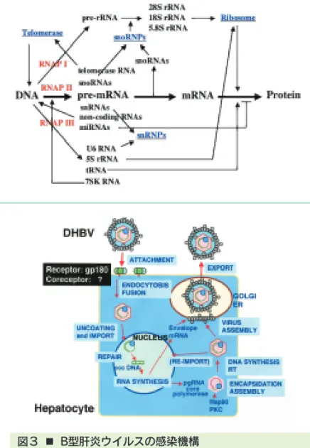
図3 ■ B型肝炎ウイルスの感染機構
B型肝炎ウイルスの感染機構を知るため,ダックB型肝炎ウイルス(DHBV)
をモデルに,DHBV蛋白質と結合する宿主蛋白質を探索している。その結
Division of Molecular Pathology
ゲノム分子病態研究分野
ゲノム分子病態分野は,発ガン剤や抗ガン剤を含む様々 なDNA損傷ストレスにたいする細胞の応答機構,特に細 胞のアポトーシス,とその異常の病態像を分子レベルで解 明し,ガンの病因の解明と予防・診断・治療への応用を図 ろうとしている。具体的には,
1)DNA損傷ストレスに対する細胞応答において司令塔 的な役割を果たしているataxia telangiectasia原因 遺伝子(ATM)ファミリーの機能の解明と,その異常に よって起こる免疫不全・小脳失調・発ガンの病態解析 2)様々なDNA損傷ストレスがどのような因子によって 認識され,ATMファミリーが活性化されるのかの解明 3)DNA損傷ストレスによって活性化されてストレス応 答に重要な役割を果たしていると考えられているc- Ablファミリー, BRCA1, Chk2等ののストレス応答 に果たす役割,特にDNA修復における役割の解明,
等の研究が現在進行している。
DNA damage is a constant threat to eukaryotic cells and defective response to this threat increases genetic instability, ultimately leading to cancer. The goal of our research is to clarify how cells recognize DNA damage and transduce signals to cell cycle checkpoint control, DNA repair and apoptosis machineries.
To achieve this goal, we are currently studying the activation and
functions of ATM (a gene mutated in ataxia telangiectasia) family
in cellular response to DNA damage, using knockout cells. We
are also studying how c-Abl family, BRCA1 and Chk2 are
activated and what roles these factors play in the response.
Division of Molecular Cell Signaling
シグナル伝達研究分野
細胞内シグナル伝達経路の異常な活性化は細胞の癌化を 誘導することが知られている。私たちは,主要な細胞内シ グナル伝達経路の一つであるMAPキナーゼ(MAPK)カス ケードに注目し,
・MAPKカスケードの in vivo における機能の解明
・MAPKカスケードの特異性を保持する分子機構の解明 を目指して研究を進めている。
Abnormal activation of intracellular signaling pathways often leads to tumors. The goal of our project is to elucidate the functions of MAP kinase (MAPK) cascades in vivo, which are major intracellular signaling pathways, and the molecular mechanisms of how the specificity of MAPK cascades is maintained.
図1 ■ MAPKカスケードの in vivo における役割,及び足 場タンパク質によるキナーゼ複合体の形成
MAPKカスケードは細胞の増殖,分化,及びアポトーシスにおいて重要な 役割を担っている。足場タンパク質は,MAPKカスケードの主要な構成成 分であるMAPK,MAPKキナーゼ(MAPKK),及びMAPKKキナーゼ
(MAPKKK)と複合体を形成することによりMAPKカスケードの特異性を保 持すると考えられる。
Fig. 1
■Function of MAPK cascade in vivo, and Formation of Multikinase Complex by Scaffold Protein
Recent studies indicate that MAPK cascades, in which major components are MAPK, MAPK kinase (MAPKK), and MAPKK kinase (MAPKKK), play important roles in cell proliferation, differentiation, and apoptosis. Scaffold proteins could contribute to the specificity determination of MAPK cascades.
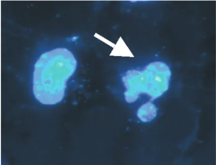
図2 ■ JNK MAPキナーゼによって誘導されたアポトー シス
アポトーシスに特徴的な核の断片化が認められる。
Fig. 2
■Apoptosis induced by a constitutively active JNK MAP kinase
Nuclear fragmentation, a characteristic feature of apoptosis is observed by
Hoechst staining (arrow).
がん病態制御研究部門
Department of Cancer Biomedicine
■ 細胞機能統御研究分野 Division of Molecular Oncology
教授佐藤 博
Professor SATO, Hiroshi
准教授滝野 隆久
Associate Professor TAKINO, Takahisa
准教授遠藤 良夫
Associate Professor ENDO, Yoshio
准教授久野 耕嗣
Associate Professor KUNO, Kouji
■ 分子生体応答研究分野 Division of Molecular Bioregulation
教授向田 直史
Professor MUKAIDA, Naofumi
助教馬場 智久
Assistant Professor BABA, Tomohisa
■ 免疫炎症制御研究分野 Division of Immunology and Molecular Biology
教授須田 貴司
Professor SUDA, Takashi
助教今村 龍
Assistant Professor IMAMURA, Ryu
助教木下 健
Assistant Professor KINOSHITA, Takeshi
Division of Molecular Oncology
細胞機能統御研究分野
目的と研究課題
正常細胞においてがん遺伝子,がん抑制遺伝子の変異が 蓄積した結果としてがんが発生し,悪性化する。悪性化し たがんは組織内へ浸潤し,遠隔臓器へ転移する。我々はが ん化,悪性化そして転移性獲得の過程を分子レベルで明ら かにすると共にその成果を診断・治療法へと応用すること を目指している。
がんの組織内への浸潤には組織・基底膜の破壊を伴う。
我々は1994年にがん転移の鍵を握るタンパク分解酵素を 発見しMT1-MMPと命名した(Nature, 1994)。MT1- MMPは細胞浸潤のみならず増殖・運動などの調節にも重 要な役割を果たしているとのデーターが蓄積しつつある。
Aim and Projects on going
Accumulation of mutation in ocogenes and tumor suppressor genes in normal cells results in malignant tumors. Malignant tumors invade into tissues and finally metastasize to distant organs. The goal of our project is to elucidate the molecular mechanism of tumor metastasis and develop diagnostic and therapeutic application.
Tumor invasion into tissue requires degradation of tissue basement membrane. We discovered a protease which is the key enzyme for tumor metastasis, and named it as MT1-MMP (Nature, 1994). Accumulating evidences indicate that MT1-MMP plays important roles in not only tumor invasion but also regulation of tumor growth and migration.
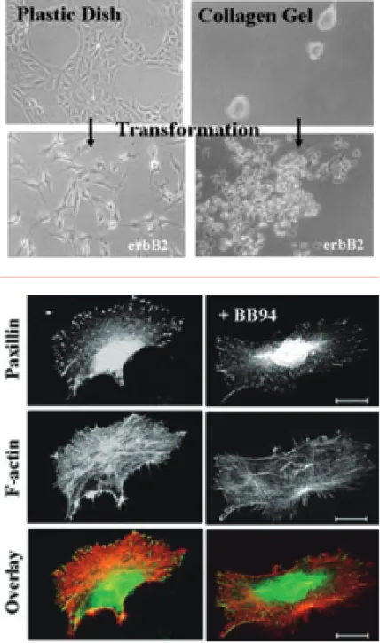
図1 ■ 上皮細胞のがん化に伴うMT1-MMPの発現と浸潤 正常上皮細胞株MDCKはがん遺伝子(erbB2)によりトランスフォームし,
がん細胞の形態を示すとともにMT1-MMPを発現する。コラーゲンゲル内 での培養では正常細胞は凝集して増殖するのに対してMT1-MMPを発現す るがん化した細胞は浸潤性の増殖をする。
Fig. 1
■Induction of MT1-MMP and Invasive Growth by Ocnogenic Transformation of Normal Epithelial Cells Normal epithelial MDCK cells were transformed with oncogne (erbB2), and showed tumor phenotype including MT1-MMP expression. Normal cells grow to form cysts in collagen gel, but transformed cells which express MT1- MMP show invasive growth.
図2 ■ 細胞運動とMT1-MMP
MT1-MMPを発現するHT1080細胞をコラーゲン上で培養するとパキシリ ンで可視化された接着斑とアクチンの走行により細胞運動の状態が見え る。BB94の添加によりMT1-MMPを阻害すると接着班の局在が変化し,
細胞は静止状態となる。
Fig. 2
■Cell Migration and MT1-MMP
HT1080 cells were cultured on collagen, which express MT1-MMP, and were
stained for paxillin to visualize focal adhesion and actin. Addition of MT1-
MMP inhibitor BB94 altered the localization of focal adhesion, and suppressed
cell migration.
Division of Molecular Bioregulation
分子生体応答研究分野
目的,研究課題,最近の主要な成果
組織障害に対して,生体は炎症反応を行い,組織障害を 軽 減 す る よ う に 働 く 。 し か し , 過 剰 な 炎 症 反 応 は , Helicobacter pyloriiの慢性感染で見られるように,組織 障害を進行させ,時にがんを発症させる。
固形がん組織中の線維芽細胞・血管内皮細胞などのいわ ゆるストローマ細胞と白血球は,がん細胞との相互作用を 通して,ケモカインを始めとする炎症性サイトカインを始 めとする生理活性物質を産生する。産生された因子は,が んの進展・転移に重要な役割を果たしている。本研究分野 では,
1)ケモカイン関連遺伝子欠損マウスを用いた解析から,
ケモカインががんの発症・進展に,種々の面から関与 していることを明らかにしつつある。
2)セリン/スレオニン・キナーゼ活性を保有する,原が ん遺伝子Pim-3の発現が,肝臓・膵臓におけるがん病 変で亢進していて,好アポトーシス分子Badの不活性 化を通して,がん細胞のアポトーシスを抑制し,がん の進展に寄与している可能性を明らかにした。このこ とは,Pim-3を分子標的とした新たな抗がん療法の可 能性を示唆している。
Aims, Ongoing Projects, and Recent Achievements
Inflammatory responses occur upon tissue injuries, to reduce tissue damage. If inflammatory responses are exaggerated and prolonged as observed in chronic infection with Helicobacter pylorii, tissue injuries continue, leading sometimes to carcinogenesis.
By interacting with tumor cells, stroma cells and leukocytes can produce various bioactive substances including chemokines.
The produced molecules can affect tumor progression and metastasis. We are elucidating the interaction between tumor cells and stroma cells and obtained the following results recently.
1) By using mice deficient in chemokine-related genes, we are showing that chemokines can contribute to tumor development and progression by exerting various activities.
2) We revealed that the expression of a serine/threonine kinase, Pim-3, was aberrantly enhanced in malignant lesions of liver and pancreas. Moreover, aberrantly expressed Pim-3 can inactivate a proapoptotic molecule, Bad by phosphorylating its serine residue, and eventually prevent apoptosis of tumor cells.
Thus, Pim-3 may be a good molecular target for cancer treatment.
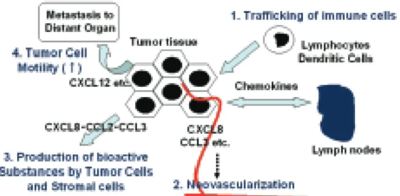
図1 ■ ケモカインのがん病態における役割
ケモカインは,①免疫担当細胞のがん病巣から所属リンパ節への移動過程 の調節,②腫瘍血管新生の誘導,③がん細胞の運動性亢進による転移能の 亢進以外に,がん細胞・ストローマ細胞からの種々の生理活性物質の産生 を誘導し,がん病態の形成に関与している。
Fig. 1
■Roles of chemokines in tumor progression and metastasis processes
Various chemokines contributes to progression and metastasis through the following functions.
a . Regulation of immune cell trafficking b. Induction of neovascularization c . Enhancement of tumor cell motility
d. Induction of production of bioactive substances by tumor and stromal cells
図2 ■ がん病変で発現亢進するセリン/スレオニン・キ ナーゼPim-3
がん病変で発現亢進するPim-3は,好アポトーシス分子,Badをリン酸化 し,不活性化することによって,がん細胞のアポトーシスを抑制している。
Fig. 2
■Aberrant expression of a serine / threonine kinase, Pim-3 in malignant lesions
Pim-3, aberrantly expressed in various malignant lesions, inactivates a pro-
Division of Immunology and Molecular Biology
免疫炎症制御研究分野
私たちの体を構成している一つ一つの細胞は,必要に応 じて自殺する能力を備えている。アポトーシス(枯死)とは,
この様な機能的,能動的細胞死の典型である。放射線など で遺伝子に多くの傷がついた時も,細胞はがん細胞になる 前に自殺することで,がんの出現を防いでいる。
一方,我々は,アポトーシス誘導蛋白Fasリガンドに対 する中和抗体が,肝炎などの炎症性疾患の動物モデルで治 療効果を示すことを明らかにした。さらに慢性肝炎から肝 癌を発症する動物モデルで,この抗体を用いて肝炎の治療 を行うと,肝癌の発症も抑制された。これらの結果から,
我々はFasリガンドのシグナル伝達経路が炎症性疾患の治 療薬,慢性炎症に伴う発がんの予防薬の標的になりうると 考え,研究を進めている。
最近の研究から,Fasリガンドに限らず,アポトーシス と炎症の双方に関わる蛋白が多数発見されている。すなわ ち,アポトーシスと炎症(の一部)の機構は共通の祖先的生 体防御システムから,機能的にも密接に関連しながら進化 したものと考えられる。この様な視点から,我々はアポ トーシスと炎症の双方に関連する蛋白因子を同定し,その 生体防御における役割とがんとの関わりを解明することを 目指している。
Each cell composing our body has an ability to kill itself when necessary. Apoptosis is a common type of such functional and active cell death. To prevent oncogenesis, cells often die by apoptosis when their genes were severely damaged.
On the other hand, we have demonstrated that a neutralizing antibody against Fas ligand (FasL), an apoptosis-inducing protein, has therapeutic potential in animal models of inflammatory diseases including hepatitis. Furthermore, using this antibody, we successfully prevented hepatic cancer development in an animal model of chronic hepatitis. Currently, we are exploring the signal transduction pathway of FasL, which is a potential target of drugs therapeutic for inflammatory diseases and/or preventive for cancer associating with chronic inflammation.
Recent studies have revealed that besides FasL, many other proteins have roles in both apoptosis and inflammation. We are exploring the function of such proteins, which could be important players in biodefense and cancer.
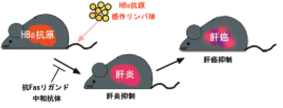
図1 ■ 慢性肝炎モデルにおける抗Fasリガンド抗 体の治療効果
肝臓にB型肝炎ウイルス抗原を発現するマウスに同抗原で免疫し たマウスのリンパ球を移植すると,慢性肝炎を発症し,一年以上 後にほぼ100%肝癌を発症する。このモデルで,抗Fasリガンド 抗体をマウスに投与すると,肝炎ばかりでなく肝癌も抑制された。
Fig. 1
■Therapeutic effect of an anti-FasL antibody in an animal model of chronic hepatitis Transplantation of HBs antigen-primed lymphocytes into transgenic mice expressing HBs antigen in the liver caused chronic hepatitis, and after one year or more, led to hepatic cancer. Administration of an anti-FasL antibody not only ameliorated hepatitis, but also prevented cancer development.
図2 ■ 抗Fasリガンド抗体を投与しなかった場合(左)と投与 した場合(右)の慢性肝炎モデルマウスの肝臓
感作リンパ球移植後15ヶ月。抗体非投与マウスの肝臓(左)は萎縮し,大小の 腫瘤(矢頭および矢印)が出来ている。これらの腫瘤が肝癌であることは組織学 的に確認した。これに対し,抗体投与マウスの肝臓(右)は大きさも組織学的に
がん幹細胞研究センター
Center for Cancer and Stem Cell Research
■ 遺伝子・染色体構築研究分野 Division of Molecular Genetics
教授平尾 敦
Professor HIRAO, Atsushi
助教仲 一仁
Assistant Professor NAKA, Kazuhito
■ 腫瘍遺伝学研究分野 Division of Genetics
教授大島 正伸
Professor OSHIMA, Masanobu
助教大島 浩子
Assistant Professor OSHIMA, Hiroko
■ 幹細胞医学研究分野 Division of Stem Cell Medicine
教授西村 栄美
Professor NISHIMURA, Emi
助教田所 優子
Assistant Professor TADOKORO, Yuko
助教三浦美和子
Assistant Professor MIURA, Miwako
Division of Molecular Genetics
遺伝子・染色体構築研究分野
生体の組織には,それを構成する細胞の源となるいわゆ る 幹細胞 が存在していることが明らかとなってきた。
それぞれの組織において幹細胞は,その居場所, ニッチ に局在し,環境因子からの制御を受けながら前駆細胞へ分 化し,細胞の供給に貢献している。同時に,自分自身を作 り出すいわゆる自己複製という幹細胞に特有の能力を持つ とされている。我々は,幹細胞においてATMが活性酸素 を抑制し,その結果,細胞周期制御因子INK4a/Rb経路を 活性化することによって幹細胞の自己複製を負に制御して いることを見いだし,活性酸素が幹細胞の自己複製に重要 な因子であることを示した。
近年,がん組織の中にも,幹細胞的な役割を持つ がん 幹細胞 の存在が示唆され,がんの治療法開発に向けた新 たな標的細胞として注目されている。我々は,これまでの 組織幹細胞の研究を展開し,がん幹細胞の制御機構の解明 を目指している。
Stem cells are defined as cells that are capable to perpetuate through self-renewal, and develop into mature cells of a particular tissue through differentiation. The hematopoietic stem cells (HSCs) are maintained in an undifferentiated quiescent state in the bone marrow niche. We demonstrated that the self-renewal capacity of HSC is dependent on Atm-mediated inhibition of oxidative stress, indicating that appropriate regulation of reactive oxygen species (ROS) is critical for maintaining stem cell function.
Recent evidence has demonstrated that the ability to proliferate
extensively and form new tumors reside only in a small proportion
of cancer cells. These tumorigenic or tumor-initiating cells,
which are called cancer stem cells, have been identified. We are
trying to understand the molecular mechanisms which regulate
normal tissue stem cells and cancer stem cells.
Division of Genetics
腫瘍遺伝学研究分野
Aim and Projects on going
Accumulating evidence has indicated that both oncogenic mutations and host reactions are responsible for tumorigenesis.
To elucidate the molecular mechanisms underlying tumor-host interactions, we have constructed genetically engineered mice and examined histopathogenesis of gastrointestinal tumors developed in these models. Our special interests are "microenvironment during tumor initiation and promotion" and "microenvironment for maintenance of tumor stem cells".
1) We have demonstrated that COX-2, an inducible enzyme for prostaglandin biosynthesis, plays a key role in intestinal tumor development (Cell, '96). Moreover, we have generated mouse model with increased production of prostaglandin E
2(PGE
2) in the stomach that develops inflammation-associated hyperplastic tumors (EMBO J., 2004) and indicated that activated macrophages and TNF-α pathway are important for tumorigenesis (Cancer Res., 2005). We also found that cooperation of Wnt and PGE
2signaling causes development of gastric adenocarcinoma in mice (Gastroenterol., 2006).
2) Tumor stem cells have not been established in the gastrointestinal cancer yet. Wnt signaling plays an important role in maintenance of normal intestinal stem cells, and its activation causes polyp development (PNAS, '95).
Accordingly, it is conceivable that Wnt pathway is also essential for tumor stem cells in the gastrointestinal cancer tissues. We are studying regulation of Wnt signaling using tumors developed in the transgenic models as well as cancer cell lines.
図1 ■ WntとPGE2の相互作用により発生する胃がん
Wnt1,COX-2,mPGES-1を同時に発現させたトランスジェニックマウス(K19- Wnt1/C2mE)の胃粘膜では,WntシグナルとPGE2経路の相互作用により胃がんの発 生が認められる。
K19-Wnt1/C2mE
mice expressing Wnt1, COX-1, mPGES-1 in gastric mucosa develop adenocarcinoma in glandular stomach, indicating that cooperation of Wnt and PGE
2pathways is responsible for gastric tumorigenesis.
図2 ■ WntとPGE2の相互作用(模式図)
消化管幹細胞の維持に必要なWntシグナルが亢進すると,未分化な上皮細胞が増殖 を始める。その時に周囲の間質細胞でCOX-2依存的に産生されるPGE2が腫瘍形成
目的と研究課題
腫瘍の発生や進行過程には,上皮細胞内の遺伝子変異と 周囲の微小環境による作用が複雑に関与している。これら の相互作用を分子レベルで解明する事を目的として,遺伝 学的な手法により様々なマウスモデルを作製し,病理学的 および生化学的な解析を行なっている。特に,腫瘍発生の 局所での微小環境の役割と,がん幹細胞維持に関わる微小 環境の研究に焦点を当てている。具体的には以下のプロジ ェクトを進めている。
1)消化器がん発生過程では,上皮細胞周囲の間質細胞で プロスタグランジン合成酵素であるCOX-2が重要な 役割を果たしている(Cell, '96)。COX-2下流で産生 されるプロスタグランジンPGE2を胃粘膜で産生させ たマウスモデルでは,炎症をともなう過形成性腫瘍が 発生し(EMBO J., 2004),マクロファージ活性化に 起因したTNF-αが腫瘍発生に重要であることを明ら かにした(Cancer Res., 2005)。現在は,Wntシグナ ルとPGE2の相互作用により胃がんを発生するマウス モデルを作製し,このモデルを使って腫瘍組織間質で の成体反応が胃がん発生に果たす役割について解析し ている(Gastroenterol., 2006)。
2)消化器がん組織でのがん幹細胞の存在についてはほと んど報告がない。Wntシグナルは胃腸管上皮の幹細胞 の維持に重要であり,Wntシグナルの恒常的亢進は消 化管での腫瘍発生の原因となる(PNAS, '95)。したが って,消化器がんにおけるがん幹細胞維持には,Wnt シグナルが関与していると考えられる。そこで,培養 癌細胞や胃がん発生モデルマウスを用いて腫瘍細胞間 のWntシグナル強度の振れについての解析を進めて いる。
Division of Stem Cell Medicine
幹細胞医学研究分野
目的と研究課題
私たちは,幹細胞システムの動作原理の解明とその破綻 によりおこる病態研究を軸として,生体組織の再生,老化,
癌化のメカニズムの解明とその臨床応用に向けて研究を 行っている。
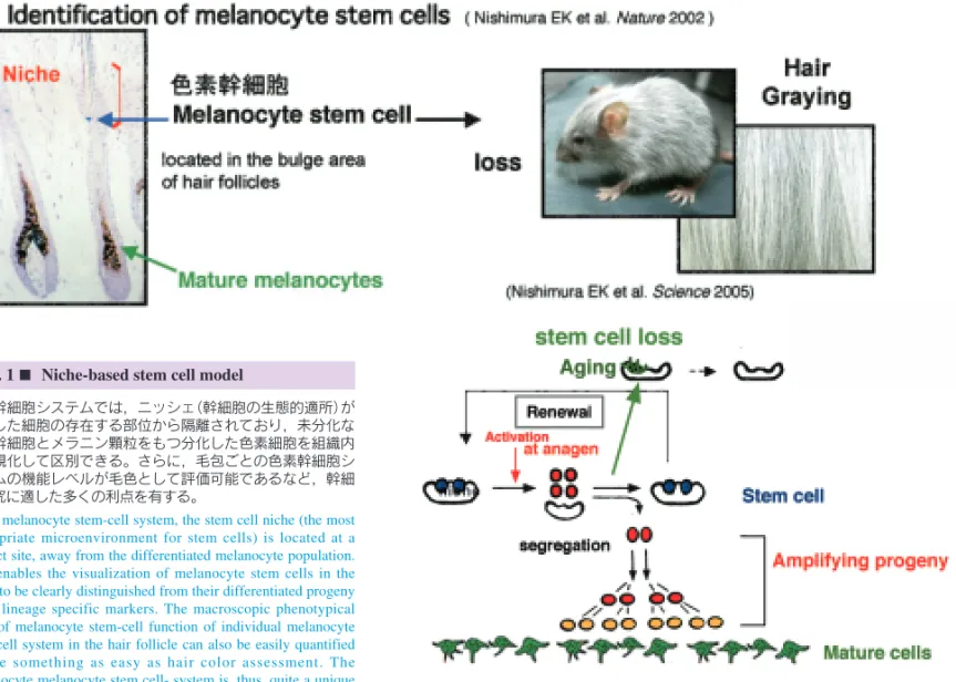
生体を構築する多くの組織や臓器は,幹細胞システムに よりその恒常性が維持されている。私たちは,皮膚毛包の バルジ領域に,色素細胞の供給源となる 色素幹細胞 を 初めて同定し,この細胞が色素細胞を供給し毛に色がつく 仕組み,および幹細胞周囲の微小環境となるニッシェ
(Niche)が幹細胞の運命決定において果たす役割を明らか にした。さらに,加齢によって色素幹細胞の維持が不完全 となり白髪を発症することを世界に先駆けて明らかにし た。いかにして生体が幹細胞を維持しているのかその仕組 みの解明を目指しており,白髪以外の多くの老化形質発現 に至る仕組みの解明にも取り組んでいる。癌も加齢に伴い 顕著に増加する。私たちは,新しく ほくろ およびメラ ノーマ(悪性黒色腫)のモデルを作製し,色素幹細胞および 癌幹細胞に着目して,その発生制御を研究しており,癌幹 細胞を標的とした癌根治療法を開発したいと考えている。
Aim and Projects on going
Recent advances in stem cell biology have revealed that a number of tissues employ stem cell systems to maintain their homeostasis. Our goal is to understand the mechanisms of tissue homeostasis driven by stem cell systems and to understand physiological or pathological conditions resulting from stem-cell system defects. Our studies started from our previous discovery of "melanocyte stem cells", which supply melanocytes (pigment cells) required for hair pigmentation (Nature,2002). We recently demonstrated that the incomplete maintenance of melanocyte stem cells causes hair graying, one of the most obvious signs of ageing (Science, 2005). We are now starting to understand multiple ageing phenotypes in different tissues using a novel mouse model of ageing. Cancer is also one of ageing phenotypes in a broad sense. We are trying to elucidate the mechanisms of cancer development and resistance to cancer treatments by focusing on "cancer stem cells".We believe that our approaches will provide clues to understand the mechanisms of tissue regeneration, ageing and cancer development.
色素幹細胞システムでは,ニッシェ(幹細胞の生態的適所)が 分化した細胞の存在する部位から隔離されており,未分化な 色素幹細胞とメラニン顆粒をもつ分化した色素細胞を組織内 で可視化して区別できる。さらに,毛包ごとの色素幹細胞シ
Fig. 1
■Niche-based stem cell model
分子標的がん医療研究開発センター
Molecular and Celluar Targeting Translational Oncology Center
■ 腫瘍制御研究分野 Division of Translational and Clinical Oncology
教授源 利成
Professor MINAMOTO, Toshinari
准教授川上 和之
Associate Professor KAWAKAMI, Kazuyuki
■ 機能ゲノミクス研究分野 Division of Functional Genomics
教授鈴木 健之
Professor SUZUKI, Takeshi
助教石村 昭彦
Assistant Professor ISHIMURA, Akihiko
■ 腫瘍動態制御研究分野 Division of Tumor Dynamics and Regulation
教授松本 邦夫
Professor MATSUMOTO, Kunio
助教中村 隆弘
Assistant Professor NAKAMURA, Takahiro
■ 腫瘍内科研究分野 Division of Medical Oncology
教授矢野 聖二
Professor YANO, Seiji
准教授渡邊 弘之
Associate Professor WATANABE, Hiroyuki
講師大坪公士郎(病院籍)
Associate Professor OHTSUBO, Koushiro
助教毛利 久継
Assistant Professor MOURI, Hisatsugu
助教土山 智也(病院籍)
Assistant Professor TSUCHIYAMA, Tomoya
■ 腫瘍外科研究分野 Division of Surgical Oncology
教授高橋 豊
Professor TAKAHASHI, Yutaka
講師安本 和生(病院籍)
Associate Professor YASUMOTO, Kazuo
助教山下 要
Assistant Professor YAMASHITA, Kaname
Division of Translational and Clinical Oncology
腫瘍制御研究分野
研究の概要と課題
消化器癌と呼吸器癌を主な対象にして,がんの多様な分 子細胞メカニズムと腫瘍外科学的特性の解明を目指した基 礎・臨床研究を行なう。
1)がん化シグナル制御の分子細胞機構
2)がんのトランスクリプトーム解析と転移の分子診断 3)遺伝薬理学的解析によるオーダーメイド化学療法 4)DNAメチル化をマーカーとしたがん診断・治療開発 5)遺伝性大腸がんの分子診断と診療
当研究所附属分子標的がん医療研究開発センターのコア 分野として,再発や転移性腫瘍を含む難治性がんへの取り 組み,制がんへの応用ならびに探索的がん医療を指向する トランスレーショナル研究に重点をおく。
Research Direction and Activities
The mission of the division centers on laboratory and clinical research to develop the novel strategies and modalities for diagnosis and treatment of cancer in the gastrointestinal and respiratory tracts. Research projects are based on molecular and cellular characteristics of individual tumor types that are relevant to metastatic potential, recurrence and outcome. Our current efforts are focused on:
1) Molecular mechanism underlying oncogenic signaling networks
2) Serial analysis of gene expression (SAGE) in cancer 3) Tailored chemotherapy by pharmacogenetics 4) Translational research of DNA methylation markers
5) Clinical and genetic approach toward molecular management of hereditary non-polyposis colorectal cancer (HNPCC) We are intending to translate as much the achievements created from these studies as possible to the fields responsible for diagnosis and treatment of cancer patients in clinical setting.
図1 ■ グリコーゲン合成酵素キナーゼ3β(GSK3β)は Wntシグナルに依存しない新しいがん標的である
Glycogen synthase kinase 3
β(GSK3
β) supports and promotes tumor cells' survival and proliferation, and protects them from apoptosis in cancers developed in the major digestive organs, the results warrant proposing this kinase as a novel target in cancer treatment (PCT/JP 2006/300160).
図2 ■ RNAトランス因子CRD-BPはmRNAの安定性を修 飾してWnt,NF-κBとc-Myc経路をリンクする
RNA trans-factor CRD-BP is a previously unrecognized transcription target of
β-catenin/Tcf complex, and stabilizes mRNA of
β-TrCP (
β- transducin
repeats-containing protein), NF-
κB and c-Myc. CRD-BP is a novel cancer
target that integrates multiple oncogenic signaling pathways
Division of Functional Genomics
機能ゲノミクス研究分野
がんの発症・悪性化のメカニズムを理解し,がんを克服 するためには,原因となる遺伝子変異の同定が極めて重要 である。しかしヒトのがんでは,多くの変異の蓄積とその ヘテロな形質ゆえに,現在でも原因遺伝子の同定が容易で はない。これに対し,レトロウイルス感染マウスでは,ウ イルスが感染細胞のゲノムに挿入し,挿入部位の遺伝子破 壊や周辺遺伝子の発現修飾によって,がんを誘発するため,
ウイルス挿入部位を解析することで,原因遺伝子を容易に 同定することができる。本研究分野では,ウイルス感染マ ウスを用いて,がん関連遺伝子を網羅的に同定し,その機 能や相互作用の解析を通して,新しいがん分子標的の探索 や先進的ながんの遺伝子診断法の確立を目指している。ま た,重要な標的遺伝子については,逆遺伝学的手法で新た な疾患モデルマウスを作製し,個体レベルでのがんの病態 解析や治療法の開発に活用することも目標にしている。現 在の主な研究テーマは次のとおりである。
1)ゲノム不安定性を示す変異マウスを利用した新しいが ん抑制遺伝子の単離と機能解析
2)ヒストンの翻訳後修飾を制御する酵素群と発がんとの 関係
3)発がんにおけるmicroRNAと特定のシグナル伝達経路 との協調性
A detailed knowledge of the genes and signaling pathways mutated in cancer will be required to develop the novel target- based cancer therapeutics. However, the heterogeneity and complexity of genomic alterations in most human cancers hamper straightforward identification of cancer-causing mutations. We use the retrovirus-infected mice as model systems for identifying new cancer genes efficiently. Retroviruses induce tumors through the activation of proto-oncogenes or inactivation of tumor suppressor genes as a consequence of retroviral integrations into host genome. Thus the viral integration sites provide powerful genetic tags for cancer gene identification. We are exploring the novel molecular targets for cancer treatment based on functional characterization of cancer genes isolated by high-throughput screens using retroviral insertional mutagenesis. Once these genes are identified, we use gene knock-out and transgenic mice to understand how these genes function in tumorigenesis, and to develop new animal models for human cancer. Our current projects are as follows.
1) Identification of novel tumor suppressor genes using retroviral insertional mutagenesis in mice with genomic instability 2) "The histone code" and cancer
3) Analysis of the cooperativity between microRNAs and specific signaling pathways on cancer development
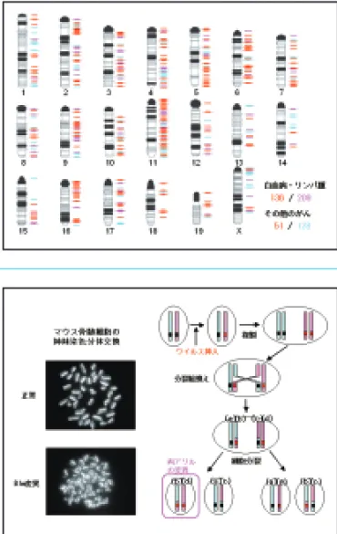
図1 ■ ヒトのがんに関わる遺伝子の多くは,ウイルス挿入変異の 標的となっている
英国サンガー研究所Cancer Gene Censusに登録されているヒトの白血病・リンパ 腫に関係する遺伝子(ピンクで示す),その他のがんに関係する遺伝子(ブルーで示 す)のマウスにおける相同遺伝子のうち,ウイルス挿入の標的となる遺伝子を赤で 示す。
Fig. 1
■Most of human cancer genes are shown to be the targets of retroviral insertional mutagenesis in mice
We mapped the mouse orthologs of human genes involved in leukemia/lymphoma (shown in pink) and in other cancers (shown in blue) registered in Cancer Gene Census Database. Among them, we found many genes (shown in red) identified as the targets of retroviral insertional mutagenesis in mice.
図2 ■ ブルーム症候群モデルマウスを利用したがん抑制遺伝子の 効率的な単離
ブルーム症候群はヒトの劣性遺伝病であり,その患者は,ゲノム不安定性により 様々ながんを発症する。原因遺伝子Blmの変異マウスは,姉妹染色分体交換,分裂
Division of Tumor Dynamics and Regulation
腫瘍動態制御研究分野
細胞増殖因子は極微量ながら細胞の増殖・分化や細胞 死,さらに遊走や形態形成など多彩な細胞機能を調節する タンパク質である。HGF(hepatocyte growth factor: 肝 細胞増殖因子)は,当初,肝細胞の増殖促進を指標として 中村ら(現大阪大)によって発見・単離・クローニングされ た増殖因子であり,Metチロシンキナーゼを受容体として 生理活性を発揮する。HGFは発生過程においては上皮−
間葉相互作用を介した器官の形態形成を担う一方,成体に おいては肝臓をはじめとする様々な組織・臓器の再生を担 っている。また,がん細胞のダイナミックな動態,すなわ ち浸潤や転移に関与している。本研究所における私達の研 究室は2007年4月にスタートし,HGFとMet受容体を中 心として組織再生(肝再生など)の制御機構の研究,がん−
間質相互作用を介したがん悪性化機構とNK4による制がん 研究などを行っている。がんは「never healing wound
(修復しない傷)」とたとえられる。多くのがんはダイナミ ックな組織の修復・再生を担う生物学的な仕組みを巧妙に 使って勢力拡大―成長や浸潤・転移―に至る。私達は生化 学・分子生物学を基盤として,HGF-Met系を分子標的と する制がん研究や再生制御の研究などオリジナルな研究成 果を発信したいと考えている。
Hepatocyte growth factor (HGF) was originally discovered by T. Nakamura (Osaka University at present) as a mitogenic protein for mature hepatocytes. HGF exerts various biological activities, including cell proliferation, migration, and morphogenesis in diverse biological processes. The receptor for HGF is Met tyrosine kinase. HGF plays critical roles in dynamic morphogenesis and regeneration of various tissues such as the liver. In cancer tissues, however, activation of the Met/HGF receptor is tightly associated with malignant behavior of cancer, i.e., invasion and metastasis, thus HGF-Met system is emerging target in the molecular target therapy of cancer. HGF is a stromal- derived mediator in tumor-stromal interaction. Our group started research from the April in 2007. We are studying on 1) regulation of tumor invasion-metastasis by the HGF-Met, 2) therapeutic approach with NK4 (HGF-antagonist and angiogenesis inhibitor), 3) negative (suppressive) mechanisms for the Met receptor function/signal transduction and their biological significance in the termination of tissue regeneration and organ homeostasis, and 4) drug discovery based on structure of HGF-Met complex. HGF- Met system makes a way for dynamic reconstruction of tissues via epithelial-stromal interactions for regeneration of wounded tissues, whereas it is utilized for acquisition of malignancy of cancers. The simile that "cancer is never-healing wound" seems pertinent from the aspect of HGF-Met.
図1 ■ HGF(肝細胞増殖因子)とNK4の生物機能
HGFは697個のアミノ酸からなるタンパク質でMetチロシンキナーゼを受容体と し,発生過程における器官形成や肝臓をはじめとする組織の再生を担う一方,多く のがん細胞の動態(浸潤・転移)を促す。私達はNK4をHGF-アンタゴニストとして 見いだすとともに,血管新生阻害作用をも有する2機能性分子であることを明らか にした。
Fig. 1
■Biological functions of HGF (hepatocyte growth factor) and NK4
HGF is a heterodimer protein composed of 697 amino acids and the receptor for HGF is Met tyrosine kinase. HGF plays key roles in morphogenesis and tissue regeneration such as the liver. In cancer tissues, HGF plays a critical role in tumor invasion and metastasis as a mediator in tumor-stromal interaction. We discovered NK4 as the antagonist against HGF-Met. NK4 has dual functions as HGF-antagonist and angiogenesis inhibitor.
図2 ■ NK4の制がん作用の概略
NK4はHGF-Met系に対するアンタゴニスト(競合的阻害分子)としてがんの浸潤・
転移を阻害すると同時に,腫瘍血管新生阻害を介してがんの成長を抑制する。すな わち,がんの動態を止める"凍結作用"と腫瘍血管新生を阻害することによってがん の成長を抑制する"休眠作用"を発揮することによって,がんの生物学的な悪性形質 を抑制する。
Fig. 2
■Anti-cancer action of NK4
Division of Medical Oncology
腫瘍内科研究分野
転移はがん治療の最大の障壁であり,がん転移の分子機 構解明には臨床を反映した動物モデルが必要不可欠であ る。我々は,ヒト肺癌細胞株を用い再現性の高い転移モデ ルを臓器別(多臓器,脳,肺,骨,がん性胸水)に確立し,
種々の分子標的薬の抗転移効果を検証している。
本研究分野では,独自の転移モデルを用いたトランスレ ーショナルリサーチを展開し,難治性固形癌である肺癌や 胸膜中皮腫,膵癌に対し新規分子標的治療によるがん転移 の克服を目指している。主要な研究課題は以下のごとくで ある。
1)肺癌の造骨性および溶骨性骨転移の分子機構解明 2)肺癌の脳転移の分子機構解明
3)胸膜中皮腫に対する分子標的治療開発 4)膵癌の早期診断および分子標的治療開発
Metastasis is the major obstacle of cancer treatment, and clinically relevant animal models are essential for elucidating the molecular pathogenesis of cancer metastasis. We have established reproducible mouse models representing multi-organ metastasis, brain metastasis, lung metastasis, bone metastasis, or malignant pleural effusion, using human lung cancer cell lines.
We are elucidating anti-metastatic effects of several molecular targeted drugs in these models.
The goal of our translational research with these metastasis models is the establishment of novel molecular targeted therapeutics for overcoming cancer metastasis produced by malignant solid tumors, such as lung cancer, pleural mesothelioma, and pancreatic cancer. Our main research projects are as follows.
1) Molecular pathogenesis of osteoblastic and osteolytic bone metastasis by lung cancer.
2) Molecular pathogenesis of brain metastasis by lung cancer.
3) Molecular targeted therapy for pleural mesothelioma.
4) Early detection and molecular targeted therapy for pancreatic cancer.
図1 ■ VEGF 発現抑制による脳転移抑制
高脳転移株はVEGFを過剰発現しており,antisense (AS)による VEGF抑制で,脳転移が抑制された
Fig. 1
■Inhibition of endogenous expression of VEGF suppresses production of brain metastasis
Highly brain metastastic cells (PC14PE6) over expressed VEGF.
Inhibition of endogenous expression of VEGF (PC14PE6/AS) suppressed vascularization and production of brain metastasis.
図2 ■ VEGF受容体阻害薬による肝転移抑制 VEGF受容体阻害薬(Vandetanib)は腫瘍血管のアポトーシスを 誘導し血管新生および肝転移を抑制した
Fig. 2
■Treatment with inhibitor of VEGF receptor
suppresses production of liver metastasis
Division of Surgical Oncology
腫瘍外科研究分野
癌を治療せしめるには,手術,化学療法,放射線療法な どの様々な治療から適したものを選ぶ必要がある。これを 達成するためには,個々の癌の生物学並びに個々の患者の 免疫応答を知る必要がある。我々の教室では,基礎的研究 として
1)癌転移の分子機構と制御−血管新生とマトリックスメ タロプロテアーゼを中心にアプローチ,
2)癌患者の免疫応答やサイトカインネットワークの解明 とOK-432などの免疫賦活剤による免疫療法の開発,
3)種々の遺伝子の検索などによる,より適切な化学療法,
臨床的研究として,
4)内視鏡機器による低浸襲性手術の確立,
5)生存期間の延長につながる新たな治療戦略(休眠療法)
の開発,
などを主に進めている。
これらの基礎的,臨床的研究から得られた成績をもとに して,個々の患者それぞれに適したオーダーメイドの治療 を行い,癌患者のQOLを保った生存期間の延長こそが 我々の目指すところである。
We should aim for adequate therapies among various ones, such as operation, chemotherapy, radiation, and others to cure cancer patients. To achieve those aims, we should know the cancer biology and host immune system of individual cancer patients. So we are focusing on following research works, 1) To clarify the mechanism of cancer metastasis especially from
angiogenesis and matrix metalloproteinase through the methods of pathology and molecular biology and to inhibit metastasis,
2) To know the immune system and cytokine network of cancer patients and to develop of immune therapy using OK-432 and other immune boosters.
3) To establish proper chemotherapy combined with other therapies through the study of various genes, and clinical works.
4) To establish less invasive operation using laparoscopic instruments.
5) To develop some combination therapies followed by prolonged survival (tumor dormancy therapy) of cancer patients.
In summary, our goal is an order-made therapy for individual cancer patients and prolongation of survival with QOL based on basic and clinical studies.
図2
共 通 施 設
Central Facilities
機器の共通利用及び共同研究等の場として中央研究室を設け ている。その一部を紹介する。
Central facilities were established, so that the equipment is accessible by anyone and collaborative research can be carried out. Below is a list of the facilities.
■ 自動セルソーター (自動細胞解析分取装置) Automated Cell Sorter
正常細胞やがん細胞は様々な多様性を有する細胞集 団です。平成17年度予算によって購入したBD FACS Ariaを用いることにより,このような細胞集団から希 望する細胞群を単離することができます。細胞は細胞 径,顆粒性,表面マーカーやDNA含量などによって 分画することが可能です。本装置を用いるメリットは 清潔な環境下,毎秒10,000細胞以上の速度で細胞を 分取でき,分画した細胞を培養することできることで す。さらに本装置は,遺伝子導入細胞のような解析し たい抗原を発現する細胞数が非常に少ない場合にも用 いることができます。本装置は幹細胞生物学,免疫学,
発生生物学やがん生物学などの様々な実験に利用され ています。
Normal or cancer cells consist of heterogeneous cell- populations. The BD FACS Aria, which was purchased in 2005, allows the isolation of defined cell subset(s) from heterogeneous mixtures. Cells can be sorted according to size, granularity, surface markers and DNA content. An
advantage of using the FACS Aria is that cells can be sorted at rates up to 10,000 cells/second in a sterile environment enabling the recovered cells to be cultured. This is also applicable to transfected cells, where only a small proportion of the cells may express the antigen of interest.
The FACS Aria has been used for a variety of experiments in stem cell biology, immunology, developmental biology, and cancer biology.
■ フルオロイメージャー Fluoro-image Analyzer
既設のバイオイメージ装置に加えて,新たにフルオ ロイメージャーtyphoonを設置したことにより,バイ オイメージ解析室の充実が図られました。アイソトー プを用いることなく目的とした蛋白や核酸の微量検出 と画像処理が行え,コンピュータによる情報解析の領 域が拡大しました。設置以来,広く研究者の共同利用 が行われています。