Sci.Bull.Fac.Educ.,NagasakiUniv.,No.39,pp.61〜71(1988)
Methionine−InducedStomata10pening
ill CO〃J〃J(イわJ〟(、()川川JJ〃六
NobutakaJINNO
BiologlCalLaboratory,Faculty ofEducation,
NagasakiUniversity,Bunkyo−Machi,Nagasaki852
(ReceivedOct.31,1987)
Abstract
Methionine significantly induced the stomatal opening of epidermal strips of Commelina communis among 17 proteinous amino acids tested. The stomatal opening induced by Met was larger in the light than in the dark. Moreover, Met could partially
overcome the Ca2+‑induced stomatal closing.
At the early stage of stomatal opening induced by Met, the opening did not
accompany the accumulation of K+ in the guard cells.
Radioactivity of non‑methy labeled Met was largely incorporated into the protein fraction, while that of methyl labeled Met was larger into the cell wall fraction.
Silver grains of methyl labeled Met distributed mainly at guard and subsidiary cells while that of non‑methyl labeled Met did not.
Key words : Commelina communis, K+, methionine, stomata.
lntroduction
Usingaphytotoxin fusicoccin whichstimulatesprotonsecretionintocellwalls
(Marrさetal.1973,Marrさ1979),JinnoandKuraishi(1982)Showedthatthestomatal openingatearlystagedidnotaccompanytheaccumulationofK+intheguardcellof
Commelina communis,andsuggestedthattheopenlngCOuldbecausedbychangesin physicochemical properties of the cell walls of the guard cells due to fusicoccin
−inducedlowacidity.
Ithasbeenshownthatcellwallsofguardcellisstaineddeeplywithrutheniumred
(Arens1969,Srivastava andSingh1972,SinghandSrivastava1973),indicatingthat
Abbreviation:Met,methionine.
62 Nobutaka JlNNo
the cell walls are filled with pectins. Auxin‑induced incorporation of methyl group of Met into pectin fraction has been repeatedly shown in a coleoptile of oat (Albersheim and Bonner 1959, Cleland 1963a, b, Jansen et al. 1960, Ordin et al. 1955, 1957) and maize (Cleland 1960a, 1963a, b). Moreover, Ordin et al. (1957) and Albersheim and Bor.ner (1959) proposed that the incorporation resulted in the changes in the mechanical properties of cell wall and has a possibility to induce the changes in cell shape. Thus, it seems possible that the changes in the mechanical properties of guard cell wall due to the incorporation of methyl groups into pectins has an effect on the stomatal opening. In the present study, Met was treated to the epidermis of Commelina communis to see the effect on the stomatal opening.
Material and Methods
Plants of Commelina communis were grown in a greenhouse under natural light conditions. In most of the experiment, plants from July to September were used. The fully expanded leaves were cut at their petiole with a razor blade, and kept the leaves for about 15 h in the dark at 30 'C by dipping the petioles in distilled water in order to close the stomata.
The lower epidermis of the dark treated leaves of about 4 x 4 mm were peeled off with a forceps. The epidermis was rinsed in 10 mM MES buffer (pH 6.5) at ・C for 30 min to remove small air bubbles attached to the stomatal cavity. After preincuba‑
tion, the epidermis was immersed in a bathing medium. The medium for opening was consisted of 10 mM MES buffer containing 100 mM KCI with or without amino acids.
The epidermis was kept under ca. 12 klx light provided by a bank of three 20 W fluorescent tube (Matsushita FL 20S・W) .
For the uniformity, epidermis was prepared from one leaf. At least three epider‑
mal strips were used for one experiment and each had at least 10‑15 stomata. Stomata were observed under a microscope, mounting on a slide glass and photographed at 100‑
fold magnification using a calibrated eyepiece at regular time intervals. The stomatal aperture was determined on a film enlarger at designated magnification. At least 10 stomata per strips were measured.
Staining for K+ in the guard cells was carried out by the method of Rogers et al.
(1979) . As shown previously (Jinno and Kuraishi 1982) , for the quantitative estimation of K+ content in the guard cells, the black cobaltous sulfide precipitate formed within the guard cells was photographed and developed on a photographic paper. After making a copy with a transparent film (NP‑DRY, Canon Sale Co., Ltd., Tokyo) , the balck area was measured with a automatic area meter (Model AAM‑7, Hayashi Denkou Co., Tokyo) .
Methionine‑Induced Stomatal Opening in Commelina communis 63
Labeling of the epidermis with radioactive methionine
Fully expanded 20‑30 Ieaves were dipped their stems in distilled water for 15 h at 30 'C in the dark. Lower epidermis peeled at middle portion of the leaves was divided into three portions and then itnmersed in a 20 ml of bathing solution, 10 mM MES buffer (pH 6.5) containing 100 mM KCI and I mM Met, using a covered petri dish (15x2.5 crn). These procedures were carried out under a dim green safely light provided by a 20 W fluorescent tube (Matsushita FL 20S・W) with 3 sheets of green cellophan paper. The area of peeled epidermis was measured with an automatic area met r. Furthermore, L‑[Methyl‑14C]methionine (methyl labeled) and/or L‑L2(n) ‑ 3H] methionine (non‑methyl labeled) were added at concentration of 11.1 pCi and 56.6 pCi, respectively, in order to observe whether only methyl group of methionine was incorporated into the guard cell walls.
After 3 h preincubation under the light of 12 klx, one port・ion of epidermis was harvested and two other were treated for three more hours in either under the light or the dark.
Extraction and determination of pectin, hemicellulose and cellulose from the cell walls of the epidermis
After the labelling treatment, fractionation of wall constituents was carreied out according to the method of Saito and Kasai (1978). The Epidermis was first washed thoroughly with distilled water, and then 3 times with 100 % methanol for 10 min. The epidermis was rehydrated for I h with distilled water, and treated for 18 h with 200 pglml Pronase P (Kaken Kagaku, Tokyo) in 50 mM Na‑phosphate buffer (pH 6.8) containing 5 % ethanol. The filtrate was used as a protein fraction.
After the pronase treatment, the epidermis was washed 3 times with distilled water and was throughly ground by a mortar and pestle with 5 ml distilled water. The homogenate was centrifuged at 1000 Xg for 10 min and the resulting precipitate was suspended twice in distilled water to obtain the crude cell wall fraction.
The pectic fraction was obtained after extracting crude cell wall fraction with hot 50 mM EDTA in 50 mM Na‑phosphate buffer (pH 6.5) at 95‑100 'C for 10 min 3 times.
The precipitate was treated 3 times with 17.5 % NaOH, neutralized with conc. acetic acid and named as a hemicellulose B fraction. The NaOH insoluble fraction was washed with 0.03 N acetic acid and ether (1:1, v/v) . The residue was dried for 15 h at 60 'C and dissolved in 0.2 ml of 72 % sufric acid, and then 2.8 ml distilled water was added. This was named as a cellulose fraction.
The neutral sugars in each fraction except the protein fraction were analyzed according to the phenol‑sulfric acid method (Dubois et al. 1956). The uronic acid concentration was determined by carbazole method (Galambos 1967). Sugar contents in each cell wall fraction were calculated into glucose equivalents.
64 Nobutaka JlNNO
Distribution of silver grains in the epidermis due to the radioactive methionie Epidermal strips taken from one leaf divided into two portions. One was immer‑
sed in 7 ml of 10 mM MES buffer (pH 6.5) containing 100 mM KCI and 16.9 pCi L‑[2 (n)‑3H] methionine and the other in 7 ml of the same buffer containing 100 mM KCl and 3.4 pCi L‑ [methyl‑14C] methionine, and then they were kept for 3 h at 30 'C under the light of 12 klx. After the treatment, the epidermis washed fully were attached to a slide glass by means of albumin/glycerol adhesive, dried and then coated with a photographic liquid emulsin (Sakura NR‑M2, Konishiroku Photo Ind. Co., Ltd.
Tokyo) under a dim red safety lamp. The preparation was kept for 7 weeks in a refrigerator at ca. 2 'C in the dark and followed by the development in Kpnidol (Konishiroku Photo Ind. Co., Ltd. Tokyo) . After washing, the epidermis was covered with a cover glass using a glycerol, and then silver were observed.
‑3H] methionine were purchased Japan Ltd..
Determinations of radioactivity incorporated into the epidermis cells
Using 10 ml of scintillator (ACS‑II, Amersham, England) , radioactivity in protein, pectin, hemicellulose B and cellulose fraction were determined with a liquid scintilla‑
tion counter (Model LSC‑900. Aroka, Co., Ltd., Tokyo).
L‑ [methyl‑ 1 4C] methionine
(2.18 GBq/mmoD and L‑[2(n) Table I Effects of 17 proteinous amino acids on the stomatal opening
(118 GBq/mmol)
from Amersham Stomatal aperture (pm) Exp. 1 Exp. 2
Results
All proteinous amino acids at the concentration of 3 mM were dissolved in 10 mM MES buffer with 100 mM KCI and applied to the epidermal strips of Commelina communis leaf. Only Met had a significant effect on the stomatal opening (Table 1) .
Fig. I shows the time course
experiment of Met‑induced.
stomatal opening in the light and dark. A concentration at 3 mM Met induced marked stimulation of stomatal opening only I h after
No amino acid Gly
Ala Val Leu
Ile
Ser Thr Asp Glu Lys A rg
Met Phe Tyr Trp His Pro
6.2 0.5 5.2i0.3
5.6:!:0.5
5.8i0.3 7.2+0.7 5.2i0.3 5.6 0.3 6.0i0.3 6.6+0.4 6.1+0.4 5.9 0.5 6.3 0.5 9.1+0.4*
5.5 0.3 6.3+0.4 5.2+0.4 6.2 0.5 6.0 0.4
6.3 ! 0.3
7.2+0.4 6.2 0.6 6.2+0.6 7.0+0.4 6.5+0.6 5.8+0.7 6.6+0.5 5.6 0.4 6.0+0.6 7.0 0.5 5.9 0.5
9.2 ! 0.6*
6.3 0.5 5.3+0.4 7.0 0.4 8.5+0.7*
5.9i0.5 Amino acid at 3 mM was applied to three epidermal strips of Commelina communis leaf at 30 'C under the light of 12 klx. Data given are the means SE after 4 h incubation.
* Significant stimulation of the stomatal opening from non‑amino acid treatment at 5 % Ievel.
Methionine‑Induced Stomatal Opening in Commelina communis 65
14
12
E
:¥ , o,
8
Jg
6
9
4
cf)
10
2
o
Light (+ Met )
Light Dark ( +
Dark
Met )
3 Ti me( h )
Flg I Effect of Met on the stomatal opening. Met at 3 mM was applied to four epidermal strips of Commelina communis leaf at 30 'C under the dark of the light (12 klx) . The size of strips was ca. 4 x 4 mm.
the application and retained its effect for another 3 h. Although Met‑induced stomatal opening under the light was larger than that under the dark, the time course was similar to that in the dark.
Table 2 Stomatal aperture and K+
Commelina communis
content in guard cells on Met treated epidermis strips of
Methionine ( mM )
After 2 h
K+ content in Stomatal
aptrture (pm) guard cells (pmm2)
After 4 h
K+ content in Stomatal
aperture (pm) guard cells (pmm2)
o 1 3 9
11.7+0.7
(21)
12.1+0.7
(21)
13.5 0.8*
(23) 14 . I + o . 7*
(22)
279 .
323 .
308 .
313 .
7i23.8
(35)
li23.8
(35)
9 21.8
(34)
0 25.0
(35)
18.8 0.7
(24)
20.5i0.5*
(24)
21.5 0.5*
(23)
22.0 0.4*
(26)
458 . 7 25 . 9 (35) 635 . Oi 31 . 2*
(47)
655.4 27.4*
(41) 661 . O i 30 . 6*
(47) Data given are the means SE. Values in parenthesis indicate the number of cells measured.
* Significant stimulation from non‑Met treated strips at 5 % Ievel.
66 Nobutaka JINNO
Effect of Met concentration and K+ content in the guard cells were shown in Table 2. After 2 h incubation, I mM Met scarecely had a siguificant effect on the stomatal opening but 3 and 9 mM did. Furthermore, Met‑induced stomatal opening did not accompany K+ accumulation in the guard cells after 2 h treatment, but significantly accumulated K+ after 4 h treatment. Table 3 shows that the treatment with I mM Ca2+ and 3 mM significantly suppressed the Ca2+̲enhanced stomatal closing. Further‑
more, stomatal aperture was correlated to K+ content in the guard cells.
Ethionine, an analog of Met, was treated to the epidermis and the opening was also enhanced after 2 and 4 h incubation (Table 4) , although the ethionine effect was a less extent when compared with Met. Ethionine‑induced stomatal opening did not show significant increase in K+ content in the guard cells.
Incorporation of the radioactivity due to non‑methyl and methyl labeled Met into
Table 3 Effects of Met on the Ca2+̲enhanced stomatal closing Treatment Stomatal
aperture (pm)
K+ content in guard cells (pmm2) No Ca2+
1 mM Ca2+
1 mM Ca2+ +3 mM Met
14.8+0.
(21)
8.4iO.
(21)
11.6+0.
(18) 8 7 9*
257 .
112 .
153 .
9i29.9
(15)
3 12.9
(16) 7 + 16 . 8*
(12) Epidermal strips were pretreated under the light 12 klx at 30 'C in MES buffer (pH 6.5) containing 100 mM KCI and 3 mM Met for 2 h and then treated I more hour in the bathing medium with or without Ca2+. Data given are the means SE. Values in parenthesis indicate the number of cells measured.
* Significantly different from the I mM Ca2+ treatment at 5 % level.
Table 4 Stomatal aperture and K+
ethionine treatment
content in guard cells on
Ethionine (mM) aperture (pm) Stomatal
K+ content in guard cells (pmm')
o 1
3 9
26.gi0.4
(20)
28.0i0.3*
(20)
29.6i0.3*
(17)
28.6i0.3*
(21)
268 . I 25 . (11) 253 . 2 29 .
(13) 276 . 9 35 .
(9)
261 . 1 19 . (17)
7
9
7
o
Epidermal strips were treated for 4 h under the light of 12 klx at 30 'C. Data given are the means :!: SE. Values in parenthesis indicate the number of cells measured.
* Significantly different from the no ethionine at 5 % Ievel.
lvlethionine‑Induced Stomatal Opening in Commelina communis 67
Table 5 Distribution of non‑methyl and methyl Met into epidermal
cells
Radioactivity"
Non‑methyl Methyl labeled labeled
Ratiob
dpm/cm2
Protein fraction Cell wall fraction
31 . 9
2.8
20 . 1
8.9
O . 63 3 . 18
Epidermai strips were treated for 3 h under the light of 12 klx at 30 ' C. Each vaiue is the average of two experiments.
* Shown as the value per area of epidermal strips.
Methy'l labe]ed to non‑methyl kabeled.
cell wall fraction were undertaken in order to see the transfer of methyl group from Met into cell wall fraction (Table 5). Although radioactivity due to non‑methyl labeled M:et was larger than that due to methyl labeled at the protein fraction, the radioactivity at the cell wall faction was reversed. The radioactivity ratio of methyl labeled Met to non‑methyl labeled Met was about 3 times larger at the cell fraction than at the protein fraction.
Photographlc liqiud emulsion on the epidermis were developed after 7 weeks of exposure to radioisotopes. The silver grains due to methyl labeled Met mainly distributed at guard cells and inner lateral subsidiary cells (Fig. 2a). On the other hand, the silver grains due to non‑methyl labeled Met was scarecely distributed at guard cells (Fig. 2b).
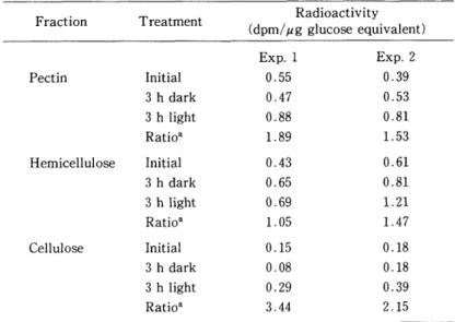
Table 6 shows the distribution of radioactivity incorporated from methyl labeled Met into pectin, hemicellulose and cellulose fraction under light and dark condition and revealed that much radioactivity was incorporated into both pectin and hemicellulose fraction. And furthermore, Iight which induces stomatal opening stimulated the
j i
a
Fig.
lr :;i't :?" ::::: :: :: +::::i:::::
: : ' !'; : :: ::: ;' :: ;' : "s :i:i‑F;'; : ::'i: s:
I'
:; ::
i: i:i:;:i:1:: S: :i ;sr't' ': i:: :F!'iii:::: :: : ::::̲ ‑ s':1*r'!:'r :( i;ii ::::
:;1:' ;::* +
' : :;:
:It'; tt'!::i::t':
: ':'i'If ::
s;
; 't f s$xl:::
t :t 'i : i
: i : :' i : :: t :i ii::::: l :::':I ;': ; ; ' ' ti F ; t'; i ' :: :: ::';"i:::' : : :': t'::' :s 's::: 1 ' :s':' $! ;ii̲ ;;{ :i i 01:: :?: i ::'s: i ' :":sl'i:' ;s:: 's ' + t';:: :i s' ' 't'
i :: :: : 'i:i : : :':' t ::: ':i" ' :Fi: :; ;'; t"+
! "' : : t; :: ' ' ' :: ;: i̲' 't;:1 t "^ : ' ;: ::i ;;;i i : : : : i;; *'
' b s' : ' ' ;; It
2 Incorporation of radioactive Met into guard and subsidiary cells of Commelina communi' " EPidermal striPs were incubated for 3 h at 30 eC under the light of 12 klx' 2i' Radioactivit}' due t( ) meth"I Iabeled Met' b' Radioactivit}T due to non‑
methvl labelecl 1et"r' inner lateral subsidiar)r cell; **1 guard cel]'
68 Nobutaka JlNNO
Table 6 Distribution of methyl labeled Met in the cell wall fraction
Fraction Treatment Radioactivity
(dpm/pg glucose equivalent)
Pectin Initial
3 h dark 3 h light Ratio' Hemicellulose Initial
3 h dark 3 h light Ratio'
Cellulose Initial
3 h dark 3 h light Ratio*
Exp. l
O . 55 O . 47 O . 88 1 . 89
O . 43 O . 65 O . 69 1 . 05
O . 15 O . 08 O . 29 3 . 44
Exp. 2
O . 39 O . 53
0.81
1 . 53
O . 61
0.81 1.21
1 . 47
O . 18 O . 18 O . 39 2 . 15
Epidermal strips were pretreated for 3 h under the light (initiaD and then incubated for another 3 h in either the dark or the light at 30 'C.
* Radioactivity in 3 h light to that in 3 h dark treatment.
incorporation of radioactivity into both pectin and cellulose fraction.
Discussion
Many workers have claimed that the accumulation of K+ into the guard cells is responsible for the stomatal opening in several plant species (Imamura 1943, Yama‑
shita 1952, Fujino 1967, Fischer 1968, Humble and Hisiao 1970, Thomas 1970, Willmer and Mansfield 1970). However, recent studies showed evidences on the stomatal opening without accumulation of K+ into the guard cells. MacRobbie and Lettau (1980a, b) and MacRobbie (1981) reported that the change in K+ content of the guard cells of Commelina communis was too small to account for their osmotic change at the early stage of opening. Jinno and Kuraishi (1982) also showed that fusicoccin‑induced stomatal opening of Commelina communis did not accompany the accumulation of K+
at early stage and emphasized the importance of the acidity in the cell walls of the guard cells.
Ca2+ suppressed stomatal opening in epidermal strips of Vicia faba (Willmer and Mansfield 1969, Fischer 1972) and of Commelina communis (Fujino 1967). Raschke and Humble (1973) presumed that Ca2+ inhibition of stomatal movement ins mediated by the inhibition of proton liberation. However, Raschke (1979) Iater claimed that the Ca2+̲induced stomatal closure should be caused by more unknown direct mechanism than by the inhibition of proton liberation.
Ca2+ has been known form the cross bridge with carboxyl groups of pectic acid, increasing in the stiffeness of the cell wall or inhibiting the extention of the cell wall
Methionine‑Induced Stomatal Opening in Commetina communis 69
(Burstr6m 1968, Preston 1979, Baydoum and Brett 1984, Demarty et al. 1984) . Based on the observation that the guard c,ell walls are filled with pectins (Arens 1968, Srivastava and Singh 1972, Singh and Srivastava 1973) , Ca2+ applied to stomata may cause the calcium bridge of pectin and result in the stomatal closure due to the decrease in the elasticity of extensibility of the guard cell wall.
In the present investigation, Met was applied to the epidermis of Commelina communis to see its effect on the stomatal opening from the view of the methylation of pectin of guard cell walls.
Auxin‑induced cell elongation was suggested that auxin complex calcium, thereby breaking the calcium bridge and rendering the wall more elastic (Bennent‑Clarke
1956) .
By the application of Met to epidermis, methylation of calcium bridge in the guard cell walls may be induced, resulting in the decrease of the amount of calcium bridge.
The methylation may have a same effect on the guard cell walls as auxin, increasing in the cell wall elasticity.
The increase in the elasticity of guard cell walls may induce the expansion of guard cell. Thus, Met‑induced stomatal opening may caused by the changes in the mechanical properties of guard cell walls.
Application of ethionine to the stomata significantly enhanced the stomatal opening although at a less extent to that of Met. Cleland (1960a, 1963b) showed ethionine inhibited the transfer of methyl groups from Met into pectin in oat coleoptiles. The experiment indicates that the transfer of ethyl groups from ethionine into pectins. On the guard cell wall of epidermis of Commelina communis, ethyl groups of ethionine also could be incorporated into the pectins, resulting in the partial decrease in cell wall stiffeness.
Thus, present results indicate that the stomatal opening can be induced by the elastic increase of the guard cell wall by Met treatment and does not always require the accumulation of K+
References
Albersheim, P. and J. Bonner (1959) Metabolism and hormonal control of pectic substances. J. Biol.
Chem. 234: 3105‑3108.
Arens, T. (1968) Radialstrukuren in den Stomata von Ouratea spectabilis (Mart.) Engl. Protoplasma 39: 403‑411.
Baydoum, E. A.‑H. and C. T. Brett (1984) The effect of pH on the binding of calcium to pea epicotyl cell walls and its implications for the control of cell extention. J. Exp. Bot. 35: 1820‑1831.
Bennet‑Clarke, J. A. (1956) In "The Chemistry and Mode of Action of Plant Growth Substances" (R.
L. Wain and F. Wightman, eds.), pp. 284‑291. Butterworth, London.
Burstrdm, H. G. (1968) Calcium and plant growth. Biol. Rev. 43: 287‑316.
70 Nobutaka JINNO
Cleland, R. (1960a) Auxin‑induced methylation in maize. Nature 185: 44.
Cleland, R. (1960b) Ethionine and auxin‑action in Avena cleoptile. Plant Physiol. 35: 585‑588.
Cleland, R. (1963a) Independence of effects of auxin on cell wall methylation and elongation. Plant Physiol. 38: 12‑28.
Cleland, R. (1963a) The occurrence of auxin‑induced pectin methylation in plant tissues. Plant Physiol. 38: 738‑740.
Demarty, M., C. Morvan and M. Thellier (1984) Calcium and the cell wall. Plant. Cell Environ. 7: 441
‑448.
Dubois, M., K. A. Gilles, J. K. Hamilton, P. A. Rebers and F. Smith (1956) Calorimetric method of determination of sugars and related substances. Anal. Chem. 28: 350‑356.
Fischer, R. A. (1968) Stomatal opening: role of potassium uptake by guard cells. Science 160: 784‑785.
Fischer, R. A. (1972) Aspects of potassium accumulation by stomata of Vicia faba. Aust. J. Biol. Sci.
25: 1107‑1123.
Fujino, M. (1967) Role of adenosinetriphosphate and adenosinetriphosphatase in stomatal movement.
Sci. Bull. Fac. Educ.. Nagasaki Univ. 18: 1‑47.
Galambos, J. T. (1967) The reaction of carbazole with carbohydrates. 1. Effects of borate and sulfamate on the carbazole color of sugars. Anal. Biochem. 19: I19‑132.
Humble, G. D. and T. C. Hisiao (1970) Light‑dependent influx and efflux of potassium of guard cells during stomatal opening and closing. Plant Physiol. 48: 447‑453.
Imamura, S. (1943) Untersuchungen uber den Mechanismus der Turgorschwankung der Spaltdffnun
‑gs.schliesszellen. Jap. J. Bot. 12: 251‑347.
Jansen, E., R. Jang, P. Albersheim and J. Bonner (1960) Pectic metabolism of growing cell walls.
Plant Physiol. 35: 87‑97.
Jinno, N. and S. Kuraishi (1982) Acid‑induced stomatal opening in Commellina communis and Vicia faba. Plant Cell Physiol. 23: 1169‑1174.
MacRobbie, E. A. C. and J. Lettau (1980a) Ion content and aperture in "isolated" guard cells of Commelina communis L. J. Memb. Biol. 53: 199‑205.
MacRobbie, E. A. C. and J. Lettau (1980b) Potassium content and aperture in "intact" stomatal and epidermal cells of Commelina communis L. J. Biol. 56: 240‑256.
Marr , E. (1979) Fusicoccin: a tool in plant physiology. Annu. Rev. Plant. Physiol. 30: 237‑288.
Marr , E., P. Lado, E. R. Caldogno and R. Colombo (1973) Correlation between cell enlargement in pea internode segments and decrease in the pH of the medium of incubation. I. Effects of fusicoccin, natural and synthetic auxin and mannitol. Plant Sci. Lett. 1: 179‑184.
Ordin, L., R. Cleland and J. Bonner (1955) Influence of auxin on cell wall metabolism. Proc. Natl.
Acad. Sci., USA 41: 1023‑1029.
Ordin, L., R. Cleland and J. Bonner (1957) Methyl esterification of cell wall constituents under the influence of auxin. Plant Physiol. 32: 216‑220.
Preston, R. D. (1979) Polysaccharide conformation and cell wall function. Annu. Rev. Plant Physiol.
30: 55‑78.
Raschke, K. (1979) Movements of stomata. In physiology of movements. Edited by W. Haupt and M.
Methionine‑Induced Stomatal Opening in Commelina communis 71
E. Feinleib. pp. 383‑441. Springer‑Verlag Heidelberg New York.
Raschke, K. and G. D. Humble (1973) No uptake of anions required by opening stomata of Vicia faba:
guard cells release hydrogen ions. Planta 115: 47‑57.
Rogers, C. A., R. D. Powell and P. J. H. Sharpe (1'979) Relationship of temperature to stomatal aperture and potassium accumulation in guard cells of Vicia faba. Plant Physiol. 63: 388‑391.
Saito, K. and Z. Kasai (1978) Convertion of labeled substrates to sugars, cell wall polysaccharides, and tartatic acid in grape berries. Plant Physiol. 62: 215‑219.
Singh, A. P. and L. M. Srivastava (1973) The fine structure of pea stomata. Protoplasma 76: 61‑82.
Srivastava, L. M. and A. P. Singh (1972) Stomatal structure in corn leaves. J. Ultrastruct. Res. 39: 345
‑363.
Thomas, D. A. (1970) The regulation of stomatal aperture in tobacco leaf epidermal strips. I. The effects of ions. Aust. J. Biol. Sci. 23 : 961‑979.
Willmer, C. M. and T. A. Mansfield (1969) A critical examination of the use of detached epidermis in studies of stomatal physiology. New. Phytol. 68: 363‑375.
Willmer. C. M. and T. A. Mansfield (1970) Further observations of cation‑stimulated stomatal opening in isolated epidermis. New. Phytol. 69: 639‑645.
Yamashita, T. (1952) Influence of potassium supply upon various properties and movement of the guard cell. Sielboldia Acta Biol. 1: 51‑70.