1
Clinicopathological and genetic characteristics associated with brain metastases from lung adenocarcinoma and utility as prognostic factors
Hiromasa Kobayashi1, 2, Makoto Hamasaki1, Takashi Morishita2, Masayo Yoshimura1, Masani Nonaka2, Hiroshi Abe2, Tooru Inoue2, Kazuki Nabeshima1
1. Department of Pathology, Faculty of Medicine, Fukuoka University, Fukuoka, Japan 2. Department of Neurosurgery, Faculty of Medicine, Fukuoka University, Fukuoka, Japan
Corresponding Author:
Makoto Hamasaki, MD., Ph.D.
Department of Pathology, Fukuoka University Faculty of Medicine Nanakuma 7-45-1, Jonan ward, Fukuoka, Japan.
81-92-801-1011
81-92-865-9901 (FAX)
E-mail: [email protected]
Abbreviations
ALK, anaplastic lymphoma kinase; BM, brain metastases; ECM, extracranial metastasis; EGFR, epidermal growth factor receptor; GPA, graded prognostic assessment; H&E, hematoxylin and eosin; KPS, Karnofsky performance score; NSCLC,
2
non-small cell lung cancer; OS, overall survival; RPA, recursive partitioning analysis;
TKI, tyrosine kinase inhibitor; TTF-1, Thyroid transcription factor – 1; WBRT, whole brain radiation therapy; WHO, World Health Organization.
Keywords: brain metastases, lung adenocarcinoma, prognostic factor, EGFR mutation, histopathological subtype
Running Title: KOBAYASHI et al: PROGNOSTIC FACTORS IN BRAIN METASTASES FROM LUNG ADENOCARCINOMA
3
Abstract
Brain metastases (BM) are common in patients with lung adenocarcinoma, and represent a significant cause of morbidity in the disease. A more comprehensive understanding of the clinicopathological characteristics that serve as prognostic factors for survival in patients with BM from lung adenocarcinoma would help inform
treatment strategies for this patient population. In the present study, we evaluated clinicopathological factors, including EGFR mutation status, in 59 patients who were diagnosed with BM from lung adenocarcinoma and underwent BM resection between 1985 and 2014 in our institution. The most frequent subtype of BM from lung
adenocarcinoma were solid adenocarcinoma (57.6%), followed by papillary adenocarcinoma (22.0%) and acinar adenocarcinoma (18.6%). Fourteen patients (23.7%) had EGFR mutations, which were significantly associated with female gender (9/14, 64.3%), never smoker status (8/14, 57.1%), BM in the frontal lobes (9/14, 64.3%), and papillary adenocarcinoma (5/14, 35.7%). Statistical analysis revealed a significant association between clinical factors, particularly never smoker status and BM in the frontal lobes, and more favorable disease prognosis. Our results suggest histological and genetic analysis of tissue from BM provides information useful for managing treatment of patients with resectable BM arising from lung adenocarcinoma.
4
Introduction
The frequency of brain metastases (BM) from non-small cell lung cancer (NSCLC) exceeds 20% at the time of diagnosis of the primary tumor, and a significant proportion of patients develop BM over the course of disease (1, 2). In recent years, the incidence of BM in patients with NSCLC has further increased, due to improved survival resulting from the molecular-targeting therapy and improvements in diagnostic neuroimaging modalities such as magnetic resonance imaging (MRI) and positron emission tomography (PET) (3). Adenocarcinoma is the most common subtype of NSCLC, occurring in approximately 40% of all patients, and is characterized by rapid progression, early distant metastasis, and a significantly higher incidence of BM compared with other subtypes of NSCLC (4–6). BM are considered to be a poor prognostic factor in NSCLC, and are associated with median overall survival (OS) times of just 4–7 weeks in patients that are not treated (6-8). In patients with BM, both recursive partitioning analysis (RPA) and graded prognostic assessment (GPA) score are still the most widely accepted models for evaluation of disease prognosis (6, 9).
In lung adenocarcinoma, mutations in the epidermal growth factor receptor (EGFR) gene have recently been identified as a driver mutation promoting tumor growth. Therapies targeting mutant EGFR protein are associated with relatively high overall response rates, and have been established as standard-of-care treatments (10-12).
Furthermore, EGFR-targeted therapies have shown efficacy in treatment of BM from NSCLC (13–15).
Histological and genetic evaluation is essential in identifying patients with
5
NSCLC that may benefit from EGFR-targeted therapies. In cases in which tissue sampling from the primary tumor is difficult, tissue samples from BM lesions may provide a useful alternative for evaluation. In most previous reports, histological analysis and genetic evaluation in patients with NSCLC have been conducted using tissue from the primary tumor only. There are a few reports describing histopathological and genetic evaluation using tissue samples from both BM and the primary lung adenocarcinoma (16, 17). Given evidence that the presence of EGFR mutations is a positive prognostic factor in patients with BM from lung adenocarcinoma, it is feasible that analysis of tissue from BM could be used to guide more aggressive and effective treatments for patients with EGFR mutation-positive NSCLC.
In the present study, we retrospectively evaluated clinical features and histopathological subtypes of BM from lung adenocarcinoma in a patient population from a single institution. We included in this analysis evaluation of EGFR mutations and association of these factors with disease prognosis. Our results suggest analysis of tissue from BM may inform treatment of patients with metastatic brain lesions arising from lung adenocarcinoma.
Materials and methods Study design and patients
We reviewed 232 BM in the histopathological file of the Department of Pathology that had been surgically resected at the Department of Neurosurgery, Fukuoka University Hospital, between January 1985 and December 2014. Tumor
6
resection was performed to improve neurological symptoms and quality of life, but did aim to prolong survival. Patients in which BM were resected included those in which:
(1) a single large (> 3 cm) or symptomatic tumor was present; (2) the primary tumor was well-controlled; (3) life expectancy exceeded 3 months; and (4) informed consent of the patient or family was obtained. We retrospectively analyzed 68 patients with BM from lung adenocarcinoma. We excluded from analysis data from 8 patients lacking clinical data and specimens, and from 1 patient in which PCR amplification of EGFR failed. Data from a total of 59 patients were included in this study. The study protocol was reviewed and approved by the Ethics Committee of the Fukuoka University Hospital (No. 15-5-14).
Pathologic evaluation
Surgically-resected specimens were fixed in 10% formalin and embedded in paraffin blocks. Tissue sections were cut at a thickness of 4 µm, and stained with hematoxylin and eosin (H&E) for histopathological examination. Histological subtyping was performed according to the 2015 World Health Organization (WHO) criteria for classification of lung tumors (18). Clinicopathological parameters considered in this study included patient age, gender, location of BM, histological subtype of BM, smoking status, Karnofsky performance score (KPS), number of BM, presence of extracranial metastasis (ECM), GPA, timing of diagnosis, tyrosine kinase inhibitor (TKI) treatment before diagnosis of BM, presence of RAS mutations or anaplastic lymphoma kinase (ALK) rearrangements, and re-operation.
7
Analysis of EGFR and KRAS mutations and ALK rearrangement
EGFR and RAS gene mutations were analyzed in paraffin-embedded tissue
sections from BM only. We detected EGFR mutations (including G719X in exon 18, exon 19 deletion, T790M in exon 20, and L858R and L861Q in exon 21) and RAS mutations (G12C and G12A in exon 2 and Q61H in exon 3) in matching formalin-fixed paraffin-embedded tissue samples using cycleave PCR (SRL Inc., Tokyo, Japan). ALK rearrangement was analyzed by immunohistochemistry.
Statistical analysis
To identify clinical features associated with EGFR mutations, categorical variables were compared using Wilcoxon signed-rank test and Fisher’s exact test. OS was defined as time from the date of BM diagnosis to the date of death or the last follow-up. The Kaplan-Meier method was used for survival curve analysis, and differences between survival curves were analyzed using the log-rank test. Multivariate analysis with the Cox proportional hazard model was used to determine independent prognostic factors. P-values of less than 0.05 were considered statistically significant.
Statistical analysis was performed with SPSS software for Windows, version 21.0.
Results
Patient characteristics
Patient characteristics are summarized in Table I. A total of 59 patients with
8
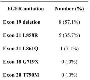
BM from lung adenocarcinoma were included in this study. The median patient age was 61.3 ± 12.2 years old (range 38 to 87 years). Forty-five patients were male (76.3%) and fourteen patients were female (23.7%). A total of 14 patients (23.7%) had EGFR mutations (Table II), of which deletion of exon 19 was the most common genetic alteration (found in 8 patients, 57.1%), followed by L858R mutation in exon 21 (5 patients, 35.7%). Only one patient (7.1%) had a L861Q mutation in exon 21. EGFR mutations occurred significantly more often in female vs male patients (64.3% vs 35.7%, p = 0.0001). EGFR mutations also occurred significantly more frequently in never smokers vs current or past smokers (57.1% vs 42.9%, p = 0.04). BM within the frontal lobe were significantly more common in patients with EGFR mutations (64.3% vs 21.4% [BM in the cerebellum] vs 7.1% [BM in the posterior lobe], p = 0.03). The rate of re-operation occurred more often in patients with wild-type vs mutated EGFR (37.8%
vs 7.1%, p = 0.04). The 2 groups did not differ in GPA measures of age, KPS, number of BM, presence of ECMs, timing of diagnosis, and frequency of RAS mutation and ALK rearrangements.
All patients received craniotomy followed by total resection of the BM. In thirty-two patients (54.2%), ECMs were detected at the time of BM diagnosis. The sites at which ECMs occurred in the patients with EGFR mutation were similar to those in which ECMs occurred in patients with WT. In patients with EGFR mutations, 6 patients had ECMs (one patient had multiple lesions). Of these ECMs, 6 occurred in the lymph nodes (100%), one occurred in bone (16.7%), and one occurred in liver (16.7%). In patients with WT, 21 patients had ECMs (4 patients had multiple lesions). Of these
9
lesions, 15 occurred in the lymph nodes (71.4%), 6 occurred in bone (28.6%), 2 occurred in liver (9.5%) and 2 occurred in the adrenal glands (9.5%). There was no statistically significant difference in the sites at which ECMs occurred in comparing patients with these two conditions. About the number of BM, it was only one in 31 patients (52.5%), two in 13 patients (22.0%), three in four patients (6.8%), four in two patients (3.4%) and five or more in nine patients (15.3%), respectively. After surgical resection of BM, most patients received local therapy (38 patients, 64.4%), consisting of either whole brain radiation therapy (WBRT; 24 patients, 40.7%,) or stereotactic
radiosurgery (SRS; 14 patients, 23.7%). After local therapy, 18 patients (30.5%)
received systemic chemotherapy. Five patients (8.5%) received systemic chemotherapy only, while 2 patients (3.4%) with EGFR mutations were treated with the EGFR-TKI gefitinib before the diagnosis of BM. Patients with poor performance status and patients that declined treatment (14 patients, 23.7%) were provided with best supportive care.
Pathological subtypes
The predominant subtypes of BM associated with lung adenocarcinoma were solid adenocarcinoma (34 patients, 57.6%), followed by papillary adenocarcinoma (13 patients, 22.0%), acinar adenocarcinoma (11 patients, 18.6%) and micropapillary adenocarcinoma (one patient, 1.8%) (Table I). EGFR mutations occurred most commonly in papillary adenocarcinoma (5 patients, 35.7%), followed by acinar adenocarcinoma (4 patients, 26.7%), solid adenocarcinoma (4 patients, 26.7%) and micropapillary adenocarcinoma (1 patient, 7.1%). There was no statistically significant
10
relationship between the occurrence of an EGFR mutation and histological subtype.
However, the wild-type EGFR gene was significantly more common in patients with solid adenocarcinoma relative to other histological subtypes. Figure 1 shows representative histological subtypes of BM from lung adenocarcinoma.
Tissue from both the primary lung adenocarcinoma and BM were available for 20 patients. Characteristics of both the primary lung adenocarcinoma and BM are summarized in Table III. The most common histological subtype in patients with primary lung adenocarcinoma who also had BM was solid adenocarcinoma (10 patients, 50.0%), followed by acinar adenocarcinoma (7 patients, 35.0%), and papillary adenocarcinoma (3 patients, 15.0%). Histopathological subtypes of lung adenocarcinoma and BM matched each other in 75% of all of cases.
Patient survival
After a mean follow-up duration of 17.9 months, 39 of the 59 patients (66.1%) had died, 10 patients (16.9%) survived, and 10 patients (16.9%) were lost to follow-up.
The median OS was 19.2 months. Table IV shows results for the univariate analysis of OS. OS was significantly longer in patients with EGFR mutations (Figure 2A; median OS of 31.5 months vs 15.6 months, p = 0.02). There was no difference in OS in patients with exon 19 deletion vs those with L858R mutations in exon 21 (p = 0.31, data not shown). In addition, OS was significantly longer in patients with BM in the frontal lobe (Figure 2B; median OS of 28.2 months vs 13.7 months [BM in the cerebellum] vs 12.7 months [BM in other brain regions], p = 0.004) and in never smokers (Figure 2C; 33.1
11
months in never smokers vs 12.5 months in current or past smokers, p = 0.001). Patients diagnosed with BM during the initial work-up after detection of the primary tumor tended to have longer OS than patients in which BM was diagnosed before detection of the primary lung adenocarcinoma (median OS of 23.4 months vs 12.5 months, p = 0.079), but this did not reach statistical significance. Female gender and high GPA score also tended to be associated with longer survival (p = 0.131 and p = 0.180), but these results were not statistically significant. Patient age, histological subtype of BM, KPS, and re-operation were not associated with statistically significant differences in OS.
Statistical analysis of patients with ALK rearrangement was not feasible because only 3 patients with this genetic change were present in our patient population.
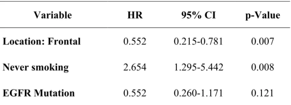
Multivariate analysis showed BM located in the frontal lobe and never smoker status were independent and significant prognostic factors (Table V; p = 0.007 and p = 0.008, respectively).
Discussion
In the present study, we evaluated clinical and histological features of BM from lung adenocarcinoma to identify prognostic factors predictive of OS in this patient population.
Previous studies have investigated the incidence of EGFR mutations in a large population of lung cancer patients (19, 20). EGFR mutations were detected in 30% of patients with adenocarcinoma; 38% of female patients; 47% of non-smokers; and 32%
of patients of East Asian origin (19, 21). It was also easy to detect in smaller primary
12
tumors (22). Deletion of exon 19 and L858R mutations in exon 21 accounted for approximately 85% of EGFRs mutation and have been recognized as important markers in determining the appropriate treatment strategy for patients with advanced lung cancer (21). BM from lung cancer were detected at the time of diagnosis of the primary tumor in 8–23% of patients with EGFR mutations, and in 24% of patients with NSCLC during follow-up care (23, 24). In patients with NSCLC in which EGFR mutations were present, 34.2% had BM 1 year after diagnosis; 38.4% at 2 years; 46.7% at 3 years;
48.7% at 4 years; and 52.9% at 5 years (25).
At the time of the initial diagnosis of primary lung adenocarcinoma, the incidence of EGFR mutation was reported to vary according to the site of BM. Lung adenocarcinoma patients with EGFR mutations had a higher incidence of BM (24, 26).
However, other studies have reported that the timeline of development of BM was not significantly longer in EGFR-mutated NSCLC (27). Therefore, the association between the presence of EGFR mutations and BM is controversial. Multiple BM (≥ 2) have been reported to occur more frequently in patients with EGFR mutations, but the size of the largest BM were not related to the presence of EGFR mutations (28).
Several studies have compared EGFR status in the primary tumor and BM in patients with NSCLC. These results showed extremely high concordance rates in EGFR mutation status (varying from 93.3 to 100%) between the primary tumor and BM (26, 29). In the current study, 5 of 20 patients tested had EGFR mutations in both the primary lung adenocarcinoma and BM. Five patients had the same EGFR mutation in both sites (3 patients had exon 19 deletions and 1 patient each had had L858R and
13
L861Q substitutions in exon 21). In our study, the frequency of EGFR mutations in patients with BM (23.7%) was lower than previously reported (19, 21). This discrepancy may be due to the characteristics of the patient population in our study.
EGFR mutations in lung adenocarcinoma are most common in female patients and
non-smokers, but our study cohort included predominantly male patients (76.3% of all patients).
It has been reported that the presence of EGFR mutations is associated with significantly higher risk of recurrence of BM (28). However, our results did not bear this out: the rate of recurrence and repeated resection was higher in patients without EGFR mutations.
Both the International Association for the Study of Lung Cancer (IASLC) / American Thoracic Society (ATS) / European Respiratory Society (ERS) criteria for classification of lung adenocarcinoma (30) and the 2015 WHO criteria for classification of tumors of the lung (18) recommend identification of pathological patterns. Primary invasive lung adenocarcinoma has five architectural growth patterns, including lepidic, acinar, papillary, solid, and micropapillary patterns. Tsuta et al. reported that the most prevalent subtype of adenocarcinoma was papillary predominant (37.1%), followed by lepidic predominant (15.1%), solid with mucin production-predominant (13.7%), acinar predominant (10.8%), and micropapillary predominant (6.7%) in a study of 757 cases of invasive lung adenocarcinoma (31). In a previous study of 119 patients with primary lung adenocarcinoma, we reported that the most common histopathological subtype was papillary predominant (58%), followed by acinar predominant (16%), micropapillary
14
predominant (9%), solid predominant (4%), and lepidic predominant (2%) (32). EGFR mutation have been reported to occur significantly more often in adenocarcinoma papillary predominant (31).
Only one report has demonstrated the clinical relevance of pathological subtypes in metastatic lung adenocarcinoma (16). In this study of 100 patients with metastatic lung adenocarcinoma, the most frequent histological pattern was solid with mucin (50%), followed by acinar (29%), micropapillary (20%) and papillary (1%). This study included 30 tissue samples from BM. The study authors surmised that this difference of predominant components in primary and metastatic sites was based on the function of the metastatic potential of each adenocarcinoma subtype. The major solid histopathologic subtype of metastatic lung adenocarcinoma was associated with poorer OS compared with other histopathologic subtypes (16). Furthermore, this histopathologic subtype was less likely to harbor EGFR mutations and occurred less frequently in never smokers (16). In a separate study, Mitual et al. reported micropapillary predominant adenocarcinoma might be more likely to metastasize (17).
This study also found that the most frequent subtype of BM from lung adenocarcinoma were solid predominant (57.6%). OS did not vary as a function of histological subtype.
However, wild type EGFR was significantly more common in patients with the solid predominant subtype. This data is consistent with previous reports, but these results require validation in larger cohorts.
In recent years, TKIs targeting mutated EGFR proteins has been established as a standard treatment for NSCLC. This treatment is associated with longer
15
progression-free survival and OS vs treatment with conventional chemotherapy (11, 33), particularly in selected populations, such as in Eastern Asian, female, and never smoker patients with adenocarcinoma (14). In addition to showing improved efficacy, TKI treatment is associated with less toxicity relative to conventional chemotherapy. Several studies have reported that the TKI treatment of NSCLC patients with BM results in intracranial response rates ranging from 26.7–83% (14, 15), and rates of 64–87% when combined with WBRT (34). TKI treatment for the patients with BM resulted in reduced tumor size and significantly improved OS (18.8 vs 11.1 months; 13-15). In this study, only 2 patients received TKI treatment before surgical resection, because most patients were treated before the development of TKI. Excluding these 2 patients, there was no statistically significant difference in OS between patients with and without EGFR mutations (17.1 vs 15.6 months, p = 0.174). The presence of an EGFR mutation prolonged OS, but improved outcome may be due to the efficacy of treatment with TKIs.
In agreement with our results, for patients with BM from lung adenocarcinoma, the presence of EGFR mutations has been associated with improved survival from the time of development of BM (15–25 months vs 7 months; 27, 35-37). On the other hand, there was no significant difference in the timeline of BM development when comparing patients treated with EGFR-TKI vs those receiving conventional chemotherapy (24).
To our knowledge, previous studies have not reported an association between EGFR mutation and the site of BM. Thus, our finding of a significant association of EGFR mutations with BM in the frontal lobe is novel. In low-grade gliomas, lesions localized in the frontal lobe could be vigorously resected without causing persistent
16
functional deficits (38). The frontal lobe, which does not contain vital brain regions controlling speech and motor areas, is a brain region in which relatively aggressive surgical treatment can be pursued with lower risk of causing lasting and serious functional deficits.
In patients with BM, RPA and GPA score are still the most widely accepted variables considered in evaluating disease prognosis (6, 9). However, our study shows that the presence of EGFR mutations, BM within the frontal lobe, and never smoker status are additional independent prognostic factors.
Limitations of this study include its retrospective nature, and we cannot exclude potential bias in treatment strategy after resection of BM. We were not able to evaluate other clinically important genetic changes, such as ALK rearrangement, due to the limited number of patients with this genetic change in our study group. Despite these limitations, to our knowledge, this is the first report to investigate histopathological subtypes of BM from lung adenocarcinoma, and the association of these subtypes with EGFR mutations and disease prognosis in a large patient cohort.
Conclusion
In the present study, we used univariate analysis to show that three factors - EGFR mutation, BM localized to the frontal lobes, and never smoker status - can be used to further refine estimates of disease prognosis. In addition, multivariate analysis confirmed these results, and showed that the presence of BM in the frontal lobes and never smoker status were the most informative prognostic factors. In adenocarcinoma,
17
obtaining tissue samples from the primary tumor is not infrequently difficult. Our study suggests that tissue from the BM may be useful as an alternative tissue for analysis, because resectable BM from lung adenocarcinoma cases had characteristics that are similar to those of the primary tumor tissue. TKI treatment for BM from lung adenocarcinoma should be considered if indicated by histopathological and the genetic examination.
Conflicts of Interest Disclosure
The authors have no conflicts of interest to declare.
Acknowledgment
We thank Ms. M. Onitsuka and Ms. H. Fukagawa for excellent technical assistance with immunohistochemistry. This work was supported in part by a grant from the Research Center for Advanced Molecular Medicine, Fukuoka University, Fukuoka, Japan.
18
References
1. Shi AA, Digumarthy SR, Temel JS, Halpern EF, Kuester LB and Aquino SL: Does initial staging or tumor histology better identify asymptomatic brain metastases in patients with non-small cell lung cancer? J Thorac Oncol 1: 205-210, 2006.
2. Heon S, Yeap BY, Britt G, Costa DB, Rabin MS, Jackman DM and Johnson BE:
Development of central nervous system metastases in patients with advanced non-small cell lung cancer and somatic EGFR mutations treated with gefitinib or erlotinib. Clin Cancer Res 16: 5873-5882, 2010.
3. Barnholtz-Sloan JS, Sloan AE, Davis FG, Vigneau FD, Lai P and SAwaya RE:
Incidence proportions of brain metastases in patients diagnosed (1973 to 2001) in the Metropolitan Detroit Cancer Surveillance System. J Clin Oncol 22:2865-2872, 2004.
4. Cox JD, Scott CB, Byhardt RW, Emami B, Russell AH, Fu KK, Parliament MIB, Komaki R and Gasper LEW: Addition of chemotherapy to radiation therapy alters failure patterns by cell type within non-small cell carcinoma of lung (NSCCL):
analysis of radiation therapy oncology group (RTOG) trials. Int Radiat Oncol Biol Phys 43: 505-509, 1999.
5. Wang SY, Ye X, Ou W, Lin YB, Zhang BB and Yang H: Risk of cerebral metastases for postoperative locally advanced non-small-cell lung cancer. Lung Cancer 64: 238-243; 2009.
6. Gaspar L, Scott C, Rotman M, Asbell S, Phillips T, Wasserman T, McKenna WG and Byhardt R: Recursive partitioning analysis (RPA) of prognostic factors in three
19
Radiation Therapy Oncology Group (RTOG) brain metastases trials. Int J Radiat Oncol Biol Phys 37: 745-751, 1997.
7. Sperduto PW, Berkey B, Gasper LE, Mehta M and Curran W: A new prognostic index and comparison to three other indices for patients with brain metastases: an anlysis of 1,960 patients in the RTOG database. Int J Radiat Oncol Biol Phys 70:
510-514, 2008.
8. Chang DB, Yang PC, Luh KT, Kuo SH, Hong RL and Lee LN: Late survival of non-small cell lung cancer patients with brain metastases. Influence of treatment.
Chest 101: 1293-1297, 1992.
9. Kepka L, Cieslak E, Bujko K, Fijuth J, Wierzchowski M. Results of the whole-brain radiotherapy for patients with brain metastases from lung cancer: The RTOG RPA intra-classes analysis. Acta Oncol 44 (4): 389-398, 2005.
10. Paez JG, Janne PA, Lee JC, Tracy S, Greulich H, Gabriel S, et al. EGFR mutations in lung cancer: correlation with clinical response to gefitinib therapy. Science 304(5676): 1497-1500, 2004.
11. Mok TS, Wu YL, Thongprasert S, Yang CH, Chu DT, Saijo N, et al. Gefitinib or carbonplatin-paclitaxel in pulmonary adenocarcinoma. N Engl J Med:
361(10)947-957, 2009.
12. Maemondo M, Inoue A, Kobayashi K, Sugawara S, Oisumi S, Isobe H, et al.
Gefitinib or chemotherapy for non-small-cell lung cancer with mutated EGFR. N Engl J Med 362(25): 2380-2388, 2010.
13. Shimato S, Mitsudomi T, Kosaka T, Yutabe Y, Wakabayashi T, Mizuno M, et al.
20
EGFR mutations in patients with brain metastases from lung cancer: association with the efficacy of gefitinib. Neuro Oncol 8(2): 137-144, 2006.
14. Kim JE, Lee DH, Choi Y, Yoon DH, Kim SW, Suh C, et al. Epidermal growth factor receptor tyrosine kinase inhibitors as a first-line therapy for never-smokers with adenocarcinoma of the lung having asymptomatic synchronous brain metastasis. Lung Cancer 65(3): 351-354, 2009.
15. Park SJ, Kim HT, Lee DH, Kim KP, Kim SW, Suh C, et al. Efficacy of epidermal growth factor receptor tyrosine kinase inhibitors for brain metastasis in non-small cell lung cancer patients harboring either exon 19 or 21 mutation. Lung Cancer 77(3): 556-560, 2012.
16. Clay TD, Do H, Sundararajan V, Moore MM, Conron M, Wright GM, et al. The clinical relevance of pathologic subtypes in metastatic lung adenocarcinoma. J Thorac Oncol 9(5): 654-663, 2014.
17. Amin MB, Tamboli P, Merchant SH, Ordonez NG, Ro J, Ayala AG, et al.
Micropapillary component in lung adenocarcinoma: a distinctive histologic feature with possible prognostic significance. Am J Surg Pathol 26(3): 358-364, 2002.
18. Travis WD BE, Burke AP, Marx A, Nicholson AG. WHO Classification of Tumors of the Lung, Pleura, Thymus and Heart. International Agency for Research on Cancer, Lyon, 2015.
19. Mitsudomi T, Yatabe Y. Mutations of the epidermal growth factor receptor gene and related genes as determinants of epidermal growth factor receptor tyrosine kinase inhibitors sensitivity in lung cancer. Cancer Science 98(12): 1817-1824,
21
2007.
20. Mitsudori T, Kosaka T, Yatabe Y. Biological and clinical implications of EGFR mutations in lung cancer. Int J Clin Oncol 11(3): 190-198, 2006.
21. Shigematsu H, Lin L, Takahasi T, Nomura M, Suzuki M, Wistuba, II, et al. Clinical and biological features associated with epidermal growth factor receptor gene mutations in lung cancers. J Natl Cancer Inst 97(5): 339-346, 2005.
22. Yano M, Sasaki H, Kobayashi Y, Yukiue H, Haneda H, Suzuki E, et al. Epidermal growth factor receptor gene mutation and computed tomographic findings in peripheral pulmonary adenocarcinoma. J Thorac Oncol 1(5): 413-416, 2006.
23. Doebele RC, Lu X, Sumey C, Maxson DA, Weickhardt AJ, Oton AB, et al.
Oncogene status predicts patterns of metastatic spread in treatment-naïve nonsmall cell lung cancer. Cancer 118(18): 4502-4511, 2012.
24. Hendriks LE, Smit EFm Vosse BA, Mellema WW, Heideman DA, Bootsma GP, et al. EGFR mutated non-small cell lung cancer patients: more prone to development of bone and brain metastases? Lung Cancer 84(1): 86-91, 2014.
25. Rangachari D, Yamaguchi N, VanderLaan PA, Folch E, Mahadevan A, Floyd SR, et al. Brain metastases in patients with EGFR-mutated or ALK-rearranged non-small-cell lung cancers. Lung Cancer 88(1): 108-111, 2015.
26. Matsumoto S, Takahasi K, Iwakawa R, Matsuno Y, Nakanishi Y, Kohno T, et al.
Frequent EGFR mutations in brain metastases of lung adenocarcinoma. Int J Cancer 119(6): 1491-1494, 2006.
27. Eichler AF, Kahle KT, Wang DL, Joshi VA, Willers H, Engelman JA, et al. EGFR
22
mutation status and survival after diagnosis of brain metastasis in nonsmall cell lung cancer. Neuro Oncol 12(11): 1193-1199, 2010.
28. Shin DY, Na, II, Kim CH, Park S, Baek H, Yang SH. EGFR mutation and brain metastasis in pulmonary adenocarcinomas. J Thorac Oncol 9(2): 195-199, 2014.
29. Sun M, Behrens C, Feng L, Ozburn N, Tang X, Yin G, et al. HER family receptor abnormalities in lung cancer brain metastases and corresponding primary tumors.
Clin Cancer Research 15(5): 4829-4837, 2009.
30. Travis WD, Brambilla E, Noguchi M, Nicholson AG, Geisinger KR, Yatabe Y, et al.
International association for the study of lung cancer/ American thoracic society/
European respiratory society international multidisciplinary classification of lung adenocarcinoma. J Thorac Oncol 6(2): 244-285, 2011.
31. Tsuta K, Kawago M, Inoue E, Yoshida A, Takahashi F, Sakurai H, et al. The utility of the proposed IASLC/ATS/ERS lung adenocarcinoma subtypes for disease prognosis and correlation of driver gene alternations. Lung Cancer 81(3), 371-376, 2013.
32. Koga K, Hamasaki M, Kato F, Aoki M, Hayashi H, Iwasaki A, et al. Association of c-Met phosphorylation with micropapillary pattern and small cluster invasion in pT1-size lung adenocarcinoma. Lung Cancer 82(3), 413-419, 2013.
33. Zhou C, Wu YL, Chen G, Feng J, Liu XQ, Wang C, et al. Erlotinib versus chemotherapy as first-line treatment for patients with advanced EGFR mutation-positive non-small-cell lung cancer (OPTIMAL, CTONG-0802): A multicenter, open-label, randomized, phase 3 study. Lancet Oncol 12(8): 735-742,
23
2011.
34. Welsh JW, Komaki R, Amini A, Munsell MF, Unger W, Allen PK, et al. Phase II trial of erlotinib plus concurrent whole-brain radiation therapy for patients with brain metastases from non-small-cell lung cancer. J Clin Oncol 31(7): 895-902, 2013.
35. Stanic K, Zwitter M, Hitij NT, Kern I, Sadikov A, Cufer T. Brain metastases in lung adenocarcinoma: impact of EGFR mutation status on incidence and survival. Radiol Oncol 48(2): 173-183, 2014.
36. Lee DW, Shin DY, Kim JW, Keam B, Kim TM, Kim HJ, et al. Additional prognostic role of EGFR activating mutations in lung adenocarcinoma patients with brain metastasis: integrating with lung specific GPA score. Lung Cancer 86(3):
363-368, 2014.
37. Sekine A, Satoh H, Iwasawa T, Tamura K, Hayashihara K, Saito T, et al. Prognostic factors for brain metastases from non-small cell lung cancer with EGFR mutation;
influence of stable extracranial disease and erlotinib therapy. Med Oncol 31(10):
228, 2014.
38. Tanriverdi T, Kemerdere R, Baran O, Sayyahmelli S, Ozlen F, Isler C, et al.
Long-term surgical and seizure outcomes of frontal low-grade gliomas. Int J Surg 33 pt A: 60-64, 2016.
24
Figure legends
Figure 1: Histopathological subtypes of BM from lung adenocarcinoma (H&E, ×200).
Solid predominant (A), papillary predominant (B), acinar predominant (C) and micropapillary predominant (D).
Figure 2: Kaplan-Meier curves of OS according to EGFR mutation status (A), location of BM (B), smoking (C).
Abbreviations: BM: brain metastases, EGFR: epidermal growth factor receptor, MT:
mutation, WT: wild type
Table I. Patient baseline characteristics (N = 59)
Total (N = 59)
EGFR MT (N = 14)
WT (N = 45)
p-Value MT vs WT
Age, median 61.3±12.2 61.6±12.0 61.1±12.4 0.80
Gender 0.0001
Male Female
45 (76.3%) 14 (23.7%)
5 (35.7%) 9 (64.3%)
40 (88.9%) 5 (11.1%) Location
Frontal lobe 23 (39.0%) 9 (64.3%) 14 (31.1%) 0.03
Cerebellum 11 (18.6%) 3 (21.4%) 8 (17.8%) 0.76
Posterior lobe 8 (15.6%) 1 (7.1%) 7 (15.6%) 0.67 Histological subtype
Solid Papillary Acinar
34 (57.6%) 13 (22.0%) 11 (18.6%)
4 (28.6%) 5 (35.7%) 4 (28.6%)
30 (66.7%) 8 (17.7%) 7 (15.6%)
0.03 0.26 0.43 Smoking
Never
Current or past KPS
<70 70-80 81-100 Number of BM 1
2-3 > 3 ECM Absent Present GPA 0-1.0 1.5-2.0 2.5-3.0 3.5-4.0
19 (32.2%) 40 (67.8%)
19 (32.2%) 15 (25.4%) 25 (42.4%)
31 (52.5%) 17 (28.8%) 11 (18.6%)
27 (45.8%) 32 (54.2%)
13 (22.0%) 24 (40.7%) 18 (30.5%) 4 (6.8%)
8 (57.1%) 6 (42.9%)
6 (42.9%) 5 (35.7%) 3 (21.4%)
5 (35.7%) 5 (35.7%) 4 (28.6%)
6 (42.9%) 8 (57.1%)
4 (28.6%) 7 (50.0%) 3 (21.4%) 0 (.0%)
11 (24.4%) 34 (75.6%)
13 (28.9%) 10 (22.2%) 22 (48.9%)
26 (45.8%) 12 (26.7%) 7 (15.6%)
21 (46.7%) 24 (53.3%)
9 (20.0%) 17 (37.8%) 15 (33.3%) 4 (8.9%)
0.04
0.51 0.48 0.12
0.22 0.74 0.43 0.80
0.71 0.54 0.52 0.56
Timing of diagnosis BM diagnosed before PT
TKI (before BM) RAS
ALK
Re-operation
27 (45.8%)
2 (3.4%) 5 (8.5%) 3 (5.0%) 18 (30.5%)
5 (35.7%)
2 (14.3%) 0 (.0%) 0 (.0%) 1 (7.1%)
22 (48.8%)
0 (.0%) 5 (11.1%)
3 (6.7%) 17 (37.8%)
0.54
0.05 0.16 0.57 0.04
Abbreviations: EGFR: epidermal growth factor receptor, MT: mutation, WT: wild type, KPS:
Karnofsky performance score, BM: brain metastasis, ECM: extracranial metastasis, GPA: graded prognostic assessment, PT: primary tumor, TKI: tyrosine kinase inhibitor, ALK: anaplastic lymphoma kinase
Table II. EGFR mutation analysis with brain metastasis from lung adenocarcinoma (N = 59)
EGFR mutation Number (%) Exon 19 deletion 8 (57.1%) Exon 21 L858R 5 (35.7%) Exon 21 L861Q 1 (7.1%) Exon 18 G719X 0 (.0%) Exon 20 T790M 0 (.0%)
Abbreviations: EGFR: epidermal growth factor receptor A total of 14 out of 59 patients had EGFR mutations.
Table III. The pathological subtype of primary lung adenocarcinoma and metastatic brain tumor (N = 20)
No Stage Size (mm) Primary BM
1 T1aN0Mx 11x10x15 Solid (solid + acinar) Solid
2 T1bN0M0 20x15x20 Solid (solid + acinar) Acinar
3 T2aNxMx 40x30x25 Acinar Acinar
4 T1cN0M0 28x26x24 Acinar (acinar + solid) Acinar 5 T1bN0Mx 20x15x15 Acinar (acinar + papillary) Papillary
6 T1bNxMx 20x10x8 Solid Solid
7 T1bNxMx 20x8x8 Solid (acinar + papillary + MPP) Solid 8 T2aNxMx 30x38x35 Solid (solid + acinar + papillary) Acinar
9 T4N2M1 40x35x35 Acinar (acinar + MPP) Solid
10 T1cN0Mx 30x25x20 Solid (solid + acinar) Solid
11 T2NxM1 15x10x10 Solid (solid + acinar + MPP) Solid
12 T3N0M1 40x35x35 Solid (solid + acinar) Solid
13 T4N0Mx 70x70x55 MPP (MPP + acinar + solid) Solid 14 T1bN0Mx 16x15x15 Solid (solid + acinar) Papillary 15 T2aN2M0 18x17x24 MPP (MPP + acinar + papillary) Solid 16 T4N2Mx 85x60x50 Solid (solid + MPP + acinar) Solid 17 T1cNxMx 22x18x10 Acinar (acinar + MPP + papillary) MPP
18 T2bN2Mx 50x50x30 Acinar (acinar + solid + papillary) Solid 19 T1cNxMx 30x20x27 Acinar (acinar + papillary) Papillary
20 T2aNxMx 35x40x25 MPP (MPP + acinar) Papillary
Abbreviations: BM: brain metastasis, MPP: micropapillary
Table IV. Univariate analysis of overall survival with brain metastasis from lung adenocarcinoma
Variable Median OS (m) p-Value
Age
<70 (N = 45) ≧70 (N = 14)
22.7 (11.8-33.7) 19.4 (9.1-29.7)
0.729
Gender
Male (N = 45) Female (N = 14)
16.9 (11.4-22.4) 27.8 (15.7-39.9)
0.131
Location
Frontal (N = 23) Cerebellum (N = 11) Others (N = 25)
28.2 (19.0-37.5) 13.7 (7.4-20.0) 12.7 (5.9-19.5)
0.004
Smoking
Never (N = 19)
Current or past (N = 40)
33.1 (22.1-33.1) 12.5 (8.3-16.7)
0.001
Histological subtype Solid (N=34) Papillary (N=13) Aciner (N=11)
17.4 (11.4-23.5) 26.9 (13.0-40.8) 11.2 (5.9-16.6)
0.236
EGFR
Mutation (N = 14) Wild type (N = 45)
31.5 (19.3-43.8) 15.6 (10.3-20.8)
0.020
KPS 0.551
<70 (N = 19) 70-80 (N = 15) 81-100 (N = 25)
18.2 (8.8-27.6) 17.1 (8.3-25.9) 21.9 (13.4-30.4) GPA
0-1.0 (N = 13) 1.5-2.0 (N = 24) 2.5-3.0 (N = 18) 3.5-4.0 (N = 4)
22.8 (10.0-35.6) 12.8 (7.8-17.8) 20.2 (11.5-28.8) 37.0 (12.7-61.2)
0.180
Timing of diagnosis BM diagnosed before PT Yes (N =27)
No (N = 32) RAS
Mutation (N = 5) Wild type (N = 54)
12.5 (8.3-16.9) 23.4 (15.6-31.3)
7.8 (4.7-10.8) 23.7 (13.8-33.5)
0.079
0.074
Re-operation
Present (N = 18) Absent (N = 41)
17.7 (8.8-26.7) 19.7 (13.3-26.1)
0.712
Abbreviations: EGFR: epidermal growth factor receptor, OS: overall survival, KPS: karnofsky performance score, GPA: graded prognostic assessment, BM: brain metastasis, PT: primary tumor
Table V. Multivariate analysis of overall survival with brain metastasis of lung adenocarcinoma
Variable HR 95% CI p-Value
Location: Frontal 0.552 0.215-0.781 0.007 Never smoking 2.654 1.295-5.442 0.008 EGFR Mutation 0.552 0.260-1.171 0.121
Abbreviations: EGFR: epidermal growth factor receptor, HR: hazard ratio, CI: confidence interval