1
Effects of initial periodontal therapy on the incidence of P. gingivalis and EBV DNA in chronic periodontitis patients
(慢性歯周炎患者における
P. gingivalis
とエプスタインバーウイルスの 検出率に対する歯周基本治療の効果)日本大学大学院松戸歯学研究科歯学専攻
池田 賴宣
(指導:小方 賴昌 教授)
2
3
Preface
This article is based on a main paper, “Quantitative changes of P. gingivalis and EBV
DNA in saliva before and after initial periodontal therapy in chronic periodontitis patients”
in the International Journal of Oral-Medical Sciences, and a reference paper, “Effects of
initial periodontal therapy on the prevalence of Epstein-Barr virus DNA and
Porphyromonas gingivalis in Japanese chronic periodontitis patients” in the International Journal of Oral-Medical Sciences.
Abstract
Background: Chronic periodontitis (CP) is a most prevalent disease consisting of chronic
inflammation of the periodontium that is caused by the accumulation of dental plaque.
Recently, Epstein-Barr virus (EBV) is thought to be involved in the pathogenesis of
periodontitis as well as Porphyromonas gingivalis which is the representative
periodontopathic bacteria. The purpose of initial periodontal therapy (IPT) is to enhance
motivation and remove calculus, periodontopathic bacteria and their byproducts, in order
to restore periodontal health. To elucidate the effects of IPT on incidence of P. gingivalis
and EBV DNA, we used whole saliva and subgingival plaque from the CP patients.
Methods: Twenty CP patients for whole saliva and 17 CP patients for subgingival plaque
4
samples were recruited and determined periodontal status by probing pocket depth (PD),
bleeding on probing (BOP) and clinical attachment level (CAL; only the patients for
subgingival plaque samples), and whole saliva or subgingival plaque samples were
collected from two periodontal sites with PD of <3 mm (healthy sites: HS) or >5 mm
(periodontitis sites: PS) at first visit and after IPT. Saliva and subgingival plaque samples
were subjected to real-time PCR to detect P. gingivalis and EBV DNA.
Results: P. gingivalis and EBV DNA were detected in 20 (100%) and 14 (70%) saliva samples from the CP patients at first visit. After IPT, number of detections of P. gingivalis
and EBV DNA were decreased to 17 (85%) and 9 (45%) saliva samples from the patients.
Coexistence of P. gingivalis and EBV DNA were detected in 14 (70%) saliva samples
from the patients at first visit, and significantly decreased to 8 (40%) after IPT. EBV DNA
and P. gingivalis were detected 9 (52.9%) and 14 (82.3%) sites within the subgingival
samples from HS, and 13 (76.5%) and 14 (82.3%) sites within the PS at first visit. After
IPT, number of detections of EBV DNA and P. gingivalis were decreased to 5 (29.4%)
and 13 (76.5%) sites within the subgingival samples from HS, and 9 (52.9%) and 10
(58.8%) sites within the PS. Coexistence of EBV DNA and P. gingivalis in the
subgingival samples from PS at first visit (12 sites; 70.6%) were significantly decreased
after IPT (6 sites; 35.3%). Significant improvements in PD and BOP were observed after
5
IPT.
Conclusion: These results suggest that the IPT was effective in improvement of clinical parameters such as PD and BOP and reducing the coexistence of P. gingivalis and EBV
in the saliva and subgingival plaque in PS from CP patients. However, IPT could not
eradicate the EBV and P. gingivalis. Further research would be necessary for improving
the periodontal treatment strategy.
Introduction
Periodontitis, an inflammatory disease, is caused by three risk factors such as bacterial,
environmental and host factors. Severe periodontitis provokes destruction of
periodontium, gingival swelling, alveolar bone resorption, and eventual tooth loss (1).
Bacterial plaque is key etiological factor in the onset and progression of periodontitis (2).
EBV is one of the most prevalent viruses in the world. It is estimated that over 90% of
adults are EBV seropositive (3, 4). Primary infections of infants with EBV are usually
asymptomatic, but the infection of adolescence and young adult with EBV causes
infectious mononucleosis, a self-limiting, lymphoproliferative disease. Spread within
families is thought to be a common route of EBV transmission by salivary contact. The
virus infects first within oropharyngeal epithelium, and later primarily within B
lymphocytes are invaded via CD21 receptors, where it establishes a lifelong latent
6
infection (5-9). EBV has been linked to the development of several malignant tumors,
including Burkitt’s lymphoma, Hodgkin’s disease certain forms of T-cell lymphoma,
lymphoproliferative disease in immunosuppressed individuals, nasopharyngeal
carcinoma and a proportion of gastric cancers (10-12). ZEBRA is an early lytic protein of
EBV encoded BZLF1 gene. In the latent state, hypoacetylation of histone in the BZLF1
promoter by histone deacetylases is involved in maintaining EBV latency. The
reactivation of EBV from latent infection occurs frequently and multiplies with the
epithelium cells of the pharyngeal and is exhausted in saliva (7, 13-15).
There were several studies describing relationship between periodontal disease and
EBV infections (16-21). Therefore, we have examined the coexistence of P. gingivalis
and EBV in the subgingival plaque from two periodontal pocket sites with probing pocket
depth (PD) of <3 mm or >5 mm. P. gingivalis and EBV DNA were detected in higher
copy numbers in the deep periodontal pockets and found higher incidence of coexistence
as compared with shallow periodontal pockets (18, 19). In these studies, we suggested
that EBV may serve as pathogenic factors lead to periodontal disease among Japanese
patients. P. gingivalis could increase the virulence of EBV via reactivation of EBV
through butyric acid (7, 18, 19). EBV and human cytomegalovirus are significantly
related to chronic periodontitis (CP) (20). Coexistence of P. gingivalis and EBV could
7
promote the progression of CP in pregnant women (21). Therefore, interactions between
P. gingivalis and EBV might be involved in the onset and progression of CP.
The purpose of this study was to examine the effects of initial periodontal therapy (IPT)
on the prevalence of P. gingivalis and EBV DNA in the saliva and subgingival plaque.
Methods
Clinical examination and characteristics of participants
Periodontal examination comprising determination of PD, bleeding on probing (BOP)
and, clinical attachment level (CAL). At first visit and after IPT, periodontal examinations
were performed by a trained periodontist using PCP11 probe (Hu-Friedy, Chicago, IL,
USA) according to the method published previously (22, 23). CP patients were defined
as the presence of at least two sites with PD >5 mm and CAL of more than 5 mm. Twenty
CP patients for whole saliva and 17 CP patients for subgingival plaque samples were
included in this study. All subjects were systemically healthy and had no history of
periodontal treatment or any type of antibiotic therapy for at least 3 months prior to the
present study. The Institutional Review Board at the Nihon University School of Dentistry
at Matsudo approved the study (EC17-16-15-005-2). Written informed consent was
obtained from each study subject after all experiments were fully explained. They
8
received IPT, such as oral hygiene instructions, scaling and root planing (SRP) and
mechanical tooth cleaning (within 12 months) at Nihon University Hospital School of
Dentistry at Matsudo, Japan.
Sampling
Saliva samples were collected from 20 CP patients at first visit and after initial periodontal
therapy. Whole saliva was collected from each subject by chewing on a gum base,
containing neither fragrance nor flavored ingredients for 5 min (24).
Seventeen subgingival plaque samples were collected from one periodontally healthy
site (HS) with PD (<3 mm), and one periodontitis site (PS) with PD (>5 mm) of 17 CP
patients at first visit and after initial periodontal therapy. Before sampling, supragingival
plaque was removed with Gracey curette. Sterile paper points were inserted to the sample
site (three times), retained for 30 sec, pooled in Eppendorf tubes, and then stored at -80 °C
(18).
DNA extraction and real-time PCR
DNA samples from the whole saliva and subgingival plaque were prepared using High
Pure Viral Nucleic Acid Kit (Roche Applied Science, Mannheim, Germany). Real-time
9
polymerase chain reaction (PCR) was used to measure the copy numbers of P. gingivalis
and EBV DNA in the samples, using the specific primer sets described previously (19,
25). The dynamic ranges of the real-time PCR assays were determined through serial
dilution of DNA extracts either as AKATA cells or P. gingivalis TDC60 of the standards
in the range of 10
9∼10
1copies/ml (26, 27).
Statistical analysis
Significant differences between baseline values of PD and BOP (for whole saliva), and
values after IPT were analyzed using paired t-test. The chi squared test for independence,
confirmed by Fisher’s exact probability test, was used to determine whether individual
pathogens and BOP (for subgingival plaque) were changed by IPT.
Results
The age, sex, PD and BOP of the patients for whole saliva samples are summarized in
Table 1. Six males and 14 females were included in this study. The mean PD at first visit
were 2.95 ± 0.77 mm, and then it was changed to 2.15 ± 0.43 mm after IPT. BOP was
detected in 46.4 ± 27.1% at first visit, and then BOP changed in 14.9 ± 16.2% after IPT.
PD and BOP at first visit were significant improved after IPT.
10
The age, sex, PD, CAL and BOP of the patients for subgingival plaque samples are
summarized in Table 2. Seven males and 10 females were included in this study. The
mean PD of the HS and PS at first visit were 2.94 ± 0.24 mm and 7.35 ± 1.54 mm, and
then they were changed to 2.82 ± 0.39 mm (HS) and 5.76 ± 1.68 mm (PS) after IPT. The
mean CAL of the HS and PS at first visit were 3.65 ± 1.37 mm and 8.76 ± 1.92 mm, and
then they were changed to 3.82 ± 1.51 mm (HS) and 7.47 ± 1.74 mm (PS) after IPT. BOP
was detected in 2 (11.8%) HS and 17 (100%) PS at first visit, and then BOP could not
detect in HS and detected in 11 (65%) PS after IPT. PD and BOP of the PS at first visit
were significant improved after IPT.
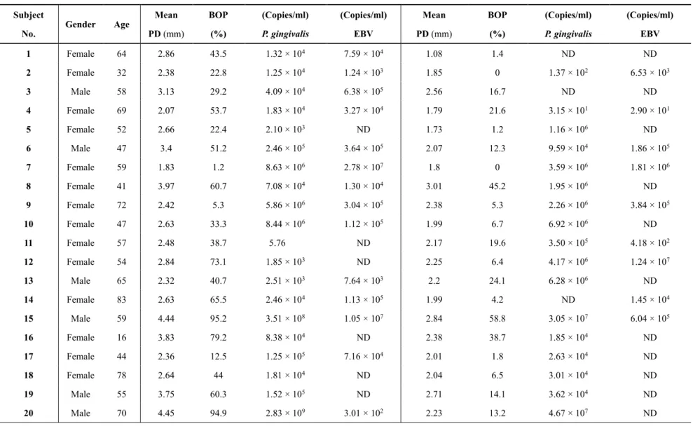
Table 3 shows gender, age, clinical data and counts of P. gingivalis and EBV DNA
(copies/ml) of each subject at first visit and after IPT. P. gingivalis and EBV DNA were
detected in saliva taken from 20 (100%, range from 5.76 to 2.83×10
9copies/ml) and 14
(70%, range from 3.01×10
2to 2.78×10
7copies/ml) participants at first visit and their
incidence decreased to 17 (85%, range from 3.15×10
1to 4.67×10
7copies/ml) and 9
participants (45%, range from 2.90×10
1to 1.24×10
7copies/ml) after IPT (Table 3).
Coexistence of P. gingivalis and EBV DNA at first visit (14; 70%) were significantly
decreased after IPT (8; 40%) (Table 4).
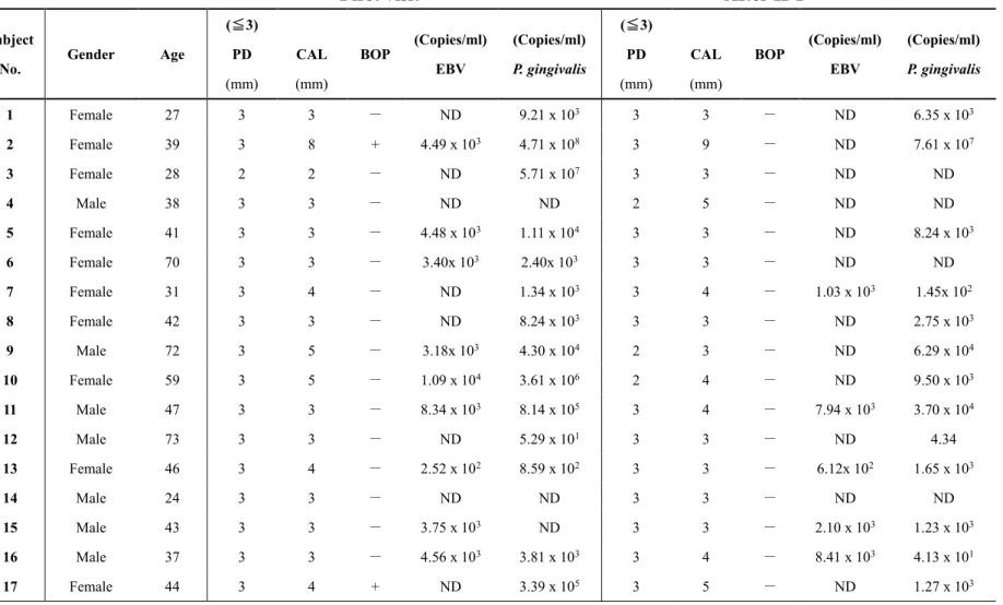
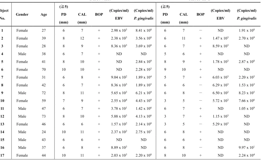
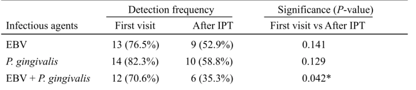
Table 5 and 6 show gender, age, clinical data and counts of EBV DNA and P. gingivalis
11
(copies/ml) of each subject in the HS and PS at first visit and after IPT. EBV DNA and P.
gingivalis were detected 9 sites (52.9%, range from 2.52×10
2to 1.09×10
4copies/ml) and
14 sites (82.3%, range from 5.29×10
1to 4.71×10
8copies/ml) in the subgingival samples
from HS at first visit and changed to 5 sites (29.4%, range from 6.12×10
2to 8.41×10
3copies/ml) and 13 sites (76.5%, range from 4.34 to 7.61×10
7copies/ml) from HS after
IPT (Table 5). EBV DNA and P. gingivalis were detected 13 sites (76.5%, range from
3.78×10
1to 2.55×10
4copies/ml) and 14 sites (82.3%, range from 2.28×10
3to 6.21×10
9copies/ml) in the subgingival samples from PS at first visit and changed to 9 sites (52.9%,
range from 6.50×10
2to 8.59×10
3copies/ml) and 10 sites (58.8%, range from 9.97×10
1to
2.70×10
9copies/ml) from PS after IPT.
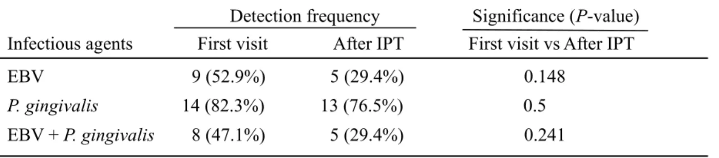
The prevalence of EBV DNA and P. gingivalis in the subgingival samples from HS or
PS are listed in Table 7 and 8. Occurrence of EBV DNA and P. gingivalis in the HS or PS
were decreased after IPT, but not statistically significant. Coexistence of EBV DNA and
P. gingivalis in the PS at first visit (12; 70.6%) were significantly decreased after IPT (6;
35.3%) (Table 8). However, coexistence of EBV DNA and P. gingivalis in the HS did not
decreased significantly after IPT (Table 7).
12
Discussion
In the present study, we showed that higher numbers of P. gingivalis,
EBV DNA andcoexistence of P. gingivalis and EBV DNA were detected in the whole saliva of CP
patients and they were decreased by IPT. It’s notable that PD, BOP and coexistence of P.
gingivalis and EBV DNA in the saliva at first visit were significantly decreased after IPT.
In the second study, we demonstrated that high incidence of EBV DNA, P. gingivalis
andcoexistence of EBV DNA and P. gingivalis were detected in the subgingival plaque from
HS and PS of CP patients and they were decreased by IPT. Especially, PD, BOP and
coexistence of EBV DNA and P. gingivalis in the PS at first visit were significantly
decreased after IPT. These results suggest that IPT is effective in improvement of PD and
BOP and reducing the coexistence of P. gingivalis and EBV in the saliva and subgingival
plaque. We wished to examine the effect of IPT on the incidence of P. gingivalis and EBV
DNA in the saliva and subgingival plaque, because several studies suggest that P.
gingivalis and EBV act synergistically to potentiate progression of periodontitis and tissue destruction of periodontium (18-21, 28, 29).
Periodontopathic bacteria is crucial risk factor for periodontal disease, it might be
associated with systemic conditions. Especially, P. gingivalis triggers changes to the
composition and amount of the oral commensal bacteria inducing inflammation and bone
13
resorption. Changes in the composition of the gut bacterium have been implicated in
several inflammatory diseases. Therefore, targeting of possible keystone bacteria, such as
P. gingivalis could help treat periodontal disease of polymicrobial etiology (30). EBV were
associated with the severity of periodontal disease and with major periodontopathic
bacteria (31, 32). These reports suggested that high copy numbers of P. gingivalis and EBV
DNA may correlate with severity of periodontitis.
We previously reported, P. gingivalis were detected in the subgingival plaque from 20
(80%) deep periodontal pockets (PS; PD >5 mm) and 9 (36%) shallow periodontal pockets
(HS; PD of <3 mm), and EBV DNA were detected in 20 (80%) PS and 10 (40%) HS of 25
Japanese CP patients (19). These results showed that detection rate of EBV DNA and P.
gingivalis in the PS at first visit (Table 8) were similar, whereas detection rate in the HS at first visit (Table 7) were higher than the results of we reported previously (19). These
results also showed that detection rate of P. gingivalis in the PS (80%) were lower than the
detection rate in the saliva (100%), whereas detection rate of EBV DNA in the PS (80%)
were similar detection rate in the saliva (70%) at first visit (Table 4). P. gingivalis and EBV
DNA coexist in the saliva of CP patient’s high frequency (70%) at first visit (Table 4). In
the subgingival plaque samples, EBV DNA and P. gingivalis coexist in the PS at first visit
at high frequency (70.6%) (Table 8). These values correlated with the data of previous
14
study that the detection rate (68%) of coexistence of P. gingivalis and EBV DNA in the
deep periodontal pockets of the CP patients (19).
We have previously reported immunostaining using B cell marker CD19 showed large
number of B cells infiltrated into the inflamed gingival connective tissues, and EBV-
encoded small RNA (EBRE) positive B cells were detected in the same location using in-
situ hybridization (18). Latent EBV might be induced into the lytic replication cycle by several inducers, such as phorbol 12-O-tetradecanoylphorbol-13-acetate, calcium
ionophores, butyric acid and anti-immunoglobulin (7, 8, 33). BamHI Z EBV replication
activator
(ZEBRA) is an early lytic protein of EBV encoded by BZLF1 gene which is
involved in converting the EBV from the latent to the lytic form. Histone deacetylase
(HDAC) induces hypoacetylation of the BZLF1 promoter, and involved in the maintaining
of EBV latency. P. gingivalis produces butyric acid which is an inhibitor of HDAC,
increased histone acetylation and induced transcription of the BZLF1 gene (7, 8). These
findings suggest that P. gingivalis is a risk factor for EBV reactivation in the periodontal
tissues.
Results of this study provides evidence for potential interactions between P. gingivalis
and EBV in the etiology of periodontitis. Periodontopathic bacteria and EBV co-existence
apparently leads to additive effects and exacerbates the progress of periodontitis (33).
15
EBV-infected periodontium tends to harbor high levels of periodontopathic bacteria.
Bacterial and viral co-existences were reported more frequently in deeper PD sites of CP
patients (28, 29).
PD, BOP and coexistence of P. gingivalis and EBV DNA in the saliva and subgingival
plaque at first visit were significantly decreased after IPT. Therefore, the results suggest
that IPT is very important to treat periodontal disease and maintain periodontal health.
References
1. Kinane DF, Attstrom R: Advances in the pathogenesis of periodontitis. Consensus
Report. J Clin Periodontol 32(Suppl. 6): 130–131, 2005.
2. Haffajee AD, Socransky SS: Microbiology of periodontal diseases: introduction.
Periodontology 2000 38: 9–12, 2005.
3. Young LS, Rickinson AB : Epstein-Barr virus: 40 years on. Nat Rev Cancer 4:757–
768, 2004.
4. Lin JC, Lin SC, De BK, Chan WP, Evatt BL: Precision of genotyping of Epstein-Barr
virus by polymerase chain reaction using three gene loci (EBNA-2, EBNA-3C, and
EBER): predominance of type A virus associated with Hodgkin's disease. Blood 81:
3372–3381, 1993.
16
5. Maeda E, Akahane M, Kiryu S, Kato N, Yoshikawa T, Hayashi N, Aoki S, Minami M,
Uozaki H, Fukayama M, Ohtomo K: Spectrum of Epstein-Barr virus-related diseases:
a pictorial review. Jpn J Radiol 27: 4–19, 2009.
6. Thorley-Lawson DA, Gross A: Persistence of the Epstein-Barr virus and the origins
of associated lymphomas. N Engl J Med 350: 1328–1337, 2004.
7. Imai K, Inoue H, Tamura M, Cueno ME, Inoue H, Takeichi O, Kusama K, Saito I,
Ochiai K: The periodontal pathogen Porphyromonas gingivalis induces the Epstein-
Barr virus lytic switch transactivator ZEBRA by histone modification. Biochimie 94:
839–846, 2012.
8. Imai K, Kamio N, Cueno ME, Saito Y, Inoue H, Saito I, Ochiai K: Role of the histone
H3 lysine 9 methyltransferase Suv39 h1 in maintaining Epsteinn-Barr virus latency
in B95-8 cells. FEBS J 281: 2148–2158, 2014.
9. Timms JM, Bell A, Flavell JR, Murray PG, Rickinson AB, Traverse-Glehen A, Berger
F, Delecluse HJ: Target cells of Epstein-Barr-virus (EBV)-positive post-transplant
lymphoproliferative disease: similarities to EBV-positive Hodgkin's lymphoma.
Lancet 361: 217–223, 2003.
10. Toussirot E, Roudier J: Epstein-Barr virus in autoimmune diseases. Best Pract Res
Clin Rheumatol 22: 883–896, 2008.
17
11. Lai KY, Chou YC, Lin JH, Liu Y, Lin KM, Doong SL, Chen MR, Yeh TH, Lin SJ,
Tsai CH: Maintenance of Epstein-Barr virus latent status by a novel mechanism, latent
membrane protein 1-induced interleukin-32, via the protein kinase Cδ pathway. J
Virol 89: 5968–5980, 2015.
12. D'Addario M, Libermann TA, Xu J, Ahmad A, Menezes J: Epstein-Barr virus and its
glycoprotein-350 upregulate IL-6 in human B-lymphocytes via CD21, involving
activation of NF-kappaB and different signaling pathways. J Mol Biol 308: 501–514,
2001.
13. Slots J, Saygun I, Sabeti M, Kubar A: Epstein-Barr virus in oral diseases. J Periodontal
Res 41: 235–244, 2006.
14. Corstjens PL, Abrams WR, Malamud D: Saliva and viral infections. Periodontol 2000
70: 93–110, 2016.
15. Ikuta K, Satoh Y, Hoshikawa Y, Sairenji T: Detection of Epstein-Barr virus in salivas
and throat washings in healthy adults and children. Microbes Infect 2: 115–120, 2000.
16. Kamma JJ, Contreras A, Slots J: Herpes viruses and periodontopathic bacteria in
early-onset periodontitis. J Clin Periodontol 28: 879–885, 2001.
17. Slots J, Kamma JJ, Sugar C: The herpesvirus-Porphyromonas gingivalis -
periodontitis axis. J Periodontal Res 38: 318–323, 2003.
18
18. Kato A, Imai K, Ochiai K, Ogata Y: Higher prevalence of Epstein-Barr virus DNA in
deeper periodontal pockets of chronic periodontitis in Japanese patients. PLoS One
8(8): e71990, 2013.
19. Kato A, Imai K, Ochiai K, Ogata Y: Prevalence and quantitative analysis of Epstein-
Barr virus DNA and Porphyromonas gingivalis associated with Japanese periodontitis
patients. Clin Oral Invest 19: 1605–1610, 2015.
20. Zhu C, Li F, Wong MC, Feng XP, Lu HX, Xu W: Association between Herpesviruses
and Chronic Periodontitis: A Meta-Analysis Based on Case-Control Studies. PLoS
One 10: e0144319,. 2015
21. Lu H, Zhu C, Li F, Xu W, Tao D, Feng X: Putative periodontopathic bacteria and
herpesviruses in pregnant women: a case-control study. Sci Rep 6: 27796, 2016.
22. Ogata Y, Nakayama Y, Tatsumi J, Kubota T, Sato S, Nishida T, Takeuchi Y, Onitsuka
T, Sakagami R, Nozaki T, Murakami S, Matsubara N, Tanaka M, Yoshino T, Ota J,
Nakagawa T, Ishihara Y, Ito T, Saito A, Yamaki K, Matsuzaki E, Hidaka T, Sasaki D,
Yaegashi T, Yasuda T, Shibutani T, Noguchi K, Araki H, Ikumi N, Aoyama Y, Kogai
H, Nemoto K, Deguchi S, Takiguchi T, Yamamoto M, Inokuchi K, Ito T, Kado T,
Furuichi Y, Kanazashi M, Gomi K, Takagi Y, Kubokawa K, Yoshinari N, Hasegawa
Y, Hirose T, Sase T, Arita H, Kodama T, Shin K, Izumi Y, Yoshie H: Prevalence and
19
risk factors for peri-implant diseases in Japanese adult dental patients. J Oral Sci 59:
1-11, 2017.
23. Nakayama Y, Ogata Y, Hiromatsu Y, Imamura K, Suzuki E, Saito A, Shirakawa S,
Nagano T, Gomi K, Morozumi T, Watanabe K, Akiishi K, Yoshie H: Clinical
usefulness of novel immunochromatographic detection device for Porphyromonas
gingivalis in evaluating effects of scaling and root planing and local antimicrobial
therapy. J Periodontol 87: 1238–1247, 2016.
24. Morozumi T, Nakagawa T, Nomura Y, Sugaya T, Kawanami M, Suzuki F, Takahashi
K, Abe Y, Sato S, Makino-Oi A, Saito A, Takano S, Minabe M, Nakayama Y, Ogata
Y, Kobayashi H, Izumi Y, Sugano N, Ito K, Sekino S, Numabe Y, Fukaya C,
Yoshinari N, Fukuda M, Noguchi T, Kono T, Umeda M, Fujise O, Nishimura F,
Yoshimura A, Hara Y, Nakamura T, Noguchi K, Kakuta E, Hanada N, Takashiba S,
Yoshie H: Salivary pathogen and serum antibody to assess the progression of chronic
periodontitis: a 24-mo prospective multicenter cohort study. J Periodontal Res 51:
768-778, 2016.
25. Kato A, Imai K, Sato H, Ogata Y: Prevalence of Epstein-Barr virus DNA and
Porphyromonas gingivalis in Japanese peri-implantitis patients. BMC Oral Health 17:
148, 2017.
20
26. Takada K, Horinouchi K, Ono Y, Aya T, Osato T, Takahashi M, Hayasaka S: An
Epstein-Barr virus-producer line Akata: establishment of the cell line and analysis of
viral DNA. Virus Genes 5: 147–156, 1991.
27. Watanabe T, Maruyama F, Nozawa T, Aoki A, Okano S, Shibata Y, Oshima K,
Kurokawa K, Hattori M, Nakagawa I, Abiko Y : Complete genome sequence of the
bacterium Porphyromonas gingivalis TDC60, which causes periodontal disease. J
Bacteriol 193: 4259–4260, 2011.
28. Chalabi M, Rezaie F, Moghim S, Mogharehabed A, Rezaei M, Mehraban B:
Periodontopathic bacteria and herpesviruses in chronic periodontitis. Mol Oral
Micobiol 25: 236–240, 2010.
29. Özcan E, Saygun NI, Serdar MA, Kubar A, Bengi VU: Porphyromonas gingivalis and
Epstein-Barr Virus are associated with increased levels of visfatin in gingival
crevicular fluid. J Periodontol 87: 443–451, 2016.
30. Hajishengallis G, Liang S, Payne MA, Hashim A, Jotwani R, Eskan MA, McIntosh
ML, Alsam A, Kirkwood KL, Lambris JD, Darveau RP, Curtis MA: Low-abundance
biofilm species orchestrates inflammatory periodontal disease through the commensal
microbiota and complement. Cell Host Microbe 10: 497-506. 2011.
31. Saygun I, Kubar A, Ozdemir A, Yapar M, Slots J: Herpesviral-bacterial
21
interrelationships in aggressive periodontitis. J Periodontal Res 39: 207–212, 2004.
32. Saygun I, Kubar A, Sahin S, Sener K, Slots J: Quantitative analysis of association
between herpesviruses and bacterial pathogens in periodontitis. J Periodont Res 43:
352–359, 2008.
33. Slots J: Herpesviral-bacterial interactions in periodontal diseases. Periodontol 2000
52: 117–140, 2010.
22
Table 1 Characteristics of patients for whole saliva samples CP patients (n =20)
First visit After IPT Age (years) 56.1 ± 15.4
Males 6 (30%) Females 14 (70%)
Mean PD (mm) 2.95 ± 0.77 2.15 ± 0.43**
BOP (%) 46.4 ± 27.1 14.9 ± 16.2**
Chronic periodontitis (CP), initial periodontal therapy (IPT),
probing pocket depth (PD), bleeding on probing (BOP),
Statistically significant; P<0.01**, mean ± SD
23
Table 2 Characteristics of participants for subgingival plaque samples CP patients (n =17)
First visit After IPT Age (years) 44.8 ± 14.9
Males 7 (41.2%) Females 10 (58.8%)
PD(mm) 2.94 ± 0.24(HS) 2.82 ± 0.39 (HS) 7.35 ± 1.54(PS) 5.76 ± 1.68 (PS)**
CAL (mm) 3.65 ± 1.37(HS) 3.82 ± 1.51 (HS) 8.76 ± 1.92(PS) 7.47 ± 1.74 (PS) BOP 2 (11.8%) (HS) 0 (0%)
17 (100%) (PS) 11(65%)**
Clinical attachment level (CAL), healthy sites (HS), periodontitis sites (PS)
Statistically significant; P<0.01**, mean ± SD
24
Table 3 Clinical data and counts of EBV DNA and P. gingivalis at first visit and after IPT for whole saliva samples First visit After IPT
Subject
No. Gender Age Mean
PD (mm)
BOP (%)
(Copies/ml) P. gingivalis
(Copies/ml) EBV
Mean PD (mm)
BOP (%)
(Copies/ml) P. gingivalis
(Copies/ml) EBV
1 Female 64 2.86 43.5 1.32 × 10⁴ 7.59 × 10⁴ 1.08 1.4 ND ND
2 Female 32 2.38 22.8 1.25 × 10⁴ 1.24 × 10³ 1.85 0 1.37 × 10² 6.53 × 10³
3 Male 58 3.13 29.2 4.09 × 10⁴ 6.38 × 10⁵ 2.56 16.7 ND ND
4 Female 69 2.07 53.7 1.83 × 10⁴ 3.27 × 10⁴ 1.79 21.6 3.15 × 101 2.90 × 101
5 Female 52 2.66 22.4 2.10 × 10³ ND 1.73 1.2 1.16 × 10⁶ ND
6 Male 47 3.4 51.2 2.46 × 10⁵ 3.64 × 10⁵ 2.07 12.3 9.59 × 10⁴ 1.86 × 10⁵
7 Female 59 1.83 1.2 8.63 × 10⁶ 2.78 × 10⁷ 1.8 0 3.59 × 10⁶ 1.81 × 10⁶
8 Female 41 3.97 60.7 7.08 × 10⁴ 1.30 × 10⁴ 3.01 45.2 1.95 × 10⁶ ND
9 Female 72 2.42 5.3 5.86 × 10⁶ 3.04 × 10⁵ 2.38 5.3 2.26 × 10⁶ 3.84 × 10⁵
10 Female 47 2.63 33.3 8.44 × 10⁶ 1.12 × 10⁵ 1.99 6.7 6.92 × 10⁶ ND
11 Female 57 2.48 38.7 5.76 ND 2.17 19.6 3.50 × 10⁵ 4.18 × 10²
12 Female 54 2.84 73.1 1.85 × 10³ ND 2.25 6.4 4.17 × 10⁶ 1.24 × 10⁷
13 Male 65 2.32 40.7 2.51 × 10³ 7.64 × 10³ 2.2 24.1 6.28 × 10⁶ ND
14 Female 83 2.63 65.5 2.46 × 10⁴ 1.13 × 10⁵ 1.99 4.2 ND 1.45 × 10⁴
15 Male 59 4.44 95.2 3.51 × 10⁸ 1.05 × 10⁷ 2.84 58.8 3.05 × 10⁷ 6.04 × 10⁵
16 Female 16 3.83 79.2 8.38 × 10⁴ ND 2.38 38.7 1.85 × 10⁴ ND
17 Female 44 2.36 12.5 1.25 × 10⁵ 7.16 × 10⁴ 2.01 1.8 2.63 × 10⁴ ND
18 Female 78 2.64 44 1.81 × 10⁴ ND 2.04 6.5 3.01 × 10⁴ ND
19 Male 55 3.75 60.3 1.52 × 10⁵ ND 2.71 14.1 3.62 × 10⁴ ND
20 Male 70 4.45 94.9 2.83 × 10⁹ 3.01 × 10² 2.23 13.2 4.67 × 10⁷ ND
Not detectable (ND)
25
Table 4 Occurrence of EBV DNA and P. gingivalis in the saliva at first visit and after IPT for whole saliva samples
Detection frequency Significance (P-value) Infectious agents First visit After IPT First visit vs After IPT
P. gingivalis 20 (100%) 17 (85%) 0.07 EBV 14 ( 70%) 9 ( 45%) 0.1 P. gingivalis + EBV 14 ( 70%) 8 ( 40%) 0.05*
Initial periodontal therapy (IPT)
26
Table 5 Clinical data and counts of EBV DNA and P. gingivalis in the HS at first visit and after IPT for subgingival plaque samples
First visit After IPT
Subject
No. Gender Age
(≦3) PD (mm)
CAL (mm)
BOP (Copies/ml) EBV
(Copies/ml) P. gingivalis
(≦3) PD (mm)
CAL (mm)
BOP (Copies/ml) EBV
(Copies/ml) P. gingivalis
1 Female 27 3 3 - ND 9.21 x 10³ 3 3 - ND 6.35 x 10³
2 Female 39 3 8 + 4.49 x 10³ 4.71 x 10⁸ 3 9 - ND 7.61 x 10⁷
3 Female 28 2 2 - ND 5.71 x 10⁷ 3 3 - ND ND
4 Male 38 3 3 - ND ND 2 5 - ND ND
5 Female 41 3 3 - 4.48 x 10³ 1.11 x 10⁴ 3 3 - ND 8.24 x 10³
6 Female 70 3 3 - 3.40x 10³ 2.40x 10³ 3 3 - ND ND
7 Female 31 3 4 - ND 1.34 x 10³ 3 4 - 1.03 x 10³ 1.45x 10²
8 Female 42 3 3 - ND 8.24 x 10³ 3 3 - ND 2.75 x 10³
9 Male 72 3 5 - 3.18x 10³ 4.30 x 10⁴ 2 3 - ND 6.29 x 10⁴
10 Female 59 3 5 - 1.09 x 10⁴ 3.61 x 10⁶ 2 4 - ND 9.50 x 10³
11 Male 47 3 3 - 8.34 x 10³ 8.14 x 10⁵ 3 4 - 7.94 x 10³ 3.70 x 10⁴
12 Male 73 3 3 - ND 5.29 x 101 3 3 - ND 4.34
13 Female 46 3 4 - 2.52 x 10² 8.59 x 10² 3 3 - 6.12x 10² 1.65 x 10³
14 Male 24 3 3 - ND ND 3 3 - ND ND
15 Male 43 3 3 - 3.75 x 10³ ND 3 3 - 2.10 x 10³ 1.23 x 10³
16 Male 37 3 3 - 4.56 x 10³ 3.81 x 10³ 3 4 - 8.41 x 10³ 4.13 x 101
17 Female 44 3 4 + ND 3.39 x 10⁵ 3 5 - ND 1.27 x 10³
27
Table 6 Clinical data and counts of EBV DNA and P. gingivalis in the PS at first visit and after IPT for subgingival plaque samples
First visit After IPT
Subject
No. Gender Age
(≧5) PD (mm)
CAL (mm)
BOP (Copies/ml) EBV
(Copies/ml) P. gingivalis
(≧5) PD (mm)
CAL (mm)
BOP (Copies/ml) EBV
(Copies/ml) P. gingivalis
1 Female 27 6 7 + 2.98 x 10³ 8.41 x 10⁸ 6 7 - ND 1.91 x 10⁸
2 Female 39 8 12 + 2.38 x 10³ 3.56 x 10⁸ 6 11 + 1.47 x 10³ 2.70 x 10⁹
3 Female 28 8 9 + 8.36 x 10³ 3.69 x 10⁸ 6 7 + 8.59 x 10³ ND
4 Male 38 6 7 + ND ND 5 6 + ND ND
5 Female 41 8 10 + ND 2.84 x 10⁹ 8 9 + 1.78 x 10³ 2.87 x 10⁸
6 Female 70 10 10 + ND 2.28 x 10³ 9 10 + ND ND
7 Female 31 6 8 + 9.04 x 10² 1.89 x 10⁴ 5 7 + 6.03 x 10³ 2.20 x 10⁵
8 Female 42 6 7 + 8.36 x 10³ 1.89 x 10⁵ 6 6 - 6.29 x 10³ 1.53 x 10³
9 Male 72 8 11 + 5.65 x 10³ 6.21 x 10⁹ 6 8 - 6.50 x 10² 8.23 x 10⁵
10 Female 59 7 9 + 2.55 x 10⁴ 4.43 x 10⁹ 3 5 - 5.72 x 10³ 7.66 x 10⁶
11 Male 47 6 7 + 3.78 x 101 1.42 x 10⁶ 6 7 + ND 1.05 x 10⁹
12 Male 73 8 10 + 5.88 x 10³ 4.13 x 10⁶ 3 7 + 1.15 x 10³ ND
13 Female 46 6 6 + 1.57 x 10² 2.14 x 10⁸ 3 5 - 5.29 x 10³ ND
14 Male 24 10 11 + 2.37 x 10² 2.75 x 10⁷ 6 8 + ND ND
15 Male 43 6 6 + ND ND 6 6 + ND ND
16 Male 37 6 8 + 8.89 x 10³ ND 6 8 - ND 9.97 x 101
17 Female 44 10 11 + 2.03 x 10³ 2.20 x 10⁸ 8 10 + ND 2.24 x 10⁴
28
Table 7 Occurrence of EBV DNA and P. gingivalis in the subgingival samples from HS at first visit and after IPT for subgingival plaque samples
Detection frequency Significance (P-value) Infectious agents First visit After IPT First visit vs After IPT
EBV 9 (52.9%) 5 (29.4%) 0.148
P. gingivalis 14 (82.3%) 13 (76.5%) 0.5
EBV + P. gingivalis 8 (47.1%) 5 (29.4%) 0.241
Healthy sites (HS)
29