Global Metabolic Regulation via Mig1 as a
Transcriptional Regulator in Thermotolerant Yeast
Kluyveromyces marxianus DMKU3-1042
PhD Thesis
Mochamad Nurcholis
Supervisor : Prof. Mamoru Yamada
A thesis submitted in partial fulfilment of the requirement for the
degree of doctor of applied molecular bioscience
Graduate School of Medicine
Yamaguchi University
CONTENTS
Pages
LIST OF TABLES ...
iv
LIST OF FIGURES ...
v
CHAPTER 1
General Introduction ...
1
CHAPTER 2
Functional analysis of Mig1 and Rag5 as expressional regulators in
thermotolerant yeast Kluyveromyces marxianus ...
4
2.1 Abstract ...
4
2.2 Introduction ...
4
2.3 Materials and methods ...
6
2.3.1 Materials ...
6
2.3.2 Strains, media, and growth conditions ...
7
2.3.3 Analytical methods ...
8
2.3.4 Construction of kanMX4-inserted disrupted mutants of
Kmmig1 and Kmrag5 by site-specific homologous
recombination ...
8
2.3.5 Preparation of cell extracts ...
11
2.3.6 Enzyme assay ...
12
2.3.7 Complementation of mig1 in K. marxianus ...
13
2.3.8 RT-PCR analysis ...
13
2.3.9 Statistical analysis ...
14
2.4 Results ...
14
2.4.1 Growth comparison of K. marxianus and S. cerevisiae
mutants and their parental strains on YP plates containing
sucrose and 2-DOG ...
14
CONTENTS
(Continued)
Pages
2.4.2 Growth and metabolic profiles of K. marxianus and S.
cerevisiae mutants and their parental strains in YP medium
containing a single sugar or mixed sugars ...
15
2.4.3 Effect of MIG1- and RAG5-disrupted mutations on
hexokinase and gluco-hexokinase activities ...
21
2.4.4 Effect of MIG1- and RAG5-disrupted mutations on
inulinase activity ...
22
2.4.5 Effect of MIG1- and RAG5-disrupted mutations on
expression of INU1, GLK1, and RAG1 ...
23
2.4.6 Effect of MIG1- and RAG5-disrupted mutations on growth
in a minimal medium containing glucose or galactose and
amino acids ...
24
2.5 Discussion ...
25
2.6 Conclusion ...
29
CHAPTER 3
MIG1 as a positive regulator for the histidine biosynthesis pathway
and as a global regulator in thermotolerant yeast Kluyveromyces
marxianus ...
31
3.1 Abstract ...
31
3.2 Introduction ...
31
3.3 Materials and methods ...
33
3.3.1 Materials ...
33
3.3.2 Strains, media and growth conditions ...
33
3.3.3 RNA preparation for RNA-Seq ...
34
3.3.4 RNA-Seq-based transcriptomic analysis ...
35
3.3.5 Increased expression of HIS4 in Kmmig1 ...
35
CONTENTS
(Continued)
Pages
3.4.1 Effect of MIG1-disrupted mutation on expression of genes
for histidine biosynthesis ...
37
3.4.2 Increased expression of HIS4 in Kmmig1 ...
39
3.4.3 Effects of MIG1-disrupted mutation on expression of
genomic genes ...
41
3.4.4 Effects of MIG1-disrupted mutation on central carbon
metabolism ...
44
3.5 Discussion ...
45
3.6 Conclusion ...
48
REFERENCES ...
49
ACKNOWLEDGEMENT ...
60
LIST OF PUBLICATIONS ...
61
APPENDIX ...
62
LIST OF TABLES
Tables
Pages
CHAPTER 2
2.1 Strains and plasmid used in this study ...
8
2.2 Primers list for construction of Kmmig1 and Kmrag5 disrupted
mutants, RT-PCR analysis of ACT1, RAG1, GLK1, and INU1 and
complementation of Kmmig1 ...
11
CHAPTER 3
3.1 Primers used in this study ...
36
APPENDIX
S1. Transcription factors (TFs) that are assumed to be under the
control of MIG1 in K. marxianus ...
64
S2. Transcription factors (TFs) of S. cerevisiae, of which orthologs in
K. marxianus are presumably located downstream of Mig1 ...
65
S3. Differentially expressed genes (DEGs) based on unique exon
reads for Kmmig1 and the parental strain of K. marxianus ...
66
S4. GO enrichment of differentially expressed genes for Kmmig1 and
the parental strain of K. marxianus ...
122
S5. KEGG mapper analysis of differentially expressed genes for
LIST OF FIGURES
Figures
Pages
CHAPTER 2
2.1 Growth comparison of K. marxianus and S. cerevisiae on YP
plates containing glucose or sucrose and 2-DOG. ...
15
2.2 Comparison of growth, sugar utilization, and metabolite
production of K. marxianus and S. cerevisiae in liquid YPD ...
16
2.3 Comparison of growth, sugar utilization, and metabolite
production of K. marxianus and S. cerevisiae in liquid YPDS ...
18
2.4 Growth, sugar utilization, and metabolite production of K.
marxianus DMKU3-1042 and its disrupted mutants in liquid
YPX ...
20
2.5 Growth, sugar utilization, and metabolite production of K.
marxianus DMKU3-1042 and its disrupted mutants in liquid
YPDX ...
21
2.6 Hexokinase, gluco-hexokinase, and inulinase activities of K.
marxianus DMKU3-1042 and its disrupted mutants ...
23
2.7 Expression of KmINU1, KmGLK1, and KmRAG1 in YPD under a
shaking condition ...
24
2.8 Effect of histidine on the growth of K. marxianus DMKU3-1042
and its disrupted mutants on YNB agar containing glucose or
galactose ...
25
CHAPTER 3
3.1 Effects of MIG1-disrupted mutation on transcription of several
genes for histidine biosynthesis and of GLK1, INU1 and RAG1 in
K. marxianus ...
39
3.2 Schematic representation of MIG1-disruption effects on the
expression of HIS genes for histidine biosynthesis in K.
LIST OF FIGURES
(Continued)
Figures
Pages
3.3 Complementation experiments by increased expression of HIS4
under the S. cerevisiae TDH3 promoter in Kmmig1 ...
41
3.4 Expressional change of genes for the central carbon metabolic
network in Kmmig1 ...
46
APPENDIX
S1. Procedure of construction of disrupted mutants of Kmmig1 and
Kmrag5 ...
62
S2. Diagram plots of differentially expresssed genes (DEGs) for
CHAPTER 1
General Introduction
Alternative sources of energy for fuels have become necessary due to the
increasing worlwide human population, global warming and increasing petroleum
prices. Unlike fossil fuels, bioethanol is classified as a renewable and
environmentally friendly fuel since it is derived from organic matter and thus has
little emission of carbon dioxide. Bioethanol is produced worldwide. The
production of bioethanol is performed by microorganisms through fermentation
processes, mostly from carbohydrates such as sugarcane juice, molasses and
starch hydrolysates. According to Naik et al. (2010), bioethanol can be produced
from sugar-rich biomass or starch-rich biomass. The former generally refers to
grass crops such as sugar cane and root crops such as sugar beet, and the latter
mainly consists of cereal grains or crops such as maize, wheat, barley, rye and rice
and tuber crops such as cassava, sweet potato and potato. Lignocellulosic biomass
can also be used as an alternative feedstock for bioethanol production. This
biomass mainly refers to non-food biomass containing cellulose, hemicellulose
and lignin, which are major components of the plant cell wall (Horn et al. 2012).
Some microorganisms including Saccharomyces cerevisiae and
Zymomonas mobilis are able to produce ethanol as a primary metabolite, which
provides NAD
+to the glycolysis pathway under the condition that the ratio of
NADH/ NAD
+increases. Kluyveromyces marxianus and S. cerevisiae, which are
closely related species of budding yeast, have many advantageous potentials for
application to the production of food biomass, hydrolytic enzymes and ethanol.
Both species exhibit rapid growth rates and their generation times are therefore
relatively short. Both species are generally recognized as being safe (GRAS). K.
marxianus
and Kluyveromyces lactis can utilize lactose as a carbon source, but
this capability is absent in S. cerevisiae. K. marxianus produces several useful
enzymes
including
inulinase,
β-galactosidase,
β-glucosidase,
and
polygalacturonase (Fonseca et al. 2008; Kango and Jain, 2011; Lertwattanasakul
et al. 2011; Zhou et al. 2013; Zhou et al. 2014). Unlike S. cerevisiae, which has
including the ability to utilize various sugars including not only hexose sugars but
also pentose sugars such as xylose and arabinose (Rodrussamee et al. 2011;
Lertwattanasakul et al. 2013). K. marxianus DMKU3-1042, which was isolated in
Thailand, can be classified as a thermotolerant and ethanologenic yeast due to its
growth ability at high temperatures up to 45
˚C at which it can produce ethanol
from various carbon sources (Limtong et al. 2007; Nonklang et al. 2008;
Rodrussamee et al. 2011). These excellent characteristics of the yeast have the
potential for application in the ethanol industry, especially in tropical countries.
In most organisms, the utilization of other sugars is delayed in the
presence of glucose, a phenomenon that is known as glucose repression.
Similarly, yeast cells down-regulate the metabolism of alternative sugars when
glucose coexists. According to Ahuatzi et al. (2004), a high glucose concentration
is a signal for repression of the Glc7/Reg1 protein phosphatase in S. cerevisiae.
This enzyme interacts with phosphorylated Mig1 (repressor) to remove the
phosphate group in order to make Mig1 an active repressor, and the active Mig1
moves from the cytoplasm to nucleus. Moreover, in the presence of a high glucose
concentration, Hxk2 (hexokinase) also moves to the nucleus and make a complex
with Mig1, Cyc8 and Tup1 (the latter two being co-repressors), and the complex
binds to histone and RNA-PII for repressing Mig1 target genes including GAL83,
SUC2, MAL62, LAC4, and LAC12 (Gancedo and Gancedo, 1986; Nehlin and
Ronne, 1990; Sun et al. 2012; Lin et al. 2014; Zou et al. 2015). On the other hand,
for the nuclear export of Mig1, phosphorylation of its serine 311 by Snf1 kinase is
required. Hxk2 prevents the phosphorylation of Mig1 by interacting both with
Mig1 and Snf1, resulting in the maintenance of repressive conditions for several
glucose-regulated genes (Ahuatzi et al. 2007).
On the other hand, in the presence of a low concentration of glucose or
absence of glucose, Hxk2 and Mig1 remain in the cytoplasm, where neither Mig1
nor Hxk2 can repress Mig1-regulated genes (Ahuatzi et al. 2004). According to
Ahuatzi et al. (2007), during a period in which there is a low concentration of
glucose, Hxk2 does not interact with Mig1 but still interacts with Snf1. Inhibition
of the interaction between Hxk2 and Mig1 facilitates the phosphorylation of Mig1
at serine 311 by Snf1. Snf1 itself is regulated by phosphorylation by Sak1, and
phosphorylated Snf1 forms a complex with Snf4 and Gal8 to become an active
state. The Snf1 complex inhibits the formation of a Mig1-Hxk2-Cyc8-Tup1
complex, in which Mig1 is phosphorylated or in an inactive state and is absent in
the nucleus, resulting in derepression of Mig1-regulated genes.
Molecular level analysis of regulators involved in glucose repression in
S. cerevisiae has been extensively performed. However, the corresponding
regulators in K. marxianus have not been investigated in detail. Mig1 and Rag5 of
K. marxianus are orthologues of Mig1 and Hxk2 of S. cerevisiae, respectively.
Only a few studies have been performed to elucidate the functions of Mig1 and
Rag5 in K. lactis (Cassart et al. 1995; Prior et al. 1993) and K. marxianus (Zhou
et al. 2013; Zhou et al. 2014; Zhang et al. 2017). The present study focused on the
molecular functions of Mig1 and Rag5, especially as expressional regulators, in
the thermotolerant yeast K. marxianus DMKU3-1042.
There were two main objectives of this study. The first objective was
analysis of Mig1 and Rag5 as expressional regulators in K. marxianus
DMKU3-1042. kanMX4-inserted mutants of Kmmig1 and Kmrag5 were constructed by
site-specific homologous recombination, and their phenotypes and metabolic
profiles were compared with those of corresponding mutant strains in S.
cerevisiae under conditions with different types of sugars. Enzyme assays of
gluco-hexokinase, hexokinase and inulinase activities and gene expression levels
of INU1 for inulinase, GLK1 for glucokinase and RAG1 for a low affinity glucose
transporter were also examined. Notably, when the growth abilities of mutants
were examined in low-glucose and high-glucose minimum media, a
histidine-auxotrophic phenotype of Kmmig1 was found. The second objective was thus
analysis of Mig1 as a positive regulator for the histidine biosynthetic pathway and
as a global regulator in K. marxianus. Genome-wide expression analysis that was
carried out to search for candidate genes for the histidine-auxotrophic phenotype
of Kmmig1 led to the discovery of global regulation by Mig1. The genome-wide
expression analysis was performed by RNA-Seq with RNAs from Kmmig1 and its
parent, and their profiles were compared. Differentially expressed genes (DEGs)
were further subjected to Gene Ontology (GO) enrichment analysis and Kyoto
Encyclopedia of Genes and Genomes (KEGG) pathway mapping.
CHAPTER 2
Functional analysis of Mig1 and Rag5 as expressional regulators in
thermotolerant yeast Kluyveromyces marxianus
2.1 Abstract
To analyze the glucose repression mechanism in the thermotolerant yeast
Kluyveromyces marxianus, disrupted mutants of genes for Mig1 and Rag5 as
orthologs of Mig1 and Hxk2, respectively, in Saccharomyces cerevisiae were
constructed, and their characteristics were compared with those of the
corresponding mutants of S. cerevisiae. MIG1 mutants of both yeasts exhibited
more resistance than the corresponding parental strains to 2-deoxyglucose
(2-DOG). Histidine was found to be essential for the growth of Kmmig1, but not that
of Kmrag5, suggesting that MIG1 is required for histidine biosynthesis in K.
marxianus. Moreover, Kmrag5 and Schxk2 were more resistant than the
corresponding MIG1 mutant to 2-DOG, and only the latter increased the
utilization speed of sucrose in the presence of glucose. Kmrag5 exhibited very
low activities for gluco-hexokinase and hexokinase and, unlike Schxk2, showed
very slow growth and a low level of ethanol production in a glucose medium.
Furthermore, Kmrag5, but not Kmmig1, exhibited high inulinase activity in a
glucose medium and exhibited greatly delayed utilization of accumulated fructose
in the medium containing both glucose and sucrose. Transcription analysis
revealed that the expression levels of INU1 for inulinase and GLK1 for
glucokinase in Kmrag5 were higher than those in the parental strain; the
expression level of INU1 in Kmmig1 was higher, but the expression levels of
RAG1 for a low-affinity glucose transporter in Kmmig1 and Kmrag5 were lower.
These findings suggest that except for regulation of histidine biosynthesis, Mig1
and Rag5 of K. marxianus play similar roles in the regulation of gene expression
and share some functions with Mig1 and Hxk2, respectively, in S. cerevisiae.
2.2 Introduction
Kluyveromyces marxianus is a budding yeast that has a prominent potential
additives (Limtong et al. 2007; Fonseca et al. 2008; Gethins et al. 2014). This
yeast can also produce volatile compounds and flavor metabolites (Gethins et al.
2014), enzymes such as β-galactosidase (Fonseca et al. 2008; Zhou et al. 2013),
β-glucosidase and polygalacturonase (Fonseca et al. 2008), and inulinase
(Fonseca et al. 2008; Kango and Jain 2011; Lertwattanasakul et al. 2011; Zhou et
al. 2014) in addition to ethanol from various primary biomasses (Limtong et al.
2007; Fonseca et al. 2008; Goshima et al. 2013) including cellulosic biomass
(Matsuzaki et al. 2012; Goshima et al. 2013). K. marxianus DMKU3-1042 which
was isolated in Thailand, is one of the most thermotolerant yeast strains that can
efficiently produce ethanol at a high temperature (Limtong et al. 2007) and can
utilize various sugars such as glucose, galactose, sucrose, arabinose and xylose
(Rodrussamee et al. 2011; Lertwattanasakul et al. 2013; Lertwattanasakul et al.
2015). However, like other microbes, K. marxianus has an intrinsic system of
glucose repression, which prevents utilization of other sugars.
Unlike S. cerevisiae, which exhibits a strong glucose repression on sucrose
utilization, K. marxianus DMKU3-1042 utilizes sucrose resistantly to glucose
repression (Lertwattanasakul et al. 2011). In S. cerevisiae, Mig1 and Hxk2 play
key roles as a regulator complex in glucose repression (Ahuatzi et al. 2004), and
Mig1 represses the expression of several genes, including GAL83, SUC2, MAL62,
LAC4 and LAC12, when glucose is present (Gancedo and Gancedo 1986; Nehlin
and Ronne 1990; Sun et al. 2012; Lin et al. 2014; Zou et al. 2015). K. marxianus
with MIG1 mutation exhibited increased inulinase production (Zhou et al. 2014)
and increased lactose hydrolysis (Zhou et al. 2013). The hexokinase protein Hxk2
in S. cerevisiae, which is an ortholog of Rag5 in K. lactis (Prior et al. 1993) and
K. marxianus, is a hexokinase in the glycolytic pathway and also a transcriptional
regulator as a complex with Mig1 and other proteins (Ahuatzi et al. 2004; Ahuatzi
et al. 2007). Hxk2 is thus a bi-functional protein that is localized in both the
cytoplasm and the nucleus (Pelaez et al. 2010; Bergdahl et al. 2013). Recently,
Zhang et al. (2017) reported that HXK1-disrupted mutant in K. marxianus
exhibited enhancements of inulin hydrolysis and fructose production.
The intracellular location of Hxk2 in S. cerevisiae is changed by the
concentration of glucose available. In the presence of a high concentration of
glucose, Hxk2 in the cytoplasm moves to the nucleus and forms a complex with
dephosphorylated Mig1 and a general co-repressor complex of Cyc8-Tup1
(Ahuatzi et al. 2004). This complex then binds to the upstream regions of
promoters of glucose-repressible genes including respiratory and gluconeogenic
genes. However, in the presence of a low concentration or absence of glucose,
Hxk2 and Mig1 remain in the cytoplasm, where neither Mig1 nor Hxk2 can
repress Mig1-regulated genes (Ahuatzi et al. 2004).
Functional analysis of regulators involved in glucose repression in S.
cerevisiae have been extensively performed, but the corresponding regulators of
K. marxianus has not been investigated well. In particular, no comparison
between MIG1 null mutant and RAG5 null mutant has been reported. In this study,
to elucidate the roles of MIG1 and RAG5 in glucose repression in K. marxianus,
we constructed disrupted mutants of both genes and compared their effects on
growth or sugar metabolism with those of disrupted mutants of the corresponding
genes, MIG1 and HXK2, respectively, in S. cerevisiae. The first extensive study
on the two regulators in K. marxianus suggests that the negative regulation by
Mig1 and Rag5 is shared with that by Mig1 and Hxk2 in S. cerevisiae. Positive
regulation by Mig1 and Rag5 were discovered, the corresponding one of which
has not been reported in S. cerevisiae. Our results also suggest that Mig1 is
essential, probably as a positive regulator, for the His biosynthesis pathway in K.
marxianus.
2.3 Materials and methods
2.3.1 Materials
Oligonucleotide primers were synthesized by Greiner Bio-One (Japan).
DNA purification kits (PCR purification kit and gel extraction kit) were purchased
from QIAgen (Japan). A DNA sequencing kit was purchased from Beckman
Coulter (Germany). EcoRV and XhoI restriction enzymes were purchased from
Biolabs (Japan) and Takara (Japan), respectively. Ex Taq and primeSTAR DNA
polymerases were purchased from Takara (Japan). A One-step RNA PCR kit
(AMV) and a DNase treatment kit were purchased from Takara (Japan).
Zymolyase, 2-deoxy-glucose and yeast extract were purchased from Nacalai
Tesque (Japan). Peptone was purchased from Kyokuto (Japan). Glucose
6-phosphate dehydrogenase, ATP and β-NADP
+were purchased from Oriental
Yeast (Japan). G418 (geneticin) sulfate was purchased from Calbiochem (Japan).
Yeast marker carrier DNA was purchased from Clontech (Japan). RNase A,
D-glucose, D-sucrose and 3,5-dinitrosalicylic acid were purchased from SIGMA
(Japan). D-xylose and D-galactose were purchased from Wako (Japan). Yeast
Nitrogen Base without amino acids was purchased from DIFCO (USA). Other
chemicals used in this study were of analytical grade.
2.3.2 Strains, media, and growth conditions
The yeast strains used in this study are listed in Table 1. S cerevisiae strains
used were isogenic to BY4741 (Brachmann et al. 1998) and thermotolerant K.
marxianus strains were isogenic to DMKU3-1042 (Limtong et al. 2007). YP
contains 1% (w/v) yeast extract and 2% (w/v) peptone. To examine growth
characteristics of yeast strains on agar plates, YP was supplemented with 1.5%
(w/v) agar and one of the carbon sources YPD (2% (w/v) glucose), YPG (2%
(w/v) galactose), YPS (2% (w/v) sucrose), and YPX (2% (w/v) xylose) with or
wihout the addition of 2-DOG. To distinguish the amino acid requirement for the
growth of yeast strains on a minimal medium, YNB (0.67% (w/v) yeast nitrogen
base without amino acids) was supplemented with 2% (w/v) glucose and 0.01%
(w/v) amino acids including histidine (Sambrook and Russell 2001).
Cells were pre-cultured in 5 ml of YPG medium at 30
˚C under a shaking
condition at 160 rpm overnight. To observe growth characteristics of yeast strains
on agar plates, cells were streaked on YP or YNB plates containing glucose and
supplemented with different types of amino acids and incubated at 30
˚C for 48 h.
To observe growth and metabolic profiles of yeast strains in liquid media, cells
were transferred to 100 ml of YP medium containing a single sugar (YPD or
YPX) or mixed sugars (YPDS, containing 2% (w/v) glucose and 2% (w/v)
sucrose; and YPDX, containing 2% (w/v) glucose and 2% (w/v) xylose) with
adjustment of the initial optical density at 660 nm (OD
660) to 0.1, followed by
incubation at 30
˚C under a shaking condition at 160 rpm for 96 h. For a spotting
test, after preculture in YPG medium, the initial OD
660was adjusted to 1 with a
fresh medium. One mililiter of the adjusted preculture was subjected to low speed
centrifugation, and the pellet of cells was washed with deionized water, suspended
in deionized water at approximately 1x10
7cells ml
-1, 10-fold sequentially diluted,
and spotted onto plates of YPD and YPS with or without 0.01, 0.05 or 0.1% (w/v)
2-DOG. The plates were incubated at 30
˚C for 48 h.
Table 2.1 Strains and plasmid used in this study
Name Relevant genotype Reference
Plasmid pUG6 pSH65 LoxP-kanMX-LoxP Cre-recombinase; ble Guldener et al. (1996) Guldener et al. (2002) Yeast strains Kluyveromyces marxianus
DMKU3-1042 Isolated from Thailand Limtong et al. (2007)
Kmmig1 MIG1-kanMX-MIG1 This study
Kmrag5 RAG5-kanMX-RAG5 This study
Saccharomyces cerevisiae
BY4741 MATa his3Δ1 leu2Δ0 met15Δ0
ura3Δ0
Brachmann et al. (1998)
YGL035C BY4741; mig1 disruption MATa library
YGL253W BY4741; hxk2 disruption MATa library
2.3.3 Analytical methods
Cell density was measured turbidimetrically at 660 nm on a
spectrophotometer (U-2000A, Hitachi, Japan). Cultures were subjected to low
speed centrifugation at 5,000 rpm. The supernatant was kept at -20
˚C until use.
Quantitative analysis of metabolic profiles (sugars, ethanol, glycerol and xylitol)
was performed by high-performance liquid chromatography (HPLC) (Hitachi
Model D-2000 Elite HPLC System Manager, Japan) as described previously
(Rodrussamee et al. 2011; Suprayogi et al. 2015). A GL-C610-S gel pack column
(Hitachi, Japan) was used together with a refractive index detector (Model
L-2490) at 60
˚C with 0.5 ml min
-1eluent of deionized water.
2.3.4 Construction of kanMX4-inserted disrupted mutants of Kmmig1 and
Kmrag5 by site-specific homologous recombination
MIG1 and RAG5 DNA fragments were amplified by PCR using the genomic
DNA of K. marxianus DMKU3-1042 as a template with a corresponding set of
primers as shown in Table 2. Genomic DNA was prepared as described
previously (Sambrook and Russell 2001). PCR amplification was performed using
primeSTAR DNA polymerase. Preparation of all fragments is shown in Appendix
Fig. S1. The 1.4-kb upstream and 1.2-kb downstream DNA fragments of MIG1
were amplified with primers of 5’-F and 5’-R and primers of
MIG1-3’-F and MIG1-3’-R, respectively. This design allowed to construct MIG1 (4.2
kb) in the final construct of a kanMX4-inserted mutant, Kmmig1. The 1.2-kb
upstream and 1.2-kb downstream DNA fragments of RAG5 were amplified with
primers of RAG5-5’-F and RAG5-5’-R and primers of F and
RAG5-3’-R, respectively. This design allowed to construct RAG5 (3.9 kb) in the final
construct of a kanMX4-inserted mutant, Kmrag5. The 1.1-kb upstream and 1.1-kb
downstream DNA fragments of kanMX4 were amplified with pUG6 plasmid
DNA as a template and primers of KmMIG1-5’-F and KmMIG1-5’-R and primers
of KmMIG1-3’-F and KmMIG1-3’-R, respectively. The two DNA fragments of
kanMX4 have a 600 bp overlapping sequence, which was expected to facilitate
homologous recombination. Without the overlapping sequence, almost no
homologous recombinants were obtained. Instead, most of the recombinants were
found to be non-homologous ones. These amplified DNA fragments were
separated by agarose gel electrophoresis and purified by using a QIAquick PCR
purification kit (QIAgen). The purified upstream DNA fragment of MIG1 was
fused with the purified upstream DNA fragment of kanMX4 by using the fusion
PCR method (Chai-am et al. 2009). The fusion PCR amplification was divided
into two steps. In the first step, amplification was performed without the addition
of primers as follows: one cycle of 1 min denaturation at 98
˚C, 5 cycles of 10 s
denaturation at 98
˚C, 5 s annealing at 50-53
˚C, 2 min extension at 72
˚C and 1
cycle of 5 min extension at 72
˚C. In the second step, amplification with the
addition of primers was performed as follows: one cycle of 1 min denaturation at
98
˚C, 25 cycles of 10 s denaturation at 98
˚C, 5 s annealing at 53
˚C, 2 min
extension at 72
˚C and 1 cycle of 5 min extension at 72
˚C. Similarly, fusion PCRs
were performed for preparation of the downstream DNA fragment of MIG1 and
the downstream DNA fragment of kanMX4, the upstream DNA fragment of
RAG5 and the upstream DNA fragment of kanMX4, and the downstream DNA
The fusion PCR fragments were separated by electrophoresis on 0.7% (w/v)
agarose gel and purified by using a QIAquick gel extraction kit. The purified
fragments were adjusted to 100 ng, followed by transformation using the lithium
acetate method (Gietz and Schiestl 2007; Abdel-Banat et al. 2010) with a slight
modification.
Transformation
solutions
used
in
this
study
were
PEG/LiAc/Tris/EDTA with single stranded carrier DNA (ssDNA). After the
transformation process with competent cells, the cells were incubated in YPD at
30
˚C with 160 rpm for 3 h before plating on plates. Screening of homologous
recombinants was performed on YPX plates that each contained 150 µg ml
-1G418
with incubation at 30
˚C for 2-4 days. Disruption of MIG1 and RAG5 was
confirmed by PCR followed by restriction enzyme analysis. PCR was performed
as follows: one cycle of 1 min denaturation at 98
˚C, 25 cycles of 10 s denaturation
at 98
˚C, 5 s annealing at 53
˚C, 4 min extension at 72
˚C and 1 cycle of 5 min
extension at 72
˚C. The PCR products were digested at 37
˚C for 1 h with XhoI or
EcoRV, respectively, which were able to cleave the inside of kanMX4. The
insertion site of kanMX4 into MIG1 or RAG5 on the genome of K. marxianus was
also confirmed by a direct sequencing method. Fourteen colonies of MIG1 and 5
colonies of RAG5 candidate mutants were grown on YPX plates containing G418
sulfate (150 µg ml
-1). To confirm positive colonies, PCR amplification and
restriction enzyme digestion were carried out. Three of the 14 MIG1 colonies
(21.4%) and one of the 5 RAG5 colonies (20%) showed expected results and they
were named Kmmig1 and Kmrag5 mutants, respectively.
Table 2.2 Primers list for construction of Kmmig1 and Kmrag5 disrupted
mutants, RT-PCR analysis of ACT1, RAG1, GLK1 and INU1 and
complementation of Kmmig1
No Primer Name Nucleotide Sequences
1. MIG1-5’-F 5’-TTCCTCATTTCTGATTGTGC-3’ 2. MIG1-5’-R 5’CGAAGCTTCAGCTGGAATGAACTTAAAGGATGGG 3. KmMIG1-5’-F 5’-TCCTTTAAGTTCATTCCAGCTGAAGCTTCGTACGC-3’ 4. KmMIG1-5’-R 5’-CACCGAGGCAGTTCCATAGG-3’ 5. KmMIG1-3’-F 5’-CTCACGTTTCGAGGCCGCG-3’ 6. KmMIG1-3’-R 5’-TAGTGGTGGTCTATCCGCATAGGCCACTAGTGGAT-3’ 7. MIG1-3’-F 5’-CTAGTGGCCTATGCGGATAGACCACCACTAACG-3’ 8. MIG1-3’-R 5’-CTCTTGTCTCTTTCACCG-3’ 9. RAG5-5’-F 5’-CAGTTGCCAGCCGTCTGTTGC-3’ 10. RAG5-5’-R 5’-CGAGGCAAGCTAAACGGTGGTATCGTTGATCAAGGC-3’ 11. KmRAG5-5’-F 5’-ATCAACGATACCACCGTTTAGCTTGCCTCGTCCCC-3’ 12. KmRAG5-5’-R 5’- CACCGAGGCAGTTCCATAGG-3’ 13. KmRAG5-3’-F 5’-CTCACGTTTCGAGGCCGCG-3’ 14. KmRAG5-3’-R 5’-AGCAACCAAGGTACCCGCATAGGCCACTAGTGGAT-3’ 15. RAG5-3’-F 5’-CTAGTGGCCTATGCGGGTACCTTGGTTGCTTCTCT-3’ 16. RAG5-3’-R 5’-GCGTCAGAGCGCTTTGGAAG-3’ 17. RT-ACT1-5’-F 5’-ACGTTGTTCCAATCTACGCC-3’ 18. RT-ACT1-3’-R 5’-AGAAGATGGAGCCAAAGCAG-3’ 19. RT-RAG1-5’-F 5’-CCATGGCAAGACACCTTCAGC-3’ 20. RT-RAG1-3’-R 5’-CGGTCGTCGTAACTGTTTGC-3’ 21. RT-GLK1-5’-F 5’-GTGACAGACAAGCCTACTGGAC-3’ 22. RT-GLK1-3’-R 5’-GCACCCGATAACAGGCTCACT-3’ 23. RT-INU1-5’-F 5’-GTACAACCCAGCAGCCA-3’ 24. RT-INU1-3’-R 5’-GCTTGGAGTCGGAGGAG-3’ 25. C-mig1-5’-F 5’-TCCGTTTGTCTGCTGGGCCGT-3’ 26. C-mig1-3’-R 5’-CATCGACCACATCTGCCTGC-3’ 27. C-ble-5’-F 5’-CAGATGTGGTCGATGTCGATGACCTCCCATTGATA-3’ 28. C-ble-3’-R 5’-CGGTACCCGGGGATCTCCGTCGAGTGGGTGGTGAG-3’ Underline indicated additional hanging 15 nucleotides for fusion PCR
2.3.5 Preparation of cell extracts
Kmmig1 and Kmrag5 and the parental strains were pre-cultured in 5 ml of
YPG at 30
˚C under a shaking condition at 160 rpm for 18 h. The preculture was
inoculated into a 300-ml flask containing 100 ml of YPD medium at an initial OD
of 0.1. Cultivation was performed at 30
˚C under a shaking condition at 160 rpm
for 7 h (KmWT and Kmmig1), for 18 h (mutant No 23), and for 66 h (Kmrag5).
Those incubation times were sufficient for each yeast strains to consume about
50% of the glucose in the medium. Cells were harvested by centrifugation at
5,000 rpm for 10 min at 4
˚C. The cells were washed with 0.85% saline followed
by pipetting. Then the cells were transferred to a steriled falcon tube and
centrifuged at 5,000 rpm for 10 min at 4
˚C. The cells were re-suspended by
pipetting in 10 ml of 10 mM potassium phosphate buffer (pH 7.0) containing
MgCl
2. The cell suspension was disrupted by a French pressure cell press
(Aminico, USA) at 1,000 psi twice and centrifuged at 9,000 rpm for 10 min at 4
˚C
to remove cell debris. The supernatant was subjected to ultracentrifugation at
44,000 rpm for 1 h at 4
˚C (Himac CS 100GXL, Hitachi, Japan). The interphase
was used as cell extracts for measurement of gluco-hexokinase and hexokinase
activities.
2.3.6 Enzyme assay
Hexokinase and gluco-hexokinase activities were determined by
measuring the formation of NADPH by coupling the phosphorylation of glucose
to the reduction of NADP
+by glucose 6-phosphate dehydrogenase (G6PDH)
(Caceres et al. 2003) at 340 nm and 25
˚C on a spectrophotometer (U-2000A,
Hitachi, Japan). The assay was performed according to the procedure described
previously (Suprayogi et al. 2015) in a 1-ml cuvette containing 0.24 M
triethanolamine (pH 7.5), 5 mM MgCl
2, 5.3 mM ATP (pH 7.0), 4 mM D-glucose
or D-fructose, 0.72 mM β-NADP
+(pH 7.0), 2 U/ml G6PDH, and crude enzymes
from cell extracts of K. marxianus strains. One unit of hexokinase or
gluco-hexokinase activity was defined as the amount of enzyme that phosphorylates 1
µmol of D-glucose or D-fructose per min at pH 7.0 at room temperature. Specific
activities of the enzymes were calculated per milligram of protein. Protein
concentration was quantitatively determined by using the Lowry method (Lowry
et al. 1951). Bovine serum albumin was used as a standard. Inulinase activity was
performed at 50
˚C as described previously (Rouwenhorst et al. 1988;
Lertwattanasakul et al. 2011) with a slight modification. The amount of reducing
sugar released was determined by the colorimetric 3,5-dinitrosalicylic acid
method (Miller 1959). Cells were grown in 100 ml of YPD at 30
˚C under a
shaking condition at 160 rpm until the consumption of about 50% of the glucose
in the medium. The cells were harvested by centrifugation at 5,000 rpm for 10
min at 4˚C. The supernatant was used for measurement of inulinase activity. Cells
as a pellet were used for determination of cell dry weight. One unit of inulinase
activity was defined as 1 µmol of fructose per min at pH 4.5 and 50
˚C. Specific
activity of the enzyme was calculated per milligram of cell dry weight.
2.3.7 Complementation of mig1 in K. marxianus
For complementation of mig1, disrupted mutation of MIG1 was performed
by introducing amplified DNA fragments of the corresponding target gene
(MIG1) and a drug-resistance gene (ble, which encodes a zeomycin resistance
protein). The MIG1 fragment was amplified by PCR using genomic DNA of the
parental strain as a template and primers C-mig1-5’-F and C-mig1-3’-R (Table 2).
The primers were designed to amplify the fragment encompassing the region from
1,783-bp upstream and 583-bp downstream of the coding region of MIG1. The ble
gene was amplified by PCR from pSH65 plasmid DNA as a template with primers
C-ble-5’-F and C-ble-3’-R (Guldener et al. 2002). The amplified fragments were
purified by using a QIAquick gel extraction kit. The MIG1 fragment was
connected to ble fragments by the fusion PCR method. This procedure allowed
construction of an MIG1-ble fragment (4,657 bp). The fragment was directly
introduced into Kmmig1 by the lithium acetate method (Gietz and Schiestl 2007;
Abdel-Banat et al. 2010). Transformants were obtained on YPD plates containing
zeomycin (100 µg ml
-1) and confirmed by PCR to check the existence of the
MIG1 gene, generating Kmmig1-MIG1-ble. A complementation test was carried
out on YNB plates containing 2% (w/v) glucose or galactose and in the absence or
presence of 0.01% (w/v) histidine.
2.3.8 RT-PCR analysis
Cells were pre-cultured in 5 ml of YPG medium at 30
˚C under a shaking condition
at 160 rpm for 18 h. The preculture was inoculated into a 300-ml flask containing
100 ml of YPD with adjustment of the initial OD
660to 0.1, followed by incubation
at 30
˚C under a shaking condition at 160 rpm for 7 h (in the case of KmWT and
Kmmig1), for 18 h (in the case of mutant No 23), and for 66 h (in the case of
Kmrag5). Total RNAs were then isolated by the hot phenol method (Aiba et al.
1981; Lertwattanasakul et al. 2007) followed by DNase treatment using a DNase
kit (Takara, Japan). RT-PCR analysis was performed as described previously
(Lertwattanasakul et al. 2007; Sootsuwan et al. 2007). Primers used for KmINU1,
KmGLK1, KmRAG1 and KmACT1 are shown in Table 2. Relative band intensities
Under applied conditions, RT-PCR was able to detect mRNA specifically because
no band was observed when the reverse transcriptase enzyme was absent.
2.3.9 Statistical analysis
All the data obtained by enzyme assay and transcriptomic analysis were
used to conduct an F-test together with Turkey’s honestly significant difference
(HSD) test. All statistical analyses were performed using SPSS statistics software
version 19.0 (IBM, New York, USA).
2.4. Results
2.4.1 Growth comparison of K. marxianus and S. cerevisiae mutants and their
parental strains on YP plates containing sucrose and 2-DOG
Mig1 and Rag5 in K. marxianus are orthologs of Mig1 and Hxk2,
respectively, involved in glucose repression in S. cerevisiae (Nehlin and Ronne
1990; Ahuatzi et al. 2004; Bergdahl et al. 2013). To examine the effect of 2-DOG
as a glucose analog on sucrose utilization, Kmmig1, Kmrag5, Scmig1, Schxk2 and
their parents (KmWT and ScWT) were grown at 30
˚C on YP agar plates
containing sucrose and 2-DOG at different concentrations (Fig. 2.1). S. cerevisiae
strains grew slowly on tested plates. When grown on YPD and YPS plates,
Kmrag5 and Schxk2 exhibited significantly and slightly slow growth, respectively,
compared to that of the coresponding parental strain, but the growth of Kmmig1
and Scmig1 was similar to that of each parental strain. On YP plates containing
sucrose and 2-DOG, they exhibited different growth phenotypes. Kmmig1 and
Scmig1 showed slightly better growth than that of each parental strain, and
Kmrag5 and Schxk2 showed much more better growth. Notably, growth
differences were observed in the presence of 0.1% 2-DOG between Kmrag5 and
Kmmig1 at 24 h and between Schxk2 and Scmig1 at 24 h and 48 h. The results
suggested that the glucose analog inhibits cell growth via a mechanism that
involves MIG1 and RAG5 or HXK2 in both yeasts.
Fig. 2.1 Growth comparison of K. marxianus and S. cerevisiae on YP plates
containing glucose or sucrose and 2-DOG. Cells were grown in 5 ml of YPG at
30°C under a shaking condition at 160 rpm for 15-18 h. The initial OD
660was
adjusted to 1 or 10
0and diluted to 10
-110
-210
-3and 10
-4. About 5 µl of each serial
dilution was spotted on YPD, YPS and YPS containing 0.01, 0.05, and 0.1%
(w/v) 2-DOG. The plates were incubated at 30
oC and the growth was observed at
24 h (a) and 48 h (b).
2.4.2 Growth and metabolic profiles of K. marxianus and S. cerevisiae
mutants and their parental strains in YP medium containing a single sugar
or mixed sugars
To examine the effects of disruption of MIG1 and RAG5 or HXK2 on
sugar utilization ability, the mutant and parental strains were grown at 30
˚C in YP
liquid medium containing a single sugar (YPD or YPX) or mixed sugars (YPDS
or YPDX). Only K. marxianus strains were tested in YPX and YPDX media
because S. cerevisiae is unable to assimilate xylose (Figs. 2.2-2.5). Metabolic
profiles that were analyzed in this study represent accumulation of fructose,
ethanol, glycerol and xylitol in the medium.
2% YPD 2% YPD a 2% YPS 2% YPS 2% YPS + 0.01% 2DOG 2% YPS + 0.01% 2DOG 2% YPS + 0.05% 2DOG 2% YPS + 0.05% 2DOG 2% YPS + 0.1% 2DOG 2% YPS + 0.1% 2DOG KmWT Kmmig1 Kmrag5 ScWT Scmig1 Schxk2 b KmWT Kmmig1 Kmrag5 ScWT Scmig1 Schxk2
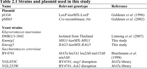
Fig. 2.2 Comparison of growth, sugar utilization and metabolite production
of K. marxianus and S. cerevisiae in liquid YPD. Cells were pre-cultured in 5 ml
of YPG at 30°C under a shaking condition at 160 rpm for 15-18 h. The initial
OD
660was adjusted to 0.1 and the cells were cultivated in 100 ml of YPD at 30°C
under a shaking condition at 160 rpm for 96 h. The parental strain of K.
marxianus DMKU3-1042 (straight line and open circles), Kmmig1 (straight line
and open squares), Kmrag5 (straight line and open triangles), the parental strain of
S. cerevisiae BY4741 (dotted line and filled circles), Scmig1 (dotted line and
filled squares) and Schxk2 (dotted line and filled triangles) were compared by
measuring OD
660(a) and the concentrations of glucose (b), glycerol (c) and
ethanol (d) in the medium. Data presented are averages of triplicate experiments,
and error bars indicate standard deviations.
In YPD medium (Fig. 2.2), Kmmig1 showed slightly higher OD
660and
about 1.5-3.0-times higher glycerol yield than those of the parental strain. The
ethanol yield of Kmmig1 at 12 h was 0.43 g/g, which was similar to that of the
parental strain (0.45 g/g). Scmig1 showed metabolic profiles similar to those of
Kmmig1, including relatively high accumulation of glycerol compared to that of
the parental strain and Schxk2. On the other hand, Kmrag5 Showed greatly
reduced growth and glucose utilization and produced small amounts of glycerol
and ethanol. Schxk2 showed almost no effect on utilization of glucose or
production of glycerol and ethanol, which were different from those of Kmrag5.
0 5 10 15 20 25 30 0 12 24 36 48 60 72 84 96 OD 660 Time (h) 0 0.5 1 1.5 2 2.5 0 12 24 36 48 60 72 84 96 G lu co se (%) Time (h) 0 0.02 0.04 0.06 0.08 0.1 0 12 24 36 48 60 72 84 96 G ly ce ro l ( %) Time (h) 0 0.2 0.4 0.6 0.8 1 1.2 0 12 24 36 48 60 72 84 96 Et ha no l ( %) Time (h) a b c d
The large defect of glucose utilization in Kmrag5 may be due to the reduction in
expression of RAG1 for a low affinity glucose transporter (Prior et al. 1993;
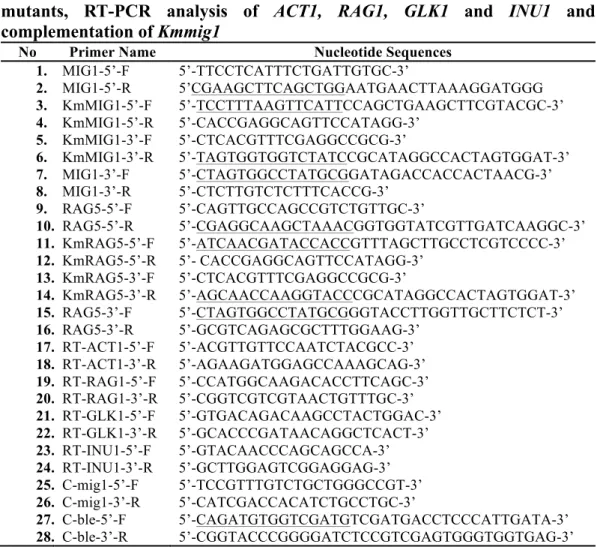
Betina et al. 2001). In YPDS medium (Fig. 2.3), Kmmig1 grew slightly better and
showed a slightly higher OD
600than did the parental strain until 48 h. However, as
in the case of YPD medium (Fig. 2.2), Kmrag5 grew very slowly and showed
very low levels of glucose utilization and ethanol production but extremely high
accumulation of fructose (0.54-0.76 g/g). Interestingly, the consumption of
sucrose in S. cerevisie was enhanced by Scmig1 and Schxk2 (Fig. 2.3C), probably
due to cancellation of glucose repression by these mutations. On the other hand,
no such difference was observed between Kmmig1 or Kmrag5 and the parental
strain. These findings suggest that S. cerevisiae is more sensitive to glucose
repression in a sucrose medium, being consistent with results in a previous report
(Lertwattanasakul et al. 2011). Moreover, Scmig1 and Schxk2 showed slow
consumption of fructose compared to that of the parental strain, probably due to
overhydrolysis of sucrose. Kmmig1 and Scmig1 accumulated relatively large
amounts of glycerol (0.05 and 0.03 g/g, respectively) compared to those of the
corresponding parental strains at 48 h.
Fig. 2.3 Comparison of growth, sugar utilization and metabolite production
of K. marxianus and S. cerevisiae in liquid YPDS. Cells were pre-cultured in 5
ml of YPG at 30°C under a shaking condition at 160 rpm for 15-18 h. The initial
OD
660was adjusted to 0.1 and the cells were cultivated in 100 ml of YPDS at
30°C under a shaking condition at 160 rpm for 96 h. The parental strain of K.
marxianus DMKU3-1042 (straight line and open circles), Kmmig1 (straight line
and open squares), Kmrag5 (straight line and open triangles), the parental strain of
S. cerevisiae BY4741 (dotted line and filled circles), Scmig1 (dotted line and
filled squares) and Schxk2 (dotted line and filled triangles) were compared by
measuring OD
660(a) and the concentrations of glucose (b), sucrose (c), fructose
(d), glycerol (e) and ethanol (f) in the medium. Data presented are averages of
triplicate experiments, and error bars indicate standard deviations.
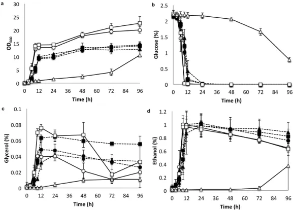
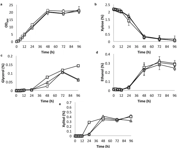
In YPX medium (Fig. 2.4), Kmmig1 showed slightly better growth,
slighlty higher turbidity, slightly faster xylose utilization at 24 h and larger
ethanol accumulation at 48 h than those of the parental strain and Kmrag5.
Kmmig1 showed increased accumulation of xylitol and glycerol at 24 h and 48 h,
respectively. These findings suggest that Mig1 represses the utilization of xylose
even in the absence of glucose and that increased utilization of xylose in Kmmig1
causes limitation of NAD
+, which leads to the accumulation of xylitol and
a c b d e f 0 5 10 15 20 25 30 35 0 12 24 36 48 60 72 84 96 OD 660 Time (h) 0 0.5 1 1.5 2 2.5 3 3.5 0 12 24 36 48 60 72 84 96 G lu co se (%) Time (h) 0 0.5 1 1.5 2 2.5 0 12 24 36 48 60 72 84 96 Su cr os e (%) Time (h) 0 0.5 1 1.5 2 0 12 24 36 48 60 72 84 96 Fr uc to se (%) Time (h) 0 0.1 0.2 0.3 0 12 24 36 48 60 72 84 96 G ly ce ro l ( %) Time (h) 0 0.5 1 1.5 2 0 12 24 36 48 60 72 84 96 Et ha no l ( %) Time (h)
glycerol as reported previously (Suprayogi et al. 2015; Suprayogi et al. 2016). In
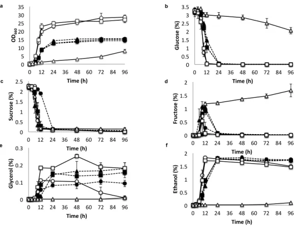
YPDX medium (Fig. 2.5), Kmmig1 showed slightly better growth than that of the
parental strain at 24 h and utilized glucose slightly faster but accumulated more
glycerol. This mutant started to utilize xylose after depletion of glucose around 12
h. Its consumption of xylose was fast at 48-72 h and it accumulated more xylitol
and glycerol than did the parent. On the other hand, the ethanol production of
Kmmig1 was similar to that of the parental strain. Kmrag5 grew slowly and
reached the same growth level as that of the parental strain after 48 h.
Consistently, glucose utilization in the mutant was slow and glucose was depleted
after 96 h. Xylose utilization was also slow in Kmrag5, but the utilization started
after 48 h when glucose still remained. Concomitantly, glycerol was dramatically
increased, but xylitol was not accumulated. Ethanol in Kmrag5 was accumulated
slowly, but after 48 h, it seemed to be produced from not only glucose but also
xylose. The growth and metabolic profiles of S. cerevisiae strains in YPDX
medium were also examined, but they could not utilize xylose (data not shown).
Enhanced accumulation of metabolites such as glycerol and xylitol was
observed in some liquid media in Kmmig1 and Kmrag5 mutants (Figs. 2.3 and
2.5). However, their accumulated amounts are very small compared to those in
other microorganisms reported (Pagliaro et al. 2007; Nolleau et al. 1993). On the
other hand, Kmrag5 was shown to accumulate fairly high levels of fructose in
YPDS liquid medium (Fig. 2.3). This ability of Kmrag5 might be useful for
bioconversion of sucrose-containing agricultural products to sweeter foods as in
the case of an HXK1 null mutant in K. marxianus NBRC 1777 (Zhang et al.
2017).
Fig. 2.4 Growth, sugar utilization and metabolite production of K. marxianus
DMKU3-1042 and its disrupted mutants in liquid YPX. Cells were
pre-cultured in 5 ml of YPG at 30°C under a shaking condition at 160 rpm for 15-18
h. The initial OD
660was adjusted to 0.1 and the cells were cultivated in 100 ml of
YPX at 30°C under a shaking condition at 160 rpm for 96 h. The parental strain of
K. marxianus DMKU3-1042 (straight line and open circles), Kmmig1 (straight
line and open squares) and Kmrag5 (straight line and open triangles) were
compared by measuring OD
660(a) and the concentrations of xylose (b), glycerol
(c) ethanol (d), and xylitol (e) in the medium. Data presented are averages of
triplicate experiments, and error bars indicate standard deviations.
a b c d e 0 5 10 15 20 25 0 12 24 36 48 60 72 84 96 OD 660 Time (h) 0 0.5 1 1.5 2 2.5 0 12 24 36 48 60 72 84 96 Xylose ( %) Time (h) 0 0.05 0.1 0.15 0.2 0 12 24 36 48 60 72 84 96 Glycerol ( %) Time (h) 0 0.1 0.2 0.3 0.4 0 12 24 36 48 60 72 84 96 Ethanol ( %) Time (h) 0 0.1 0.2 0.3 0.4 0.5 0.6 0.7 0 12 24 36 48 60 72 84 96 Xylitol ( %) Time (h)
Fig. 2.5 Growth, sugar utilization and metabolite production of K. marxianus
DMKU3-1042 and its disrupted mutants in liquid YPDX. Cells were
pre-cultured in 5 ml of YPG at 30°C under a shaking condition at 160 rpm for 15-18
h. The initial OD
660was adjusted to 0.1 and the cells were cultivated in 100 ml of
YPDX at 30°C under a shaking condition at 160 rpm for 96 h. The parental strain
of K. marxianus DMKU3-1042 (straight line and open circles), Kmmig1 (straight
line and open squares) and Kmrag5 (straight line and open triangles) were
compared by measuring OD
660(a) and the concentrations of glucose (b), xylose
(c) glycerol (d), ethanol (e), and xylitol (f) in the medium. Data presented are
averages of triplicate experiments, and error bars indicated standard deviations.
2.4.3 Effects of MIG1- and RAG5-disrupted mutations on hexokinase and
gluco-hexokinase activities
Since Kmrag5 and Kmmig1 grew much more slowly and sliglthty faster,
respectively, than the parental strain in YPD and YPDS media (Figs. 2.2 and 2.3)
and since RAG5 encodes hexokinase, the effects of MIG1- and RAG5-disrupted
mutations on hexokinase and gluco-hexokinase activities were determined (Fig.
2.6A). Kmmig1 exhibited specific activities of hexokinase and gluco-hexokinase
that were 1.1- and 1.3-times higher, respectively, than those of the parental strain.
Surprisingly, Kmrag5 showed almost no activity of either enzyme, indicating no
Fig. 5 Nurcholis et al.
a b c d e f 0 5 10 15 20 25 0 12 24 36 48 60 72 84 96 OD 660 Time (h) 0 0.5 1 1.5 2 2.5 0 12 24 36 48 60 72 84 96 G lu co se (%) Time (h) 0 0.5 1 1.5 2 2.5 0 12 24 36 48 60 72 84 96 Xy lo se (%) Time (h) 0 0.05 0.1 0.15 0.2 0 12 24 36 48 60 72 84 96 G ly ce ro l ( %) Time (h) 0 0.2 0.4 0.6 0.8 1 1.2 0 12 24 36 48 60 72 84 96 Et ha no l ( %) Time (h) 0 0.2 0.4 0.6 0.8 0 12 24 36 48 60 72 84 96 Xy lit ol (%) Time (h)
activity of glucokinase, which is encoded by GLK1. Therefore, it is likely that
Rag5 is involved in the expression of not only RAG1 but also GLK1. Interestingly,
a mutant, called No 23, having a RAG1 mutation of G270S by a single nucleotide
substitution seems to retain glucokinase activity but to have almost no hexokinase
activity (Suprayogi et al., 2015). The mutation of No 23 thus affects glucokinase
activity but not GLK1 expression regulation activity of Rag5. On the other hand,
the small influence of the disrupted mutation of Kmmig1 on both activities
suggests that Mig1 has a weak negative effect on the expression of either RAG1 or
GLK1.
2.4.4 Effects of MIG1- and RAG5-disrupted mutations on inulinase activity
To further examine the regulatory functions of Rag5 and Mig1, the effects
of MIG1- and RAG5-disrupted mutations on inulinase activity were determined
(Fig. 2.6) since INU1 encoding inulinase has been proposed to be under the
control of Mig1 (Lertwattanasakul et al. 2011). Kmrag5 showed about 80-times
higher inulinase activity than the parental strain. This finding suggests that Rag5
is a negative regulator for INU1 as well as RAG1 and GLK1. The mutation of
Kmmig1, however, had almost no effect on inulinase activity. Notably, the
mutation of No 23 increased inulinase activity, though the activity was about half
of that in Kmrag5, suggesting that the mutation of No 23 causes reduction of
INU1 expression regulation activity of Rag5 (Fig. 2.6B). These results including
results of experiments on hexokinase and gluco-hexokinase activities indicate that
MIG1 and RAG5 in K. marxianus play different roles in gene expression
Fig. 2.6 Hexokinase, gluco-hexokinase and inulinase activities of K.
marxianus DMKU3-1042 and its disrupted mutants. Cells were pre-cultured in
5 ml of YPG at 30°C under a shaking condition at 160 rpm for 15-18 h.
Hexokinase (dot and black bar) and gluco-hexokinase (backslash and gray bar)
activities (a). The initial OD
660was adjusted to 0.1 and the cells were cultivated in
100 ml of YPD at 30
oC under a shaking condition at 160 rpm until 50% of the
glucose remained. Bars represent ±SD of values from experiments performed in
triplicate. Inulinase activity (gray bar) (b). The initial OD
660was adjusted to 0.1
and the cells were cultivated in 100 ml of YPD at 30
oC under shaking condition at
160 rpm until 50% of the glucose remained. Supernatants were collected by
centrifugation at 5,000 rpm for 10 min and then used for examination of inulinase
activity by using the DNS method. Bars represent ±SD of values from
experiments performed in triplicate. Each single asterisk indicates significantly
higher or lower enzyme activity than that of the parental strain (P<0.05).
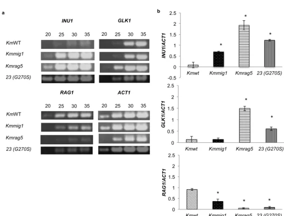
2.4.5 Effects of MIG1- and RAG5-disrupted mutations on expression of INU1,
GLK1, and RAG1
To further examine the regulatory functions of MIG1 and RAG5 at the
transcription level on the expression of INU1, GLK1 and RAG1, RT-PCR was
carried out (Fig. 2.7). The expression of INU1 has been shown to be enhanced by
MIG1 and RAG5. RAG5 disruption caused a dramatic increase in INU1
expression, being consistent with its effect on inulinase activity, while MIG1
disruption caused a slight increase in INU1 expression, though its effect on
inulinase activity was negligible (Figs. 2.6B and 2.7B). The expression levels of
INU1 and GLK1 in Kmrag5 were 22-times and 11-times higher, respectively, than
those of the parental strain. However, RAG1 expression levels in Kmmig1 and
Kmrag5 was about 2.5-times and 18-times lower, respectively, than that of the
parental strain. Disruption of MIG1 had almost no effect on GLK1 expression but
increased INU1 expression to a level about 8-times higher than that of the parental
0 20 40 60 80 100 120 KmWT Kmmig1 Kmrag5 23 (G270S)
Relative inulinase activity
0.0 0.3 0.6 0.9 1.2 1.5 1.8 KmWT Kmmig1 Kmrag5 23 (G270S)
Specific activity (U/mg)
a b * * * * * * *
strain. Therefore, these results suggest that RAG5 is a negative regulator for INU1
and GLK1 and a positive regulator for RAG1 and that MIG1 might be involved as
a minor factor in the negative regulation of INU1 and the positive regulation of
RAG1.
Fig. 2.7 Expression of KmINU1, KmGLK1 and KmRAG1 in YPD under a
shaking condition. Cells grown in YPG medium for 18 h were cultivated in YPD
at 30
oC under a shaking condition at 160 rpm for 7 h (KmWT and Kmmig1), for
18 h (Mutant No 23) and for 66 h (Kmrag5). Total RNA was then isolated and
subjected to RT-PCR with primers specific to corresponding genes that amplify
an approximately 500-bp DNA fragment. (a) After reverse transcriptase reaction,
PCR products of 20, 25, 30 and 35 cycles were subjected to 1.2% agarose gel
electrophoresis and stained with ethidium bromide. (b) Relative band intensities
were determined using scanned images and IMAGE-J software. Each single
asterisk indicates significantly higher or lower gene expression level than that of
the parental strain (P<0.05).
2.4.6 Effects of MIG1- and RAG5-disrupted mutations on growth in a
minimal medium containing glucose or galactose and amino acids
To further examine the effects of mutations of MIG1 and RAG5 on hexose
sugar utilization, growth rates in a minimal medium containing glucose or
galactose were compared (Fig. 2.8). As expected, Kmrag5 grew much more
a KmWT Kmmig1 Kmrag5 INU1 ACT1 GLK1 KmWT Kmmig1 Kmrag5 RAG1 23 (G270S) 23 (G270S) 20 25 30 35 20 25 30 35 20 25 30 35 20 25 30 35 0 0.5 1 1.5 2 2.5 Kmwt Kmmig1 Kmrag5 23 (G270S) RAG1 /ACT1 0 0.5 1 1.5 2 2.5 Kmwt Kmmig1 Kmrag5 23 (G270S) GLK1 /ACT1 -0.5 0 0.5 1 1.5 2 2.5 Kmwt Kmmig1 Kmrag5 23 (G270S) INU1 /ACT1 b * * * * * * * *
slowly than the parental strain in glucose, being consistent with data shown in Fig.
2.1. Surprisingly, Kmmig1 exhibited almost no growth in a glucose or galactose
medium. We thus examined the requirement of nutrients for Kmmig1 and found
that histidine is essential for the mutant (Fig. 2.8). Since this is the first indication
of involvement of Mig1 in histidine biosynthesis, complementation experiments
were performed by introduction of MIG1 from the parental strain. It was found
that the MIG1-complemented strain grew in glucose minimal medium without
histidine. Therefore, these findings suggest that Mig1 is involved in the
expression of some genes related to histidine biosynthesis in K. marxianus. On
the other hand, the reqirement of Mig1 in S. cerevisiae is not clear because the
strain used in this study has genotypes of his, met, leu and ura.
Fig. 2.8 Effect of histidine on the growth of K. marxianus DMKU3-1042 and
its disrupted mutants on YNB agar containing glucose or galactose. Cells
were pre-cultured in 5 ml of YPG at 30°C under a shaking condition at 160 rpm
for 15-18 h. The cells were streaked on YPD and YPG as a control and on a Yeast
Nitrogen Base (YNB) plate containing 2% (w/v) glucose or 2% (w/v) galactose or
2% (w/v) glycerol and supplemented with 0.01% (w/v) histidine. The plates were
incubated at 30
oC and observed at 48 h.
2.5 Discussion
In order to understand the physiological functions of Mig1 and Rag5
including their contribution to glucose repression in K. marxianus DMKU3-1042,
we constructed two kanMX4-inserted mutants, Kmmig1 and Kmrag5, and
examined the effects of the mutations on sucrose utilization in the presence of
YPD YNB + 2% Glc YNB + 2% Glc + 0.01% His
YNB + 2% Gal YNB + 2% Gal + 0.01% His YPG
KmWT Kmmig1
Kmrag5 Kmmig1 +