会 期:平成 25年 6月 6日(木) ・7日(金) ・8日(土)
年会長:工藤 奨(九州大学大学院工学研究院)
会 場:九州大学 西新プラザ
第36回日本バイオレオロジー学会年会
プログラム・抄録集
日バイレオ誌(B & R ,電子版)第27巻 第2号
J . J pn. Soc. Bi or heol . 27( 2) ( 2013)
第 36 回
日本バイオレオロジー学会年会 プログラム・抄録集
会期:平成 25 年 6 月 6 日(木),7 日(金) ,8 日(土)
年会長:工藤 奨
九州大学 大学院 工学研究院 機械工学部門 会 場:九州大学 西新プラザ
問い合わせ先:九州大学大学院工学研究院 機械工学部門 年会実行委員会 工藤 奨・中嶋和弘
実行組織
年会長 工藤 奨 九州大学
実行委員長 中嶋 和弘 九州大学
実行委員 高松 洋 九州大学
澤江 義則 九州大学
山口 哲生 九州大学
日本バイオレオロジー学会年会のあゆみ
回 年会長 所 属 会 場 会 期
1 深田 栄一 理化学研究所 東京慈恵会医大学 高木会館講堂 1978/6/19 2 岡 小天 国立循環器病センター 国立循環器病センター 講堂 1979/6/30~7/1 3 東 健彦 信州大学 信州大学 医学部第一講義堂 1980/6/28~29 4 谷口 興一 東京医科歯科大学 東京医科歯科大学 5号館 1981/6/20~21 5 梶谷 文彦 川崎医科大学 川崎医科大学 現代医学教育博物館 1982/6/26~27 6 稲垣 義明 千葉大学 千葉県文化会館 小ホール 1983/6/18~19 7 神谷 瞭 北海道大学 北海道自治会館 自治ホール 1984/6/16~17 8 浅野 牧茂 国立公衆衛生院 国立公衆衛生院 講堂 1985/6/15~16 9 志賀 健 愛媛大学 愛媛県医師会館 ホール 1986/6/11~13 10 磯貝 行秀 東京慈恵会医科大学 東京慈恵会医大学 高木会館講堂 1987/6/13~16 11 松田 保 金沢大学 金沢大学 医学部十全講堂 1988/6/2~4 12 大島 宣雄 筑波大学 筑波大学 大学会館国際会議室 1989/7/5~7 13 峰下 雄 帝塚山短期大学 奈良県新公会堂 1990/6/21~23 14 品川 嘉也 日本医科大学 日本医科大学 大講堂 1991/6/20~22 15 平川 千里 岐阜大学 岐阜市文化センター 1992/6/25~27 16 菅原 基晃 東京女子医科大学 東京女子医大学 弥生記念講堂 1993/6/16~17 17 松信 八十男 清和大学 エーザイホール 1994/6/17~18 18 貝原 学 帝京大学 TEPCO地球館 1995/6/15~16 19 辻 隆之 国立循環器病センター 千里ライフサイエンスセンター 1996/6/6~7 20 増田 善昭 千葉大学 千葉大学 けやき会館 1997/6/5~6 21 前田 信治 愛媛大学 エスポワール愛媛文教會舘 1998/6/11~13 22 貝原 真 理化学研究所 理化学研究所 鈴木梅太郎記念ホール 1999/6/10~11 23 辻岡 克彦 川崎医科大学 倉敷公民館 2000/6/8~9 24 谷下 一夫 慶應義塾大学 慶應義塾大学 創想館マルチメディアホール 2001/6/7~8 25 大橋 俊夫 信州大学 信州大学 旭会館大会議室 2002/6/6~7 26 西成 勝好 大阪市立大学 大阪市立大学学術情報総合センター 2003/6/5~6 27 内村 功 東京医科歯科大学 東京医科歯科大学 特別講堂 2004/6/10~11 28 佐藤 正明 東北大学 東北大学 マルチメディア教育研究棟 2005/7/7~8 29 丸山 徹 九州大学 九州大学医学部 コラボステーション 2006/6/12~13 30 佐々木 直樹 北海道大学 北海道大学 学術交流会館 2007/6/14~15 31 安藤 譲二 東京大学 東京大学理学部 小柴ホール 2008/6/5~6 32 土橋 敏明 群馬大学 桐生市民文化会館 2009/6/4~5 33 氏家 弘 東京労災病院 理化学研究所 鈴木梅太郎記念ホール 2010/6/3~4 34 関 眞佐子 関西大学 関西大学 100周年記念会館 2011/6/3~4 35 佐藤 恵美子 新潟県立大学 朱鷺メッセ 新潟コンベンションセンター 2012/5/31~6/2 36 工藤 奨 九州大学 九州大学 西新プラザ 2013/6/6~8
年会,総会,理事会,リサーチ・フォーラム会場へのアクセス
年会,フォーラム,理事会,各編集委員会会場:九州大学 西新プラザ
〒814-0002 福岡市早良区西新2-16-23 TEL 092-831-8104 FAX 092-831-8105 福岡市営地下鉄「西新」駅下車、⑦番出口より徒歩約10分
(ホテル日航新潟)
年会,総会,理事会,リサーチ・フォーラム,各委員会会場 2階 見取り図
年会会場 第1会場 :大会議室
第2会場 :中会議室 ポスター発表 :多目的室
総会 :大会議室
理事会評議員会合同会議 :中会議室 電子版B&R編集委員会 :小会議室 Vascular Engineering出版打ち合わせ会 :小会議室 リサーチ・フォーラム会場 :中会議室
大会本部 :多目的室
懇親会会場へのアクセス
懇親会会場: ヒルトン福岡シーホーク 34F タワーペントハウス
〒810-8650 福岡市中央区地行浜2-2-3 TEL:092-844-8111 年会会場より貸切バスの送迎があります.
年会会場より徒歩20分
福岡市営地下鉄 西新・唐人町駅より徒歩約19分・タクシー約6分・バス約6分 ヒ ル ト ン 福 岡
シーホーク
九州大学 西新プラザ
交通のご案内
・九州大学西新プラザまでのアクセス
□鉄道を利用
JR博多駅から,
○福岡市営地下鉄利用の場合
西唐津,筑前前原,姪浜,西新行きいずれかに乗車,「西新」駅下車(約13分,250円). ○タクシー利用の場合 JR博多駅駅博多口タクシー乗り場より約20~30分
□飛行機を利用 福岡空港から
○福岡市営地下鉄「福岡空港」駅より乗車,「西新」駅下車(約19分,290円). ○タクシー利用の場合 福岡空港タクシー乗り場より約30~40分
ご 案 内
ご参加の皆様へ
受 付 時 間 :6月7日(金)8:40より
場 所 :九州大学 西新プラザ 多目的室
参加費 :5,000円(会員) (参加費には,講演抄録集1冊の代金が含まれます.)
7,000円(非会員) (参加費には,講演抄録集1冊の代金が含まれます.)
3,000円(学生)
講 演 抄 録 集 :1,000円
懇 親 会 日 時 :6月7日(金)18:00~20:00
場 所 :ヒルトン福岡シーホーク 34階 タワーペントハウス 参加費 :正会員5,000円
学生会員 2,000円(受付にてお申し込み下さい.)
会員の皆様へ 総会
開 催 日 時 :6月8日(土)11:00~11:30 場 所 :第1会場(大会議室)
名誉顧問・名誉会員・理事・監事・評議員の皆様へ 理事会評議員会合同会議
開 催 日 時 :6月6日(木)13:00~15:30 場 所 :第2会場(中会議室)
バイオレオロジー・リサーチ・フォーラムご参加の皆様へ 開 催 日 時 :6月6日(木)16:00~18:00 場 所 :第2会場(中会議室)
電子版B&R編集委員の皆様へ 電子版B&R編集委員会
開 催 日 時 :6月7日(金)11:50~12:40 場 所 :小会議室
Vascular Engineering執筆者の皆様へ Vascular Engineering出版打ち合わせ会
開 催 日 時 :6月8日(土)11:30~12:30 場 所 :小会議室
口頭発表セッションの座長の皆様へ
ご担当されるセッション開始時間の10分前までには次座長席にお着き下さい.活発な討論とな りますようにお願い申し上げます.
オーガナイズドセッション,シンポジウム,学会賞受賞講演 発表者の皆様へ
(1) オーガナイズドセッション,シンポジウムおよび学術奨励賞応募講演の発表時間は8分,討論
は4分です.学会賞受賞講演につきましては,別途ご連絡いたします.
(2) 各演者は,発表時間の30分前までに受付をお済ませ下さい.
(3) 会場には,液晶プロジェクターとレーザーポインター,およびマイクをご用意いたします.
(4) 各演者は前演者が発表している間に次演者席にお着き下さい.
機器の使用について
1. 各演者はご自身のコンピュータを接続して下さい.会場にはコンピュータを用意致しませんので,
各自でコンピュータをご用意ください.
2. コンピュータの操作は,発表者自身または共同演者で行ってください.
3. 会場には,プロジェクターとの接続コードとしてミニD-Sub 15ピン(オス)のケーブルを用意
します.(下図参照)出力端子がこれに合わない場合はアダプタをご持参下さい.また音声は会 場スピーカーへの直接出力は準備しておりません.音声ご使用の際は,演者用マイクを近づけ るなどして下さい.
4. ノートパソコンは機種によって,端子の形状や操作の異なる場合があります.ご自身のパソコン を熟知した上でお越し下さい.
5. 接続したコンピュータは,電源を切ったりサスペンドの状態などにしたりはしないで,すぐに発 表ができる状態にしておいてください.
6. 接続トラブル等による発表時間の延長は認められません.講演開始前の休憩時間に,予め試写を していただきますようお願いいたします.
プロジェクター・パソコン接続用ケーブル端子(D-sub 15ピン)
ポスター発表者の皆様へ
(1) ポスターの掲示は,6月8日(金)12:00までに,指定場所(多目的室)に貼りつけてください.
(ピンなどは年会にて用意します.)
(2) ポスターセッションは,6月8日(金)14:15~14:45です.セッション中は,ポスターの前に立
ち,聴衆に対して説明と質疑応答を行ってください.
(3) ポスター発表では,登壇しての発表はありませんが,ポスターセッション中に,演題毎に座長 を割り当て,コアタイムを設定します.コアタイムになれば,座長の指示に従い,ポスターの 前で5分程度の説明と2分程度の質疑応答を行ってください.なお発表時間および座長につい ては,ポスター掲示用パネル上部に貼り付けてある演題番号下にも記載してあります.
(4) ポスター前でPCなどを使いながら説明をすることも可能ですが,電源は用意していません.
(5) ポスターは,6月8日(土)の15:00以降に撤去してください.なお6月8日(土)の17:00を
過ぎて放置されているポスターは,年会事務局にて処分します.
ポスターの作成について
1. ポスターを貼り付けるパネルのサイズは,ヨコ110cm×タテ 210cmです.このパネルのサイズ
に収まるよう,ポスターを作成してください.演題番号は年会でパネル左上端に貼っておきま す.(下左図参照)
2. 文字や図表のサイズ,レイアウトを工夫し、離れたところからでもわかりやすいポスターを作成 することを心がけてください.
3. 「目的」・「方法」・「結果」・「結論」などを明確にし,目的は左上部に,結論は右下部になるよう
に配置してください.(下右図参照)
4. タイトル・発表者氏名・所属に続いて代表者の連絡先メールアドレスを記入してください.
○中嶋和弘*,**,隅井干城***,荒井雅隆***,工藤 奨*
*九州大学 大学院 工学研究院 機械工学部門
**九州大学 バイオメカニクス研究センター
***九州大学 大学院 工学府 機械工学専攻 連絡先:[email protected]
第36回バイオレオロジー学会年会 ポスターセッション
-ポスターの作成について(例)-
以下,発表内容を 1.緒言・目的 2.実験装置および方法 3.結果
4.考察 5.結論
等,項目に分けて記載
(できる限り緒言・目的は左上部,結語は右 下部になるように配置する)
フォントサイズ等については,特に指定しま せんが,聴講者が読みやすいように比較的 大きめのサイズにしておいてください.
講演タイトル
筆者(発表者に○を付す)
所属
連絡先(代表者のメールアドレス)
これら情報はかならず記載すること
ポスターの作成例
バイオレオロジー・リサーチ・フォーラムのご案内
6月6日(木)16:00~18:00 第2会場(中会議室)
司会:工藤 奨(九州大)
「細胞操作技術の新展開」
1.「細胞操作メカノバイオマテリアル」 木戸秋 悟
(九州大学 先導物質化学研究所)
2.「電界誘起気泡メスによる細胞加工・インジェクション技術」 山西 陽子
(芝浦工業大学 工学部 機械工学科)
特別講演のご案内
6月7日(金)13:50~14:50 第1会場 司会:工藤 奨(九州大)
「生体関節におけるバイオレオロジー」 村上輝夫(九州大学)
Japan – Korea Joint Symposiumのご案内
6月7日(金)15:00~17:30 第1会場 Chair:Toshiaki Dobashi (Gunma University) Keynote Lecture 1
INTEGRATED MICROFLUIDIC SYSTEM FOR HEMORHEOLOGICAL MEASUREMNETS: PLATELET AGGREGATION
Sehyun Shin (Korea University) Keynote Lecture 2
BIORHEOLOGICAL ASPECT OF MICROVESSEL FORMATION
Kazuo Tanishita (Waseda University) Chair:Masako Seki (Kansai University)
Keynote Lecture 3
THE ROLE OF CRITICAL SHEAR STRESS ON ACUTE CORONARY SYNDROME
Byoung Kwon Lee (Yonsei University College of Medicine) Keynote Lecture 4
VASCULAR MECHANOBIOLOGY: FLOW DETECTION AND CALCIUM SIGNALING IN ENDOTHELIAL CELLS
Kimiko Yamamoto (The University of Tokyo)
岡小天賞・論文賞受賞講演のご案内
6月8日(土)12:30~14:00 第1会場(大会議室)
司会:大橋俊朗(北海道大),山本 希美子(東京大)
岡小天賞受賞講演
学術奨励賞応募講演のご案内
6月7日(金)10:20~11:44 第1会場(大会議室)
座長: 佐々木直樹(北海道大),工藤 奨(九州大)
優れた研究発表をした若手の日本バイオレオロジー学会会員を顕彰します。応募者の本セッションで の発表に対する審査員の評点で,学術奨励賞受賞論文を決定します.年会 3 日目の総会で受賞論文名を 発表し,受賞者には賞状および副賞として金一封を授与します.
第36回日本バイオレオロジー学会年会プログラム
○印:口頭発表登壇者,またはポスター発表の発表者
第 1 日目 6 月 6 日(木)
第2会場
16:00~18:00 バイオレオロジー・リサーチフォーラム
司会: 工藤 奨(九州大)
「細胞操作技術の新展開」
1 細胞操作メカノバイオマテリアル
○木戸秋 悟(九州大学 先導物質化学研究所)
2 電界誘起気泡メスによる細胞加工・インジェクション技術
○山西 陽子(芝浦工業大学 工学部 機械工学科)
第 2 日目 6 月 7 日(金)
第1会場
9:10~10:10 OS7:ヘルスケア食品レオロジー p.27~31 座長:市川 寿(長崎大),吉村美紀(兵庫県立大),佐藤恵美子(新潟県立大)
OS7-1 しめ鯖づくりと体積相転移現象 川口美咲(長崎大),○市川 寿
OS7-2 ゲル強度に優れた魚鱗由来熱水抽出ゼラチンのキャラクタリゼーション
岡 智子(長崎大),Wang Xi,○市川 寿,関口拓磨(群馬大),近藤信吾,槇 靖幸,土 橋敏明
OS7-3 スケトウダラすり身の低温ゲル化過程のレオロジー特性
○金田 勇(酪農学園大), 青沼航平, 舩津保浩
OS7-4 コーンスターチゲルのレオロジー特性に及ぼす米粉添加の影響
○佐藤恵美子(新潟県立大),佐竹妙子
OS7-5 トマトゼリーの食味と物性に関する研究 -高齢者と若年者の比較-
○筒井和美(愛知教育大),深川琴恵,田村朝子(新潟県立大),荒井冨佐子,宮西邦夫,
佐藤恵美子,廣神里奈,早瀬和利(愛知教育大),西成勝好(湖北工業大),金巻栄作(金 巻屋),二瓶英子(有明ハイツ),秋間美也子
10:20~11:44 学術奨励賞応募講演 p.32~38 座長:佐々木直樹(北海道大),工藤 奨(九州大)
A-1 単一赤血球変形能診断システム開発へ向けた実験装置の試作とその動作検証
○都築達也(芝浦工業大),渡邉宣夫
A-2 プリオンタンパク質におけるアミロイド性集合体を防止する作用機序の解明
○佐伯政俊(山口東京理科大),森井尚之(産業技術総合研究所),橋本慎二(山口東京理 科大)
A-3 光学顕微鏡による熱拡散現象の直接的観測法の開発
○村田飛鶴(東海大),喜多理王,新屋敷直木,八木原晋
A-4 小型FALLING NEEDLE RHEOMETERを用いた血液の流動特性の解析
○鈴木貴雅(関西大),山本秀樹,川村公人(アサヒグループホールディングス株式会社), Dominik Bernitzky(Medical University of Vienna),Roberto Plasenzotti
A-5 温度勾配による糖およびエチレングリコール水溶液の輸送現象
○前田晃作(東海大),篠原春香,喜多理王,新屋敷直木,八木原晋
A-6 局所的に非一様な粒子群中の透過流動
○大友涼子(関西大),原田周作(北海道大)
A-7 摩擦負荷培養法による再生軟骨組織形成とその力学特性に関する研究
○福田圭祐(九州大学),澤江義則,小俣誠二
12:40~13:16 OS1:血管障害と流体力学 p.39~41 座長:田地川勉(関西大)
OS1-1 Flow Diverter(FD)を留置した脳動脈瘤内の血流シミュレーション
○鈴木貴士(東京理科大),高尾洋之(東京慈恵会医科大),石橋敏寛,増田俊輔,門倉 翔
(東京理科大),Ashraf Mohamed(東京慈恵会医科大),銭 逸(マッコーリ大),山本 誠
(東京理科大),村山雄一(東京慈恵会医科大)
OS1-2 拍動流中にある弾性壁動脈瘤モデルの壁せん断応力の変動
○山口隆平(芝浦工業大),氏家 弘(東京労災病院)
OS1-3 脳動脈瘤内の血流に及ぼすステントの影響
○増田俊輔(東京慈恵会医科大,サイバネットシステム),高尾洋之(東京慈恵会医科大), 鈴木貴士,門倉 翔(東京理科大),モハメドアシュラフ(東京慈恵会医科大),銭 逸(マ ッコーリ大),山本誠(東京理科大),村山雄一(東京慈恵会医科大)
13:50~14:50 特別講演 p.22 司会:工藤 奨(九州大)
生体関節におけるバイオレオロジー
○村上輝夫(九州大)
15:00~17:30 日韓シンポジウム Japan-Korea Joint Symposium p.23~26 Chair:Toshiaki Dobashi (Gunma University)
S-1 INTEGRATED MICROFLUIDIC SYSTEM FOR HEMORHEOLOGICAL MEASUREMNETS: PLATELET AGGREGATION
○Sehyun Shin (Korea University)
S-2 BIORHEOLOGICAL ASPECT OF MICROVESSEL FORMATION
○Kazuo Tanishita (Waseda University), Yoshinori Abe (Keio University), Mariko Ikeda (Keio University)
Chair:Masako Seki (Kansai University)
S-3 THE ROLE OF CRITICAL SHEAR STRESS ON ACUTE CORONARY SYNDROME
○Byoung Kwon Lee (Yonsei University College of Medicine), Sehyun Shin (Korea University)
S-4 VASCULAR MECHANOBIOLOGY: FLOW DETECTION AND CALCIUM SIGNALING IN ENDOTHELIAL CELLS
○Kimiko Yamamoto (The University of Tokyo), Joji Ando (Dokkyo Medical University)
第2会場
9:10~10:10 OS5:ティッシュエンジニアリング・人工臓器(1) p.42~46 座長:岩崎清隆(早稲田大)
OS5-1 血液凝固能および血小板凝集能からみた抗血栓性コーティングの特性に関する研究
○水野敏秀(国立循環器病研究センター研究所),山岡哲二,築谷朋典,武輪能明,巽 英 介
OS5-2 低侵襲体外循環を目指した低充填量回路の作製 -ラット体外循環モデルを用いた評価-
○藤井 豊(国立循環器病研究センター研究所),巽 英介,白井幹康,武輪能明,妙中義 之
OS5-3 空気圧駆動式人工心臓用小型駆動装置のための空気流量を利用した拍出流量モニタに関す る実験的検討
○大沼健太郎(国立循環器病研究センター研究所),本間章彦(東京電機大),住倉博仁(国 立循環器病研究センター研究所),築谷朋典,武輪能明,水野敏秀,向林宏(株式会社イワ キ),小嶋孝一,片野一夫(東京電機大),妙中義之(国立循環器病研究センター研究所),
巽 英介
OS5-4 表面粗さを付加した回転せん断負荷装置内全体の血流解析
○西田正浩(産業技術総合研究所),工藤大樹(茨城大),丸山 修(産業技術総合研究所), 山根隆志(神戸大),松田健一,増澤 徹(茨城大)
OS5-5 循環器系人工臓器の粘弾性学的血液適合性評価
○丸山 修(産業技術総合研究所),可児裕基(東京理科大),小阪 亮(産業技術総合研 究所),西田正浩,山根隆志(神戸大),巽 英介(国立循環器病研究センター研究所),妙 中義之
12:40~13:28 OS5:ティッシュエンジニアリング・人工臓器(2) p.47~50 座長:西田正浩(産業技術総合研究所)
OS5-6 自己組織心臓代用弁(バイオバルブ)の生体外機能評価
○住倉博仁(国立循環器病研究センター研究所),中山泰秀,大沼健太郎,武輪能明, 巽 英介
OS5-7 浮遊撹拌培養法によるES細胞の未分化維持培養に関する検討
○水本博(九州大),導寺香奈,網本直記,梶原稔尚
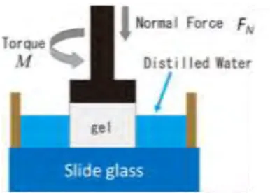
OS5-8 PVAハイドロゲルの変形特性と摩擦特性
○中嶋和弘(九州大),鎗光清道,澤江義則,村上輝夫
OS5-9 石灰化マルチチャネルコラーゲンゲルを用いた多階層構造を持つ再生骨組織の構築 安井 憲(北海道大),○古澤和也,福井彰雅,佐々木直樹
第 3 日目 6 月 8 日(土)
第1会場
9:00~9:48 OS6:生体物質の構造形成と機能発現・制御(1) p.52~55 座長:喜多理王(東海大)
OS6-1 低摩擦ハイドロゲルにおける応力-拡散結合
○山口哲生(九州大),村上輝夫
OS6-2 リン脂質二分子膜リポソームの変形とリン脂質単分子膜の凝集状態 横尾麻衣(佐賀大),黒田修未,○成田貴行,大石祐司
OS6-3 醤油-アルギン酸ペーストの粘弾性挙動
○松田夏希(佐賀大),井上侑子,Saphwan Al-Assaf(Glyndwr Univ), 大石祐司(佐賀 大), 成田貴行
OS6-4 ゼラチンゲルのレオロジーとヘリックス形成
○槇 靖幸(群馬大),渡部 翔,矢島一樹,松尾光一(広島大)
10:00~10:48 OS6:生体物質の構造形成と機能発現・制御(2) p.56~59 座長:山口哲生(九州大)
OS6-5 マルチチャネルコラーゲンゲルの階層構造に与える温度の影響
○古澤和也(北海道大),花崎洋平,増元淳一,福井彰雅,佐々木直樹
OS6-6 DNA-陽イオン界面活性剤複合体フィルムの構造と物性
○佐々木直樹(北海道大),E. C. Ossai,岡田昴樹,大木翔太,古澤和也,福井彰雅 OS6-7 蠕動運動による腸内気泡の運動
○多羅尾範郎(聖隷クリストファー大)
OS6-8 気管支分岐付近での痰塊表面の剥離
○多羅尾範郎(聖隷クリストファー大)
12:30~14:00 岡小天賞・論文賞受賞講演 司会:大橋俊朗(北海道大),山本 希美子(東京大)
15:00~16:12 OS4:細胞・分子のメカノバイオロジー p.60~65 座長:大橋俊朗(北海道大),安達泰治(京都大)
OS4-1 マイクロチャネルデバイスを用いた細胞遊走時の牽引力計測
○大橋俊朗(北海道大),菅原章人, Justin Cooper-White(The University of Queensland)
OS4-2 AFMを用いたアクチンフィラメントの曲率依存的なARP2/3との相互作用変化測定
○韓 成雄(京都大),森田恭平,安達泰治 OS4-3 細胞のマクロ形態とナノ構造の力学的制御
○出口真次(東北大),松井 翼,齋藤 明,佐藤正明
OS4-4 間質流制御による3次元血管ネットワークの形成
阿部順紀(慶應義塾大),谷下一夫(早稲田大),○須藤 亮(慶應義塾大)
OS4-5 低酸素環境下でのニューロン活性化が脳血管へ与える影響
○荒井雅貴(九州大),佐藤竜也(芝浦工業大),工藤 奨(九州大)
OS4-6 肝星細胞の遊走における力学刺激と受容体の関連性
○隅井干城(九州大),藤田陵佑(芝浦工業大),岩下 洸,工藤 奨(九州大)
第2会場
9:00~9:48 OS2:循環器ダイナミクスと疾患 p.66~69 座長:築谷朋典(国立循環器病研究センター研究所)
OS2-1 内臓循環系に及ぼす脊髄損傷の影響と再生医療について
○飯田紀子(北海道文教大)
OS2-2 骨粗鬆症による脊椎の変形と腹部大動脈瘤の関係 ―共同研究者・研究支援者を求めていま
す―
○本田 透(香川県立中央病院)
OS2-3 粥状動脈硬化斑の伸展試験
○山田 宏(九州工業大),坂田則行(福岡大)
OS2-4 多孔薄膜カバードステントの開発(生体外模擬実験による瘤塞栓性能の予測)
○田地川勉(関西大),紅林芳嘉,西 正吾(札幌東特洲会病院),中山泰秀(国立循環器 病研究センター)
10:00~10:48 OS3:血液レオロジーと微小循環(1) p.70~73 座長:渡邉宣夫(芝浦工大)
OS3-1 大きさの異なる液滴のマージネーションのシミュレーション
○牧野真人(山形大),関眞佐子(関西大)
OS3-2 円管内流れ中の粒子の分布
○名村和平(関西大),三浦和真,板野智昭,関眞佐子
OS3-3 マイクロチャンネル法によるヒト赤血球変形能の定量評価(形状回復時定数に対する糖尿 病指標の影響)
○能田卓弥(関西大),久保田麻紀,田地川勉,池本敏行(大阪医科大),田窪孝行(大阪 医科大)
OS3-4 マイクロチャネル内流れ中の血小板模擬粒子の運動
○田中慎之介(関西大),関 淳二,板野智昭,関眞佐子
15:00~15:48 OS3:血液レオロジーと微小循環(2) p.74~77 座長:丸山 徹(九州大)
OS3-5 赤血球集合に与える赤血球表面糖鎖の影響
○外山吉治(群馬大),須田 巧,窪田健二
OS3-6 非接触レーザー血流計による生体顕微鏡下での実質臓器微小血管血流計測
○林浩大(芝浦工業大),柴田政廣
OS3-7 マクロおよびマイクロ血液循環解析手法によるポアズイユの法則の実験的検証
○横川和弘(芝浦工業大),柴田政廣 OS3-8 骨格筋微小循環における酸素ダイナミクス
○柴田政廣(芝浦工業大),濱島早紀,川村彩智
ポスター発表会場
14:15~14:45 ポスターセッションコアタイム(1) p.78~81 座長:須藤 亮(慶應義塾大)
P-1 高イオン強度下でのフィブリン凝集に対する糖鎖切除効果
○窪田健二(群馬大),外山吉治,行木信一,落合正則(北海道大)
P-2 DYNAMIC SHEAR RESPONSES OF POLYMER-POLYMER INTERFACES
○名嘉山祥也(九州大),梶原稔尚
P-3 血管内皮細胞内PKCの局在変化におよぼす局所引張の影響
○隅井干城(九州大),木村恭彰(芝浦工業大),島田知弥,土屋宏紀,工藤奨(九州大)
P-4 せん断応力負荷時の血管内皮細胞内アクチン局在特性
○荒井雅貴(九州大),寺田麻理枝(芝浦工業大),工藤奨(九州大)
14:15~14:45 ポスターセッションコアタイム(2) p.82~85 座長:出口真次(名古屋工業大)
P-5 チョウザメ由来アテロコラーゲンのゲル化に伴う粘弾性変化
○関口拓磨(群馬大),槇 靖幸,近藤信吾,土橋敏明,元村まみ(長崎大),市川 寿,小 柳佳子(北海道大),岸村栄毅,安部智貴,足立伸次,都木靖彰
P-6 豆乳・米粉混合ゲルの酸凝固
○吉村美紀(兵庫県立大),江口智美,東羅あかね,中川究也
P-7 うどん麺のテクスチャーに及ぼす投入温度とタピオカ澱粉配合の影響
○江口智美(兵庫県立大),山本美晴,吉村美紀 P-8 飲料中亜硝酸の還元によるNO産生
小川武人(川崎医療福祉大),○望月精一
- 19 -
年会タイムテーブル
第1日目 6月6日(木)
第1会場(大会議室) 第2室(中会議室)
13:00~15:00
理事会評議員会合同会議 休憩
16:00~18:00
バイオレオロジー・リサーチフォーラム
「細胞操作技術の新展開」
第2日目 6月7日(金)
第1会場(大会議室) 第2室(中会議室)
9:10~10:10 OS7 p.27~31
ヘルスケア食品レオロジー
9:10~10:10 OS5 p.42~46
ティッシュエンジニアリング・人工臓器(1) 休憩(10分)
10:20~11:44 p.32~38
学術奨励賞応募講演 11:44~12:40
昼食
11:50~12:40
電子版B&R編集委員会
12:40~13:16 OS1 p.39~41
血管障害と流体力学
12:40~13:28 OS5 p.47~50 ティッシュエンジニアリング・人工臓器(2) 休憩(22分)
13:50~14:50 p.22
特別講演「生体関節におけるバイオレオロジー」
休憩(10分)
15:00~17:30 p.23~26
日韓シンポジウム
移動(30分)
18:00~20:00
懇親会(ヒルトン福岡シーホーク)
第3日目 6月8日(土)
第1会場(大会議室) 第2会場(中会議室)
9:00~9:48 OS6 p.52~55
生体物質の構造形成と機能発現・制御(1)
9:00~9:48 OS2 p.66~69
循環器ダイナミクスと疾患 休憩(12分)
10:00~10:48 OS6 p.56~59
生体物質の構造形成と機能発現・制御(2)
10:00~10:48 OS3 p.70~73
血液レオロジーと微小循環(1) 休憩(12分)
11:00~11:30
総会 11:30~12:30
昼食
11:30~12:30
Vascular Engineering出版打ち合わせ会
12:30~14:00
学会賞受賞講演
休憩(15分)
14:15~14:45 p.78~85
ポスターセッション (多目的室)
コアタイム 14:15~14:43 休憩(15分)
15:00~16:12 OS4 p.60~65
細胞・分子のメカのバイオロジー
15:00~15:48 OS3 p.74~77 血液レオロジーと微小循環(2)
第 2 日目
6 月 7 日(金)
生体関節におけるバイオレオロジー
村上輝夫
** 九州大学 バイオメカニクス研究センター[〒819-0395 福岡市西区元岡744]
1.緒言
生体関節(図1)は,筋骨格系における円滑な 運動や長期にわたる荷重支持を可能とする重要な 役割を担っている.骨端部の表層は高含水の軟骨 組織により被覆され,潤滑性を有する関節液によ る潤滑作用を受けている高機能の軸受に相当する.
「生体および生体を構成する物質の流動と変形の科学」
と定義されるバイオレオロジーの視点から考察すると,
流動と変形という現象を巧みに活かした器官とみなさ れる.生体関節を流動と変形という視点から紹介する.
2.生体関節における流動現象
2.1 関節液の流動現象 関節液は,高分子量ヒ アルロン酸と血清希釈成分を含有しており,顕著な非 ニュートン性を有する粘性液体として知られ,いわゆ る流体潤滑膜の形成や境界潤滑に寄与し,栄養補給媒 体としても機能している.低せん断速度時には高粘性 (1 s-1で1 Pa・s以上)により粘性膜を維持し,歩行時な どの高せん断速度時には粘度を低減させ(105~6 s-1で数
m Pa・s程度),粘性抵抗を低減し低摩擦の維持に寄与
している.一方で,粘弾性効果を呈し,法線応力の発 生も観測されている.多成分系である関節液では,成 分間の相互作用が粘性挙動に強く影響する場合があり,
模擬関節液の選定などでは留意する必要がある.
流体潤滑では関節液によるくさび膜やスクイズ膜 による動圧発生が荷重支持と低摩擦に寄与する.
2.2 固液二相軟骨組織における流動 生体 関節軟骨はコラーゲン線維とプロテオグリカン,
軟骨細胞から構成され,70~80%の水分を含むため 固液二相性材料に相当し,軟骨内の液体流動をと もなう複雑な粘弾性挙動を示す.流体の保持と,
流出入が後述の変形挙動にも大きい影響を有する.
3.生体関節における変形現象
関節軟骨は,骨端部海綿骨の表層を被覆する高 含水性の固液二相軟質層でありコラーゲン線維で 強化された傾斜構造組織(図2)である.軟骨は 圧縮荷重を受けながら摩擦運動を行うが,軟質で あり負荷時には海綿骨と相まった弾性変形により 面圧を低下するとともに弾性流体潤滑膜形成に寄 与する.また,とくに動的な圧縮荷重に対しては,
間隙液体圧力が荷重の大部分を支持するために軟 骨組織の変形は最小限に抑えられ,摩擦も低く維 持され,その機構は固液二相潤滑と称される.そ こでは,コラーゲン線維によるばね作用や深さ方 向の弾性率の変化(表層は深層の 1/20 程度の圧縮 弾性率)や透過率の圧密特性が液相荷重支持率の 維持に寄与する.
4.関節における潤滑機構3)
体重の数倍の荷重を受ける下肢関節でも,摩擦 係数 0.01 以下の低摩擦を維持し,優れた低摩耗性 により生涯にわたり機能を維持する.そこでは,
多様な動作条件に対応して種々の潤滑機能を維持 する多モード適応潤滑機構が重要な役割を果たし ている.生体軟骨を規範にしたハイドロゲル人工 軟骨の開発に関する科研特別推進研究(23000011) の事例を含めて研究動向を紹介する.
文 献
1) 岡正典編集:人工関節・バイオマテリアル,メ ジカルビュー社,1990,p.ii(口絵)(一部加筆).
2) Mow, V.C. et al, : Biphasic and Quasilinear Viscoelastic Theories for Hydrated Soft Tissues, In Biomechanics of Diarthrodial Joints, Vol.1, V.C.
Mow et al (Ed), Springer-Verlag, 1990, 215-260.
3) 村上輝夫:生体関節と人工関節のバイオトライ ボロジー, 「バイオメカニクスの最前線」,
佐藤正明,出口真次,安達泰治,村上輝夫,
廣川俊二著,共立出版,2013, pp.167-226.
Fig.1 Model of hip joint1)
Joint capsule
Cancellous bone
Joint cavity
Articular cartilage Synovial
fluids
Fig.2 Model of articular cartilage structure2)
Calcified cartilage Deep zone (30%) Superficial tangential zone (10-20%)
Middle zone (40-60%)
Articular surface
Tide mark Subchondral bone Cancellous bone (Trabecular bone)
INTEGRATED MICROFLUIDIC SYSTEM FOR HEMORHEOLOGICAL MEASUREMNETS: PLATELET AGGREGATION
Sehyun Shin*
*School of Mechanical Engineering, Korea University [Anam-dong Seongbuk-gu Seoul, 136-713, KOREA]
1. Introduction
Platelet aggregation and adhesion, both of which play critical roles in hemostasis and thrombosis at sites of vascular injury occur through complex signaling mechanisms associated with vascular subendothelial matrix proteins (i.e., collagen), biochemical activators in blood (i.e., thrombin, adenosine diphosphate [ADP], and von Willebrand factor [vWF]), and hemodynamic factors (i.e., shear stress and shear rate).6-8 Among these factors, shear stress influences all processes directly or indirectly and therefore should be accounted for in any in vitro platelet analysis. This has long presented a challenge in the development of new testing methods for examining platelet function.
In this study, we developed a microfluidic system that provides a simple and rapid assay for shear-induced platelet aggregation and adhesion. As a proof of principle, we demonstrated shear-dependent platelet aggregation and adhesion by measuring the difference in migration distance (MD) of blood samples in a 2-channel microchip. The system performance was evaluated and optimized at room temperature and vacuum pressure using varying agonist concentrations and stirrer rotation speeds.
2. Methods
The present study adopts a microfluidic system with rotating-stirrer for the platelet function assay. The system consists of a micro-chamber coated with ADP, entrance microchannels coated with collagen, the main microchannel section, a solenoid vale, and a syringe pump. The ADP-coated microchamber includes a stirrer that can be remotely rotated by a magnet mounted on a rotor. The rotating speed of the stirrer, which is carefully controlled by the motor, ranges from 700 to 3500 rpm (the corresponding shear rates are 900–4500 s-1). Because of the characteristics of a rotating stirrer, the generated shear rates are intermittently applied on platelets. Therefore, the stirring time is extended to 3 min so that all platelets are fully exposed to each shear rate and activated.
3. Results
We demonstrated migration distance (MD) with varying rotational speed. The present results show the shear-dependence of platelet activation and adhesion via the MD through the microchannel. MDs for blood exposed to various shear rates between 0 and 4500 s-1 were monitored results. As the shear rate increased from 0 to 1800 s-1, MD rapidly decreased until it reached an asymptotic value between 1800 and 3600 s-1. Further increase in shear rate resulted in an increase in MD.
4. Discussion
The shear-dependent characteristics of MD can be delineated with degree of platelet activation. In other words, extent of platelet activation may be proportional to the magnitude of shear rates, in which case, the gradual decrease in MR would be due to the intrinsic shear-dependence of the platelets. This hypothesis was verified with FACS analysis with counting activated platelets. Thus, the present rotating stirrer in a microchamber generated shear rates, which can effectively activate platelets. Also, activated platelets were adhered and aggregated on the collagen-coated surface and resulted in shear-dependent migration distance.
References
1) W. S Nesbitt, E. Westein, F. J. Tovar-Lopez, E.
Tolouei, A. Mitchell, J. Fu, J. Carberry, A. Fouras, S. P. Jackson: A shear gradient-dependent platelet aggregation mechanism drives thrombus formation, Nature Medicine, 15, 665-675, 2009 2) Y. Miyazaki, S. Nomura, T. Miyake, H. Kagawa,
C. Kitada, H. Taniguchi, Y. Komiyama, Y.
Fujimura, Y. Ikeda, S. Fukuhara: High Shear Stress Can Initiate Both Platelet Aggregation and Shedding of Procoagulant Containing Microparticles, Blood, 88, 3456-3464, 1996.
3) F. J. Tovar-Lopez, G. Rosengarten, E. Westein, K.
Khoshmanesh, S.P. Jackson, A. Mitchell, W.S.
Nesbitt: A microfluidics device to monitor platelet aggregation dynamics in response to strain rate micro-gradients in flowing blood, Lab Chip, 10, 291–302, 2010.
BIORHEOLOGICAL ASPECT OF MICROVESSEL FORMATION
Kazuo Tanishita*, Ryo Sudo**, Yoshinori Abe** and Mariko Ikeda**
* Institute for Nanoscience and Nanotechnology,Waseda University, [Shinjyuku-ku, Tokyo, 162-0041,JAPAN]
** faculty of science and technology, Keio University,[ Kohoku-ku, Yokohama, 223-8522, JAPAN]
1. Introduction
The vessel network formation is essentially required to achieve the function of reconstructed 3D cellular structure. Here we focused on the procedure of the tube formation of blood vessels induced with biomechanical environments, including the external mechanical stimuli to the cells and mechanical surroundings such as the mechanical properties of ECM. Thus we clarified the effect of shear stress stimulus on three-dimensional microvessel formation in vitro. Furthermore we focused on the effects of the mechanical properties of collagen gel on the formation of 3D capillary-like networks within these gels.
2. Methods
Bovine pulmonary microvascular endothelial cells (ECs) were seeded onto collagen gels to make a microvessel formation model. The shear stress was applied on the surface of confluent layer of ECs and 3D structure of microvessel formation was observed by the phase-contrast microscopy, confocal laser scanning microscopy and electron microscopy.
Furthermore bovine pulmonary microvacular ECs were cultured on a series of collagen gels of different stiffness but the same collagen concentration. The 3D structures of the networks were examined using confocal laser scanning microscopy.
3. Results and Discussion
The results show that cells invaded the collagen gel and reconstructed the tubular structures, containing a clearly defined lumen consisting of multiple cells.
Furthermore the shear stress applied to the surfaces of endothelial cells on collagen gel promotes the growth of microvessel network formation in the gel and
expands the network because of repeated bifurcation and elongation. We also found that spatial distribution of lamellipodia formed in the tip of the 3-D networks promoted the network extension under the influence of shear stress applied.
Imaging technique revealed that cells cultured in rigid and flexible gels formed 3D networks via different processes. In the flexible gel, most cells invaded the gel beneath the cell monolayer and formed thin dense networks. In contrast, cells in the rigid gel invaded into the gel as clumps and formed thick networks that were sparsely distributed. Cells in the rigid gel formed much deeper networks than those formed in the flexible gel, with some reaching about 120 µm beneath the gel surface.
Cross-sections of the networks revealed that those formed within the rigid gel had large lumens composed of multiple cells, whereas those formed within the flexible gel had small, intracellular vacuoles.
Our results indicate that the mechanical properties of adhesion substrates play an important role in
regulating 3D network formation in vitro. In particular, our findings are important when ECs are cultured with other cells types such as hepatocytes and
cardiovascular cells in 3-D substrates.
References
1) A. Ueda et al., Am J Physiol Heart Circ Physiol, vol.287, PP. H994-H1002, 2004.
2) Y. Abe et al., Trans. JSME, Vol.76, pp.1061-1067, 2010
3) N. Yamamura et al. Tissue Engineering, Vol. 13, PP.1443-1453, 2007
THE ROLE OF CRITICAL SHEAR STRESS ON ACUTE CORONARY SYNDROME
Byoung Kwon Lee*
,Sehyun Shin
***Department of Internal Medicine, Gangnam Severance Hospital, Yonsei University [211 Eonju-ro, Gangnam-gu, Seoul 135-720, Korea.]
**Department of Mechanical Engineering, Korea University [Anam-dong 5ga, Seongbuk-gu, Seoul 136-713, Korea]
1. Introduction
Red blood cell (RBC) aggregation is major determinant of whole blood viscosity at a low shear rate. Increased aggregation of RBC is associated with many vascular diseases [1]. To understand RBC aggregation mechanism and its physiological importance, a variety of indexes have been proposed. Recently, critical shear stress (CSS) by simultaneous measurement of electrical impedance, capacitance and light backscattering has developed, and is suggested as an index of RBC aggregability [2]. CSS is defined as either the minimum shear-stress required dispersing the aggregates. Therefore, we hypothesized that higher value of CSS might be associated increased thrombosis in ACS. The objective of this study is to investigate CSS and other hemorheologic parameters in ACS and to evaluate the correlation with usual biomarkers for atherosclerosis such as fibrinogen, hs-CRP, and ESR.
2. Methods
Subjects presenting with typical angina who were scheduled coronary angiography. Patients were diagnosed with stable angina (SA), unstable angina (UA) and acute myocardial infarction (AMI) based on the clinical presentation, specific electrocardiogram (ECG) alterations and serum cardiac enzyme levels. 392 subjects were finally enrolled this study consisted of 169 SA, 153 UA and 70 AMI subjects.
3. Results
Compared to the stable angina group, laboratory results revealed elevated systemic inflammatory markers in ACS:
(1) White blood cell counts (WBC) were 19.4% higher in AMI subjects (p < 0.001); (2) hs-CRP values were 2.6-fold greater in the UA and 3.7-fold higher in the AMI group. So, WBV is much higher in ACS than SA.
CSS, in unit of mPa, was 25.7% higher in ACS (333.8±147.8) than in SA (265.4±149.9) (p < 0.001).
Interestingly, CSS between UA and AMI were not significantly different. Mean value of ESR in patients groups were not significantly different, but showed increasing tendency having the rank order of AMI > UA > SA.
CSS was analyzed the correlation with all available clinical laboratory variables. CSS was highly correlated with inflammatory biomarkers, such as, WBC, hs-CRP, and the parameters related RBC aggregation, such as, fibrinogen, ESR
4. Discussion
The results of this study strongly support the hypothesis of a linkage between ACS and RBC aggregation indices, such as CSS, ESR, and low shear viscosity. And, those aggregation indices are strongly correlated with widely accepted risk factors for coronary artery disease, such as WBC, fibrinogen.
CSS which can be directly measured by the present transient, microfluidic aggregometer, were higher value in ACS than SA. CSS might provide the useful information on the characteristics of RBC aggregation-disaggregation and hemorheological risk for CAD. The CSS may be used in clinicalfield as an easily measurable novel risk marker for ACS.
Acknowledgments
This work was partly supported by the R&Dprogram of MKE/KEIT (KI001810030039), Republic of Korea.
References
1) B. K. Lee, A. Durairaj, A. Mehra, R. Wenby, H.
Meiselman and T. Alexy, Hemorheological abnormalities in stable anginaand acute coronary syndromes,Clin Hemorheol Microcirc 39, 43–45, 2008
2) B.K. Lee, J. Ko, H. Lim, J. Nam, and S. Shin, Investigation of critical shear stress with simultaneous measurement of electrical impedance, capacitance and light backscattering, Clin Hemorheol Microcirc 51, 203–212, 2012
VASCULAR MECHANOBIOLOGY: FLOW DETECTION AND CALCIUM SIGNALING IN ENDOTHELIAL CELLS
Kimiko Yamamoto*, Joji Ando**
*Laboratory of System Physiology, Graduate School of Medicine, The University of Tokyo [7-3-1 Hongo, Bunkyo-ku, Tokyo 113-0033, JAPAN]
**Laboratory of Biomedical Engineering, School of Medicine, Dokkyo Medical University [880 Kitakobayashi, Mibu, Tochigi 321-0293, JAPAN]
1. Introduction
Vascular endothelial cells (ECs) change their morphology, function and gene expression in response to shear stress generated by flowing blood, and although their responses play important roles not only in the homeostasis of the circulatory system but in blood-flow-dependent phenomena such as anigogenesis, aneurysm formation and atherosclerosis, how ECs recognize shear stress and transmit signals into the cell interior is poorly understood. Our previous studies demonstrated that ATP-mediated calcium signaling is crucial to shear stress mechanotransduction.1 Shear stress evokes a rapid, dose-dependent increase in intracellular calcium concentration by causing an influx of extracellular calcium through ATP-operated P2X4 ion channels. P2X4 activation requires ATP, which is supplied in the form of endogenous ATP released by ECs. In addition, our study in P2X4-knockout mice revealed that the absence of the calcium signaling triggered by shear stress leads to a reduction in endothelial NO production and impairs vascular functions in vivo, such as control of blood pressure, flow-induced vasodilation, and flow-mediated vascular remodeling.2 The mechanism of ATP release as an early resopnse to shear stress, however, is still unknown.
To analyze the dynamics of ATP release, we developed a novel chemiluminescence imaging method by using cell-surface attached firefly luciferase and a CCD camera.3 2. Methods
The biotin-luciferase protein produced using a genetic engineering method was attached to the biotinylated cell surface of cultured human pulmonary artery ECs with streptavidin. The cells were placed in a parallel-plate-type flow chamber, and the flow chamber was placed on the stage of an inverted microscope. The ATP-free PicaGene reagent was perfused at a constant flow rate through the chamber as a luciferase substrate solution. Luminescence emitted as a result of the ATP-triggered luciferase breakdown of luciferin was detected with a water-cooling electron multiplier CCD camera, and extracellular ATP levels at the cell surface were visualized by using the Aquacosmos software program.
The same cells were subjected to Ca2+ imaging. Cells were loaded with the Ca2+-sensitive dye Fluo-4 and changes in intracellular Ca2+ concentration under shear stress were monitored with a CCD camera.
3. Results and discussion
As soon as the cells were exposed to shear stress, ATP was released from the entire surface of the cell membrane, and the ATP signals were particularly strong at localized regions at the edge of the cell, thereby indicating the existence of two distinct
manners of ATP release, a diffuse manner and a highly concentrated, localized manner. The ATP concentration reached more than 10M in the localized ATP release, but it remained below 1 M in the diffuse ATP release.
Immunostaining of cells with an antibody to caveolin-1, a marker protein for caveolae, revealed that the localized ATP release occurred at caveolin-1-rich regions of the cell membrane. Caveolin-1 knockdown with siRNA markedly suppressed the localized ATP release, indicating involvement of caveolae in localized ATP release.
Ca2+ imaging combined with ATP imaging showed that shear stress evoked a rapid increase in Ca2+ concentration and the subsequent Ca2+ wave that originated from the same site as the localized ATP release. Comparison between the start of localized ATP release and increase in Ca2+
concentration showed that ATP release always preceded the Ca2+ increase. Treatment of cells with angiostatin, a known blocker of ATP release, almost completely abolished both the shear-stress-induced ATP release and Ca2+ increase.
These results suggest that the localized release of ATP at caveolae triggers the increase in Ca2+ concentration by activating nearby purinoceptors.
4. Conclusions
The novel ATP imaging method allowed us to visualize ATP release at the cell surface in real time, and demonstrated that exposure of cells to shear stress causes the ATP to be released in two distinct ways:
diffusely and in a highly localized pattern. This localized ATP release occurs in caveolin-1-rich regions of the membrane and ATP release at caveolae results in a rapid increase in intracellular Ca2+
concentration at these specific locations and propagation of this signal throughout the cell in form of a Ca2+ wave. Clarification of the mechanisms of ATP release should lead to a better understanding of the mechanotransduction of shear stress.
References
1) Ando J. and Yamamoto K.: Vascular Mechano- biology: Endothelial Cell Responses to Fluid Shear Stress. Circ. J. 73, 1983-1992, 2009.
2) Yamamoto K., Sokabe T., Ando J. et al.: Impaired flow-dependent control of vascular tone and remodeling in P2X4-deficient mice. Nat. Med.
12:133-137, 2006.
3) Yamamoto K., Furuya K., Ando J. et al.:
Visualization of flow-induced ATP release and triggering of Ca2+ waves at caveolae in vascular endothelial cells. J. Cell Sci. 124, 3477-3483, 2011.
しめ鯖づくりと体積相転移現象
川口美咲
*,市川 寿
***長崎大学水産学部
**長崎大学大学院水産・環境科学総合研究科 [〒852-8521 長崎市文教町1-14]
1.緒言
しめ鯖は,酢と塩の相乗効果を利用して魚肉を 締めて食感の形成を図り,同時に保存性を向上さ せる伝統的な魚肉加工食品である.先に我々は,
魚肉を酢に浸けると著しい膨潤(体積,質量増加)
が生じる事,塩の共存はそれを抑制する事を見出 した 1).すでに酢じめと塩溶性タンパク質や酵素 作用との関連等が報告されているが 2),新たに,
しめ鯖製法と魚肉の体積相転移現象との関連性を 指摘した 1).そこで,しめ鯖製造工程に沿って魚 肉の体積と物性変化の検討を行い,酢じめが引起 こされる要件を明らかにしたいと考えた.
2.実験方法
マサバ(Scomber japonicas)生鮮魚(平均体長 33 cm,平均体重 530 g)の剥皮フィレを,体軸に直交 して 5 mm 厚に切断した魚肉片を得た.これを,蒸 留水または 5〜26%の NaCl 水溶液 100 ml に 24hr 浸漬し,続いて 4% CH3COOH 溶液 100 ml に 24hr 浸 漬し,浸漬過程で魚肉片の体積(V),質量(W)お よび破断強度(BS)の経時変化を測定した.体積は,
レーザー変位計(OMRON,ZX2-LD50L)で計測した厚 み と 投 影 面 積 の 積 と し , 質 量 は , 電 子 天 秤
(SHIMADZU, AUW220)で精秤した.破断強度は,
レオメーター(FUDOH,NRN-2010J-CW)を用いて,
幅1cm に切断した魚肉片の厚み方向から定速 6 cm/min でワイヤープランジャーを押当て測定した。
3.実験結果と考察
マサバ魚肉片(浸漬実験前体積 V0,質量 W0,破断 強度 BS0)を,蒸留水に浸漬後 4% CH3COOH 溶液に 浸漬した結果,酢酸浸漬 3hr 後, V/V0比 1.86,W/
W0比 1.75 となって大きく膨潤し,BS/BS0比は 0.57 となって軟化した.その後,魚肉は顕著に溶解し始 める事がわかった.一方,NaCl 水溶液に浸漬後酢 酸に浸漬した場合の経時変化は異なり,Fig.に 26%
NaCl 水溶液 に浸漬した結果を示 した が,特に BS/BS0比(◆)は塩水浸漬中 2.5 まで上昇した上,
酢酸浸漬後 3hr で 4.67 まで上昇し,それ以降は低 下した.魚肉片をすり潰して測定した pH(▼)並
びに塩分濃度(▲)の変化(Fig.)は,酢酸浸漬後,
魚肉の pH が 4,塩分が 2%に低下するまで,やや収 縮してしかも強度が高い状態,すなわち‘酢じめ’
状態が保たれる事を示していた.
Fig. Changes of volume and breaking strength of chub mackerel meat in salt- and acid-solutions
この結果及びマサバ魚肉片を pH 2〜9 の 0.1 M Na-Phosphate 緩衝液に浸漬した時の V/V0比,W/ W0 比,BS/BS0比の検討結果,さらにその系に NaCl を 加えた時の挙動から考察すると,魚肉の酢じめは,
最初の塩水浸漬時に引起こされる「塩による浸透 圧効果」と,酢浸漬時に起こる「酸性下で生じる 体積相転移現象の塩による抑制」の相乗効果によ って誘起される現象である事が示唆される。2つ の物理現象,浸透圧変化,体積相転移と塩溶性タ ンパク質の溶解という化学現象 2)を制御すること で,魚肉の酢じめ具合が調節できると考えられる.
文 献
1) 市川寿,土橋敏明,近藤信吾:魚肉食品加工 とバイオレオロジー,日本バイオレオロジー 学会誌,19(2),60-68,2005
2) 下村道子:酢漬け魚肉の調理,調理科学,19(4), 276-280,1986
ゲル強度に優れた魚鱗由来熱水抽出ゼラチンのキャラクタリ ゼーション
岡
智子
*,Wang Xi
**,市川 寿
**,関口拓磨
***,近藤信吾
***,槇靖幸
***,土橋敏明
****長崎大学水産学部
**長崎大学大学院水産・環境科学総合研究科 [〒852-8521 長崎市文教町1-14]
***群馬大学大学院工学研究科 [〒376-8515 桐生市天神1-5-1]
1.緒言
海洋生物由来ゼラチンは,プロリンやヒドロキ シプロリン含量が1割程低く,冷却時に形成され るゲルは低融点で,しかも高強度ゲルを得難いと されていた 1)。しかし、我々は、適切な酸・アル カリ処理を施した後の魚鱗から,短時間の熱水抽 出により市販の牛/豚由来食用ゼラチン同等以上 の高強度ゲルを得る事が可能な事を報告した 2)。 この魚鱗由来ゼラチンについて,タンパク質化学 的特性とレオロジー特性の検討を行った。
2.実験方法
シログチ鱗から,前報 2)に従ってゼラチン溶液 を得た。80 ℃,30min の抽出で最も高いゲル形成 能を示したことから,この溶液の紫外線吸収スペ クトル及び凍結乾燥物の FT-IR 吸収スペクトルを 測定した。また,タンパク質溶液を昇温・冷却し た時の動的粘弾性とその周波数依存性を測定し,
さらに,φ2.5 cm の円筒形ガラス容器に高さ 2 cm 注ぎ,5 ℃で 24hr 冷却して得たゲルの破断強度を 計測した。また、得られたゼラチンのタンパク質 組成を SDS-PAGE 像のデンシトメトリーにより分 析した。
3.実験結果と考察
シログチ鱗から得られたゼラチンは,115 kDa のα1− 鎖を 26%,110 kDa のα2− 鎖を 7%,190 kDa のβ− 鎖を 35%含んだものが最も高いゲル破断強 度 3.80×10⁴ N/m2を示した。この溶液の UV-スペク
トルは 237 nm に強い吸収を示し,FT-IR スペクト ル(Fig.1)は 1,659 cm-1,1,534 cm-1, 1,242 cm-1 にそれぞれアミドⅠ,Ⅱ,Ⅲに帰属される吸収が 確認されたことから、一般に知られるゼラチンの 特徴を備えることが分かった。さらに 2,949 cm-1 にはアミド B に帰属すると思われる明瞭な吸収が 観察され、コラーゲンでは認められるもののゼラ チンで失われるとされる構造が保存されている事 も示唆された。動的粘弾性の測定結果(Fig.2,タ ンパク質濃度;17.5 mg/ml)から,ゲル化温度は 10.1 ℃,ゲルの融点は 21.0 ℃と求まり,また,
5 ℃で 2hr 冷却して形成されたゲルの G’,G”は,
それぞれ 270 Pa,11 Pa であった。ゲル状態では周 波数依存性が認められた。
なお,熱水抽出時間の経過と共にゲル形成能は低 下したが,SDS-PAGE 像から求めたα-鎖含量とβ- 鎖含量が特有の比の場合にゲル強度が高まる事が 示唆された。
Fig.2 Rheological property of white croaker scale gelatin Shere modulus G’ were acquired in the order of cooling (◆) and heating (■) processes
文 献
1) 市川寿,野崎征宣,大迫一史:藻場再生のた めの食害動物対策技術開発− 加工残滓からの コラーゲン抽出,長崎県水産部,74-83,2005.
2) 為田,岡智子,市川寿:ゲル強度に優れた魚鱗 由来熱水抽出ゼラチンの調製,第 35 回日本バ イオレオロジー学会,日本バイオレオロジー 学会誌,26(2),98 ,2012.
Fig.1 FTIR spectrum of white croaker scale gela n obtained by heat-extrac on