Correspondence to: Dr. Yoichiro Taguchi, Department of Periodontology, Osaka Dental University, 8-1, Kuzuha-hanazono-cho, Hirakata, Osaka, Japan; Tel: +81-72-864-3084; Fax: +81-72-864-3184; E-mail:
Original
Effects of High Glucose for Hard Tissue Formation on Type II Diabetes Model Rat Bone Marrow Cells In Vitro
Makiko Okuda1), Yoichiro Taguchi1), Saitatsu Takahashi1), Akio Tanaka2) and Makoto Umeda1)
1) Department of Periodontology, Osaka Dental University, Osaka, Japan
2) Department of Oral Pathology, Osaka Dental University, Osaka, Japan (Accepted for publication, December 12, 2014)
Abstract: Diabetes mellitus (DM) is one of the main etiologies and risk factors for periodontal disease, and is an important concern in periodontal medicine. It has been reported that high glucose concentrations mediate proliferation, differentiation and production of inflammatory cytokines on mesenchymal cells, but there are few reports on periodontal regenerative therapy in type II DM patients. The aim of this study was to examine the biological effects of high glucose conditions on GK rat (type II model) bone marrow mesenchymal cells (GK rat BMMSC). Cell culture experiments were performed with GK rat BMMSC. The effects of glucose at four concentrations (5.5, 8, 12 and 24 mM) were determined by examining cell proliferation, differentiation and production of inflammatory cytokines; the latter three concentrations are higher than the normal physiological glucose concentration, represented by 5.5 mM. High concentrations of glucose promoted proliferation and inhibited hard tissue differentiation and calcification of GK rat BMMSC. Differentiation correlated inversely with the expression of inflammatory cytokines, represented by IL-6. Our data suggest that high extracellular glucose concentrations promote proliferation and inhibit hard tissue differentiation and calcification in periodontal regeneration by causing an inflammatory response dependent on cytokines including IL-6.
Key words: Hard tissue differentiation; High glucose; Inflammatory cytokine
Introduction
Diabetes mellitus (DM) is characterized by high blood glucose, which results from defects in insulin secretion, insulin action, or both. DM has become one of the main threats to human health in the 21st century. The two main types of DM are classified on the basis of their underlying pathophysiology; Type 1 (T1) results from an autoimmune-mediated destruction of the insulin producing β-cells, while type 2 (T2) results from insulin resistance, rather than from a total absence of insulin production. Although the pathogenesis of DM involves complex interrelationships between genetic and environmental or acquired factors, the recent proposal that chronic inflammation or infection can provoke insulin resistance, and thereby contribute to the development of diabetes and its complications, has gained interest1).
Diabetic complications include bone disorders such as periodontal disease and osteoporosis. In particular, T2 DM is said to have a bidirectional relation with periodontal disease and
“periodontal medicine”2). Periodontal tissue wound healing is poor
in patients with diabetes. Moreover, much evidence suggest that diabetes is associated with an increased prevalence (3 to 4-fold), extent and severity of periodontal disease3,4). It has been argued that diabetes-enhanced inflammation may affect the oral environment and increase the severity of periodontitis by provoking a wider destruction of tissue4) and by delaying wound- healing5). Many in vitro studies of diabetic wound healing are undertaken6). Periodontal ligament cells, as undifferentiated mesenchymal cells, are often used as models for periodontal disease. Many recent studies have investigated the relationships between high glucose, diabetes and hard tissue differentiation. Li et al.7) reported on the effects of high glucose on mesenchymal stem cell proliferation and differentiation. García-Hernández8) et al. investigated the relationship between high glucose concentration and mineralization in human osteoblastic cells. Their data suggested that hyperglycemia may directly affect biomineralization and inflammatory processes in osteoblasts, but they noted that DM is a complex disease. Despite many studies on hard tissue formation from mesenchymal stem cells in high glucose mediated culture, cells used for these experiments are normal undifferentiated cells such as bone marrow and periodontal ligament cells.
Journal of Hard Tissue Biology 24[1] (2015) 77 - 84
© 2015 The Hard Tissue Biology Network Association Printed in Japan, All rights reserved.
CODEN-JHTBFF, ISSN 1341-7649
In the present study, we investigated undifferentiated mesenchymal cells isolated from a type II diabetes mellitus model when cultured in high concentrations of glucose. We examined the mineralization effects at four different glucose concentrations, as well as analyzing cell proliferation and gene expression related to osteoblastic differentiation and inflammatory cytokines.
Materials and Methods Cell culture
GK rat bone marrow mesenchymal cells (GK rat BMMSC) were isolated from the femurs of 8-week-old GK rats, a model type II diabetes rat. This study was performed under the Guidelines for Animal Experimentation at Osaka Dental University (Approval No. 14-08001). Briefly, rats were euthanized using 4% isoflurane, and bones were aseptically excised from the hind limbs. The proximal end of the femur and the distal end of the tibia were clipped. A 21-gauge needle (TERMO, Tokyo, Japan) was inserted into the hole in the knee joint of each bone, and the marrow was flushed from the shaft with culture medium (Eagle’s minimal essential medium, MEM; Wako Pure Chemical Industrials, Ltd., Osaka, Japan) supplemented with 10% fetal bovine serum (FBS;
Invitrogen, Life Technologies Corp., Carlsbad, CA, USA), penicillin (500 U/ml) (Cambrex Bio Science Walkersville Inc., Walkersville, MD, USA), streptomycin (500 g/ml) (Cambrex Bio Science Walkersville Inc.) and fungizone (1.25 g/ml) (Cambrex Bio Science Walkersville Inc.). The resulting marrow pellet was dispersed by trituration, and the cell suspensions from all bones were combined in a centrifuge tube. GK rat BMMSC were cultured in 75 cm2 cultureflasks (Falcon, Becton Dickinson Labware, NJ, USA) in culture medium as above. The cells were cultured at 37
°C in a humidified 5 % CO2/95 % air atmosphere. At confluence, cells were removed from flasks by trypsinization and washed twice in phosphate bufferd saline (PBS), resuspended in culture medium and seeded. Cells were used in experiments after 3–5 passages.
Normal and high glucose conditions
The glucose concentrations for this study were chosen to reflect normal, postprandial and high glucose values, similar to those seen in DM. Specifically, the normal glucose concentration of 5.5 mM is equivalent to 99 mg/dl, the postprandial concentration of 8 mM corresponds to 144 mg/ml and the high glucose concentrations of 12 and 24 mM are approximately equal to 216 and 432 mg/dl, respectively.
Cell Proliferation Assay
GK rat BMMSC were harvested and seeded at a density of 4×10³ cells/well into 96-well microplates (Falcon). After 24 h, the medium was replaced by each glucose condition medium, consisting of MEM supplemented with 5.5, 8, 12 or 24 mM glucose (Nacalai Tesque Inc., Kyoto, Japan), 10% FBS, penicillin
(500 U/ ml), streptomycin (500 g/ml) and fungizone (1.25 g/
ml). Microplates were washed with PBS and cell proliferation was determined using the CellTiter-Blue™ Cell Viability Assay (Promega, Madison, WI, USA) as MTS assay after 3, 24, 48 and 72 hours of incubation. Assays were conducted according to the manufacturer’s protocol. Briefly, following aspiration of supernatant, 100 l of CellTiter-Blue™ Reagent diluted 6-fold in PBS was added to each well, followed by 1 h of incubation at 37
°C. Fluorescence intensity (excitation 560 nm, emission 590 nm) was measured using a multi-microtiter reader (SpectraMax M5, Molecular Devices Inc., Sunnyvale, CA, USA).
Cell differentiation
At confluence, cells were removed from flasks by trypsinization, washed twice with PBS, resuspended in culture medium and seeded at a density of 4×10⁴ cells/cm2 into 24-well tissue culture plates (Falcon). The cells were incubated until they reached confluence. The medium was then removed and replaced with differentiation medium containing 10 % FBS, osteogenic supplements (10 mM -glycerophosphate (Wako), 80 mg/mL ascorbic acid (Nacalai Tesque Inc., Kyoto, Japan), and 10nM dexamethasone (Nacalai Tesque Inc.)) and glucose (at one of the four concentrations that were tested). This differentiation medium was replaced every second day.
RNA preparation and Real-time PCR Analysis
After 7, 14 and 21 days of culture, total RNA was isolated using an RNeasy Mini Kit (Qiagen, Venlo, the Netherlands). 10 µL of RNA from each sample were reverse transcribed into cDNA using a PrimeScript RT Reagent Kit (Takara Shuzo Co, Otsu, Shiga, Japan).
mRNA expression was investigated by real-time reverse transcriptase-polymerase chain reaction (RT-PCR), using a StepOne Plus™ Real-Time RT-PCR System (Applied Biosystems, Foster city, CA, USA). Ten microliters of Taqman Fast PCR Master Mix, 1 mL of the each primer (Taqman Gene Expression Assays), 2 ml of sample cDNA, and 7 ml of DEPC water (Nippongene, Tokyo, Japan) were added to each well in a Fast 96-well Reaction Plate (0.1 ml well volume; Applied Biosystems). The plate was subjected to 40 reaction cycles of 95 °C for 1 s, and 60 °C for 20 s. Gene expression levels were calculated employing the Ct method9), relative to the expression of control genes. Expression of genes including alkaline phosphatase (ALP), osteocalcin (OCN), runt-related transcription factor 2 (RUNX2), collagen type 1 (COL-1), interleukin-1 (IL-1 ) and interleukin-6 (IL-6) was quantified. ALP after 14 days of culture and OCN after 21 days of culture were measured as markers of osteogenic differentiation.
RUNX2 was measured after 7 days of culture as a marker of transcription factors. COL-1 was measured after 7 days of culture as a marker of the extracellular matrix. IL-1 and IL-6 were
△△
measured as markers of proinflammatory cytokines in GK rat BMMSC.
Mineralization
Calcium deposited in the extracellular matrix after 21 days of culture was measured after dissolution with 10% formic acid. The amount of calcium was quantified using a Calcium E-test Kit (Wako Pure Chemical Industrials Ltd). 1 ml Calcium E-Test reagent and 2 ml kit buffer were added to 50 ml of collected medium, and the absorbance of the reaction products was measured at 610 nm using a 96 well microplate reader (SpectraMax M5). The concentration of calcium ions was calculated from the absorbance value relative to a standard curve.
GK rat BMMSC were plated in 24 well plates and cultured in normal medium until they reached confluence. The culture medium was then switched to osteogenic medium at each glucose concentration and cells were cultured for 21 days. The medium was removed, cells were washed with PBS, and GK rat BMMSC were fixed in 70 % ethanol for 10 min at -20 °C. The GK rat BMMSC were next stained with a solution of 1 % alizarin red S for 5 min at room temperature and washed three times with distilled water. The calcified nodules were observed using a fluorescence microscope (BZ9000, Keyence, Osaka, Japan).
Statistical analysis
All experiments were performed in triplicate. Data were analyzed using SPSS 19.0 software (SPSS; IBM, Chicago, IL,
USA). One-way analysis of variance (ANOVA) followed by Tukey’s post hoc test was used to determine significance. Values of P < 0.05 were considered significant
Results Cell proliferation
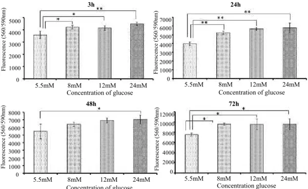
Cell proliferation during the first 3 days of incubation was assessed (Fig. 1). There were significant differences in cell proliferation between 5.5 mM glucose and the other glucose groups after 3, 24 and 72 h. High glucose concentrations tended to promote the proliferation of GK rat BMMSC.
Osteogenic gene expression
Fig. 2 shows the expression of osteogenesis-related genes including RUNX2, ALP, COL-1 and OCN, assessed by quantitative RT-PCR. Similar results were obtained for RUNX-2 and OCN mRNA expression levels; generally, high glucose inhibited mRNA expression for both genes, but 24 mM glucose induced more mRNA expression than any other concentration.
Expression of ALP and COL-1 mRNA followed similar profiles;
as glucose concentration increased, expression of ALP and COL- 1 mRNA significantly decreased.
Extracellular matrix (ECM) mineralization
ECM mineralization was assessed by deposition of calcium and alizarin red staining (Fig. 3). Calcium deposition showed the same glucose concentration dependent pattern as expression of Figure 1. Cell proliferation after 3, 24, 48 and 72 hours of culture (* p < 0.05; ** p < 0.01).
3h 24h
48h 72h
5000 4000 3000 2000 1000 0
7000 6000 5000 4000 3000 2000 1000 0
8000 7000 6000 5000 4000 3000 2000 1000 0
12000 10000 8000 6000 4000 2000 0
5.5mM 8mM 12mM 24mM 5.5mM 8mM 12mM 24mM
5.5mM 8mM 12mM 24mM 5.5mM 8mM 12mM 24mM
Concentration of glucose
Concentration glucose Concentration of glucose
Concentration of glucose Fluorescence (560/590nm)Fluorescence (560/590nm)
Fluorescence (560/590nm)Fluorescence (560/590nm)
Figure 2. Expression of osteogenesis-related genes. RUNX2, ALP, COL-1 and OCN mRNA levels were assessed by Real-time PCR (* p < 0.05; ** p < 0.01).
Figure 3. The graph indicates the quantity of extracellular calcium deposition (* p < 0.05; ** p < 0.01).
The photograph shows an image of the Alizarin red staining.
5.5mM 8mM 12mM 24mM 5.5mM 8mM 12mM 24mM
5.5mM 8mM 12mM 24mM 5.5mM 8mM 12mM 24mM Concentration of glucose
Concentration of glucose Concentration of glucose
Concentration of glucose
Expression of Type I collagenmRNA Expression ofosteocalcin mRNA
Expression of Runx2 mRNA Expression of ALP mRNA
1.2 1 0.8 0.6 0.4 0.2 0
1.4 1.2 1 0.8 0.6 0.4 0.2 0
3.5 3 2.5 2 1.5 1 0.5 0 1.6 1.4 1.2 1 0.8 0.6 0.4 0.2 0
25 20 15 10 5 0
5.5mM 8mM 12mM 24mM
Concentration of glucose
Deposition of Ca
Alizarin red staining
5.5 mM 8mM 2 mM 24 mM
RUNX 2 and OCN mRNA (Fig. 2).
High concentrations of glucose increase IL-1β and IL-6 mRNA expression
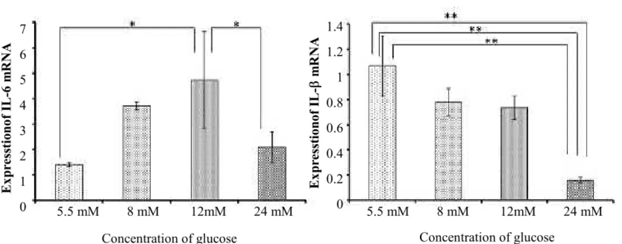
Fig. 4 shows the expression of inflammatory cytokine genes IL-1 and IL-6, assessed by quantitative RT-PCR. The expression of IL-1 mRNA was significantly inhibited as glucose concentration increased. The expression of IL-6 mRNA was opposite to that of RUNX2 and OCN mRNA; as glucose concentration increased to 12 mM, expression of the IL-6 mRNA rose, but the mRNA expression level decreased on exposure to 24 mM glucose.
Discussion
In the present study, we assessed hard tissue differentiation of undifferentiated mesenchymal cells from a type II diabetes mellitus model when cultured in high concentrations of glucose. Typical physiological glucose concentration is 5.5 mM; three of the glucose concentrations we tested were higher (8-24 mM). Highly- concentrated glucose promotes cell proliferation and inhibits hard tissue differentiation and calcification of GK rat BMMSC. The observed effect on differentiation depends on production of inflammatory cytokines such as IL-6.
Glucose in the microenvironment markedly affects gene regulation, proliferation, and differentiation of mesenchymal cells.
Fig. 1 shows that GK rat BMMSC proliferation is proportional to glucose concentration. Li et al.7) reported that in human mesenchymal stem cells, no consistent effect on proliferation was seen on short-term (4 day) exposure to 25 mM glucose. In long- term cultures (4 weeks) of cells from individual donors, variable trends were observed, and the overall analysis showed a slight but significant decrease in cell proliferation after 25 mM glucose treatment. Well et al.10) suggested that high glucose concentrations in cell culture medium do not acutely affect human mesenchymal
stem cell growth factor production or proliferation. Zhao et al.11) reported that diabetic bone marrow stromal cells demonstrated reduced proliferation ability. Because they described the proliferation of normal rat cells, we cannot compare our results with theirs. The growth speed of the cells extracted from DM seems to tend to decrease, we suggest cell proliferation is proportional to glucose concentration. Li et al.7) suggested that proliferation and osteogenic differentiation are stimulated by high glucose. However cell proliferation and differentiation are inverse events in general, and all these reports are performed at 25 mM as high glucose concentration.
High glucose in the body is associated with inflammatory conditions. The inflammatory pathways of Toll-like receptors (TLRs) have been widely studied due to their relationship with proinflammatory cytokines. TLRs recognize pathogen-associated molecular patterns (PAMPs) such as lipopolysaccharide (LPS), lipoproteins and peptidoglycans. The expression levels of TLR-2 and -4 in gingival tissue of patients with chronic periodontitis seem to correlate with the severity of the disease and with an increase in bacterial plaque12). Moreover, the expression levels of TLR-2, -4 and -9 are increased in gingival tissue in patients with type 2 diabetes and chronic periodontitis13). Recent findings have shown increased TLR-2 and -4 expression, signaling, ligands, and functional activation in monocytes from T1 DM subjects compared with controls; these levels were further accentuated in T1 DM with microvascular complications14). It has been suggested that inflammation in individuals with diabetes is exacerbated by nonmicrobial mechanisms. In addition to exogenous microbial products, TLRs can recognize endogenous ligands such as necrotic cells15 ), h eat sh o ck p ro teins Hsp 70 an d gp 9616 ), th e oligosaccharides of soluble hyaluronate17), and advanced glycation end products (AGEs)18). Causes of diabetes-related periodontitis becoming severe include neutrophil dysfunction in hyperglycemia, microangiopathy, composition suppression of collagen, and 7
6 5 4 3 2 1 0
1.4 1.2 1 0.8 0.6 0.4 0.2
5.5 mM 8 mM 12mM 24 mM 0 5.5 mM 8 mM 12mM 24 mM Concentration of glucose
Expresstionof IL-6 mRNA Expresstionof IL- mRNA
Concentration of glucose
Figure 4. Expression of proinflammatory cytokine genes. IL-6 and IL-1β mRNA expression levels were assessed by Real-time PCR (* p < 0.05; ** p < 0.01).
increase in the oxidative stress response19-21). One oxidative stress response, production of AGEs, is considered a nonmicrobial inflammatory factor. Takagi et al.22) reported that AGEs promote expression of IL-6 in human bone-derived cells. AGEs have been considered to induce inflammatory reaction in osteoblasts. We reported that hard tissue differentiation from mesenchymal stem cells decreases in inflammatory conditions caused by Porphyromonas gingivalis LPS23). We therefore suggest that the hard tissue differentiation response decreases in conditions of inflammation, particularly increased expression of IL-6. Fig. 2 and 4 show that the expression of Runx2 and osteocalcin mRNA correlated inversely with the expression of IL-6 mRNA.
Conversely, Nakajima et al.24) reported that AGEs promote hard tissue differentiation in the pulp cell, which is a type of undifferentiated mesenchymal cell. The cells we used in this work are undifferentiated mesenchymal cells obtained from GK rat thighbone.
Our markers of hard tissue differentiation all decreased on exposure to glucose concentrations of up to 12 mM. However, the expressions of Runx2 and osteocalcin mRNA and deposition of calcium increased at a glucose concentration of 24 mM. We suggest that low expression of ALP and COL-1 at 24 mM glucose indicate the weakening of a formed hard tissue. García-Hernández et al.8) reported high concentrations of extracellular glucose decreased the quality of mineral deposited in culture with a result that was similar to the mineral deposit induced by LPS stimulus.
Ding et al.25) reported that IL-1β inhibits expression of Runx2 and collagen in human mesenchymal stem cells. Consistent with this, our results show that IL-1β expression decreased with increasing glucose concentration, while expression of Runx2 increased in 24 mM glucose (Fig. 4).
The results of this study suggest that proliferation of GK rat BMMSC isolated from T2 DM model rats can be elevated by glucose concentration, however osteogenic differentiation and mineralization from such cells, which play an important role in hard tissue regeneration, can be inhibited. We speculate that this phenomenon is caused by formation of an inflamed state as extracellular glucose concentration increases; osteogenic differentiation is inversely related to expression of the inflammatory cytokine IL-6. These findings will help establish guidelines in periodontal medicine for regenerative therapy in T2 DM patients.
Acknowledgments
We are grateful to the members of the Department of Periodontology, Department of Oral Pathology for their kind advice and assistance. This work was supported by a Grant-in- Aid for Scientific Research (24792345) from the Japan Society for the Promotion of Science, and a Research Promotion Grant (14-01) and Oral Implant Research Grant (14-03) from Osaka
Dental University.
References
1. Herder C, Illig T, Rathmann W, Martin S, Haastert B, Müller- Scholze S, Holle R, Thorand B, Koenig W, Wichmann HE and Kolb H. Inflammation and type 2 diabetes: results from KORA Augsburg. Gesundheitswesen 67 (Suppl 1): S115- 121, 2005
2. Williams RC and Offenbacher S. Periodontal medicine: the emergence of a new branch of periodontology. Periodontol 2000 23: 9-12, 2000
3. Nelson RG, Shlossman M, Budding LM, Pettitt DJ, Saad MF, Genco RJ and Knowler WC. Periodontal disease and NIDDM in Pima Indians. Diabetes Care 13: 836-840, 1990 4. Taylor GW, Burt BA, Becker MP, Genco RJ, Shlossman M, Knowler WC and Pettitt DJ. Noninsulin dependent diabetes mellitus and alveolar bone loss progression over 2 years. J Periodontol 69: 76-83, 1998
5. Salvi GE, Collins JG, Yalda B, Arnold RR, Lang NP and Offenbacher S. Monocytic TNF alpha secretion patterns in IDDM patients with periodontal diseases. J Clin Periodontol 24: 8-16, 1997
6. Goova MT, Li J, Kislinger T, Qu W, Lu Y, Bucciarelli LG, Nowygrod S, Wolf BM, Caliste X, Yan SF, Stern DM and Schmidt AM. Blockade of receptor for advanced glycation end-products restores effective wound healing in diabetic mice. Am J Pathol 159: 513-525, 2001
7. Li YM, Schilling T, Beniosch P, Zeck S, Meissner-Weigl J, Schneider D, Limbert C, Seufert J, Kassem M, Schutze N, Jakob F and Ebert R. Effects of high glucose on mesenchymal stem cell proliferation and differentiation.
Biochem Biophys Res Commun 363: 209-215, 2007 8. García-Hernández A, Arzate H, Gil-Chavarría I, Rojo R and
Moreno-Fierros I. High glucose concentrations alter the biomineralization process in human osteoblastic cells. Bone 50: 276-288, 2012
9. Finke B, Luethen F, Schroeder K, Mueller PD, Bergemann C, Frant M, Ohl A and Nebe BJ. The effect of positively charged plasma polymerization on initial osteoblastic focal adhesion on titanium surfaces. Biomaterials 28: 4521-4534, 2007
10. Weil BR, Abarbanell AM, Herrmann JI, Wang Y and Meldrum DR. High glucose concentration in cell culture medium does not acutely affect human mesenchymal stem cell growth factor production or proliferation. Am J Physiol Regul Integr Comp Physiol 296: 1735-1743, 2009
11. Zhao YF, Zeng DL, Xia LG, Zhang SM, Xu LY, Jiang XQ and Zhang FQ. Osteogenic potential of bone marrow stromal cells derived from streptozotocin-induced diabetic rats. Int J Mol Med 31: 614-620, 2013
12. Sarah SM, Tamilselvan S, Kamatchiammal S and Suresh R.
Expression of Toll-like receptors 2 and 4 in gingivitis and chronic periodontitis. Ind J Dent Res 17: 114-116, 2006 13. Rojo-Botello NR, Garcia-Hernandez AL and Moreno-Fierros
L. Expression of toll-like receptors 2, 4 and 9 is increased in gingival tissue from patients with type 2 diabetes and chronic periodontitis. J Periodontal Res 47: 62-73, 2012 14. Devaraj S, Tobias P and Jialal I. Knockout of toll-like receptor-
4 attenuates the proinflammatory state of diabetes. Cytokine 55: 441-445, 2011
15. Wong FS and Wen L. Toll-like receptors and diabetes. Ann N Y Acad Sci 1150: 123-132, 2008
16. Retzlaff C, Yamamoto Y, Hoffman PS, Friedman H and Klein TW. Bacterial heat shock proteins directly induce cytokine mRNA and interleukin-1 secretion in macrophage cultures.
Infect Immun 62: 5689-5693, 1994
17. Termeer C1, Benedix F, Sleeman J, Fieber C, Voith U, Ahrens T, Miyake K, Freudenberg M, Galanos C and Simon JC.
Oligosaccharides of Hyaluronan activate dendritic cells via toll-like receptor 4. J Exp Med 195: 99-111, 2002 18. Tsan MF and Gao B. Endogenous ligands of Toll-like
receptors. J Leukoc Biol 76: 514-519, 2004
19. Lalla E, Lamster IB, Drury S, Fu C and Schmidt AM.
Hyperglycemia, glycoxidation and receptor for advanced glycation endproducts: potential mechanisms underlying complications, including diabetes-associated periodontitis.
Periodontol 2000 213: 50-62, 2000
20. Ryan ME, Camu O and Kamer A. The influence of diabetes on the periodontal tissues. J Am Dent Assoc 134: 34S-40S, 2003
21. Gurav A and Jadhav V. Periodontitis and risk of diabetes mellitus. J Diabetes 3: 21-28, 2011
22. Takagi M, Kasayama S, Yamamoto T, Motomura T, Hashimoto K, Yamamoto H, Sato B, Okada S and Kishimoto T.
Advanced glycation endproducts stimulate interleukin-6 production by human bone-derived cells. J Bone Miner Res 12: 439-446, 1997
23. Kato H, Taguchi Y, Tominaga K, Umeda M and Tanaka A.
Porphyromonas gingivalis LPS inhibits osteoblastic differentiation and promotes pro-inflammatory cytokine production in human periodontal ligament stem cells. Arch Oral Biol 59: 167-175, 2014
24. Nakajima Y, Inagaki Y, Hiroshima Y, Kido J and Nagata T.
Advanced Glycation End-products Enhance Calcification in Cultured Rat Dental Pulp Cells. J Endod 39: 873-878, 2013
25. Ding J, Ghali O, Lencel P, Broux O, Chauveau C, Devedjian JC, Hardouin P and Magne D. TNF-alpha and IL-1beta inhibit RUNX2 and collagen expression but increase alkaline phosphatase activity and mineralization in human mesenchymal stem cells. Life Sci 84:499-504, 2009