• 検索結果がありません。

Oral squamous cell carcinoma (OSCC) is locally in­

N/A
N/A
Protected

Academic year: 2021

シェア "Oral squamous cell carcinoma (OSCC) is locally in­"

Copied!
10
0
0

読み込み中.... (全文を見る)

全文

(1)

INTRODUCTION

Oral squamous cell carcinoma (OSCC) is locally in­

vasive

1, 2

and its rate of metastasis correlates with the clinical stage,

3

which is a factor related to its low survival rates. Thus, novel therapeutic stra­

tegies are needed. As most oral cancers are squamous cell carcinomas (SCCs), one feature is

progressive local invasion.

1, 2

Therefore, it is neces­

sary to improve treatments for OSCC by elucidating its invasion mechanisms. The processes of tumor invasion include cell migration, interaction between the tumor and stroma at the invasive front, and the involvement of growth factors and external sub­

stances, all of which influence the invading cells.
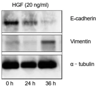
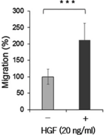
4­8

Hepatocyte growth factor (HGF) has been identi­

Effect of HGF/c­Met pathway in oral squamous cell carcinoma on EMT and metastatic potential

Kazutaka Kimura

1

, Yuichi Ohnishi

2

, Tomoharu Okamura

3

, Kazuya Tominaga

3

and Masahiro Nakajima

2

1

Graduate School of Dentistry (Second Department of Oral and Maxillofacial Surgery),

2

Second Department of Oral and Maxillofacial Surgery, and

3

Department of Oral Pathology, Osaka Dental University, 8-1 Kuzuhahanazono-cho, Hirakata-shi,

Osaka 573-1121, Japan

Oral squamous cell carcinoma (OSCC) is characterized by highly invasive tumor cells.

Hepatocyte growth factor (HGF) signaling plays an important role in the induction of epithelial­mesenchymal transition (EMT), a process that converts immotile epithelial cells into motile mesenchymal cells and is involved in cancer metastasis by promoting tumor cell scattering. The c­Met, which is a transmembrane tyrosine kinase receptor that may be activated by HGF, regulates the associated downstream gene expression. This proc­

ess is important to cell migration in normal and cancerous conditions. This study ex­

plored the potential effects of c­Met on HGF­induced EMT in an OSCC cell line.

We investigated the function of c­Met in the process of EMT, and its molecular mecha­

nism in oral cancer. OSCC cell line, HSC3 cells, were treated with HGF for varying dura­

tions. EMT­associated proteins, including E­cadherin and vimentin, were examined by western blot analysis. The role of c­Met in the mediation of EMT­like changes was investi­

gated using western blot analysis and knockdown by c­Met inhibitor. Moreover, we car­

ried out investigations using immunohistochemical and immunofluorescence staining.

We found that treatment with HGF induced EMT­like changes and enhanced the migra­

tive potential of HSC3 cells. Furthermore, HGF­mediated EMT­like changes were associ­

ated with c­Met activation, and these changes could be blocked by c­Met knockdown. In particular, immunohistochemical and immunofluorescence staining of c­Met strongry ex­

pressed in the invasion front of the tumor cells. This study clearly demonstrated a crucial function for c­Met in EMT development in oral cancer. c­Met­targeted treatment may be an effective therapy for oral cancer. We believe that our data demonstrated that c­Met in­

hibitor could reduce HGF­induced EMT and cell motility via c­Met blockade and down­

regulation of the pro­survival extracellular signal­regulated kinases pathway. (J Osaka Dent Univ 2020 ; 54 : 135­144)

Key words : HGF ; c­Met ; Oral squamous cell carcinoma ; SU11274

(2)

fied as one of the molecules that induces tumor an­

giogenesis, and is a potent hepatotrophic factor re­

sponsible for regeneration of the liver.

9, 10

HGF is now recognized as a pluripotent cytokine that medi­

ates tumor­stromal interaction with mitogenic, moto­

genic, and morphogenic activities.

11, 12

Moreover, HGF induces potent angiogenic activity in vascular endothelial cells.

13

HGF is a ligand for the c­Met proto­oncogene of the receptor tyrosine kinase.

12, 14

HGF and c­Met are upregulated in several types of cancers, such as colorectal, gastric, esophageal, breast, and lung cancers.

15­17

The upregulation of c­

Met has been correlated with mortality in cancer patients.

18­20

HGF induces degradative adhesion of epithelial cancer cells in a process called epithelial­

mesenchymal transition (EMT), thereby increasing migration and invasiveness. EMT is important in cancer metastasis because it promotes the release of cancer cells from the primary tumor, and induces vasculature and lymphatic vessel invasion as well as secondary tumors in distant organs.

21

Although EMT induced by HGF has been investigated in dif­

ferent cancers,

22, 23

the tumorigenic association be­

tween EMT and c­Met remains unclear in oral can­

cer. The present study investigated the effects of the HGF/c­Met signaling pathway on EMT and the metastasis potential of OSCC.

MATERIALS AND METHODS

Clinical and pathological data (Table 1)

We used 50 OSCC samples (39 well­differentiated, 5 moderately­differentiated, and 6 poorly­diffe­

rentiated) obtained from biopsies at Osaka Dental University Hospital. The subjects were 31 males, aged 18 to 86 (mean 63) years, and 19 females, aged 38 to 85 (mean 64) years, at the time of diag­

nosis. The primary OSCC location was the tongue.

Sixteen of the cases were T1, 19 were T2, 10 were T3, and 5 were T4 ; the ages ranged from 38 to 85 (mean 62) years for T1, from 39 to 74 (mean 61) years for T2, from 18 to 82 (mean 68) years for T3, and from 43 to 86 (mean 71) years for T4. Twenty of the 50 cases had cervical lymph node metasta­

sis (N(+)), which was treated by neck dissection.

Metastasis was confirmed by histopathology.

On the other hand, 30 of the cases had no cervi­

cal lymph node metastasis (N(−)). Thirteen were in males and 7 in females in the N(+) group, whereas 18 were in males and 12 in females in the N(−) group. The ages ranged from 38 to 86 (mean 66) years in N(+), and from 18 to 85 (mean 62) years in N(−). Eight cases were clinical stage I, 13 were stage II, 16 were stage III, and 13 were stage IV ; the ages ranged from 42 to 85 (mean 58) years for stage I, from 39 to 74 (mean 60) years for stage II, from 18 to 82 (mean 63) years for stage III, and from 60 to 86 (mean 71) years for stage IV.

This study was approved by the ethics committee of Osaka Dental University (Approval No.110985).

Cell culture and reagents

The OSCC cell line HSC3 was purchased from RIKEN Bioresource Center (Ibaraki, Japan). Cells were cultured in Dulbecco’s modified Eagle medium (DMEM) supplemented with 10% (v/v) fetal bovine serum (FBS) at 37°C in a humidified atmosphere of

Table 1 Patient characteristics at primary surgery Characteristic Patients Sex

Male 31

Female 19

Median age in years (range)

Male 63 (18­86)

Female 64 (38­85)

T stage

T1 16

T2 19

T3 10

T4 5

N stage

N0 30

N1 9

N2 11

C Stage

Ⅰ 8

Ⅱ 13

Ⅲ 16

Ⅳ 13

Differentiation

Well 39

Moderate 5

Poor 6

(3)

5% CO 2. DMEM and FBS were purchased from Gibco

(Life Technologies, Tokyo, Japan). Antibod­

ies used included anti­c­Met (Cell Signaling Tech­

nology Japan, Tokyo, Japan), anti­phospho­c­Met (Tyr 1234/1235) (Cell Signaling Technology Japan), and anti­α­tubulin (Sigma­Aldrich, Tokyo, Japan).

SU11274 was obtained from Sigma­Aldrich.

Wound healing assay

Cell migration was also assessed using an in vitro scratch wound healing assay. HSC3 cells were cul­

tured with DMEM supplemented with 10% FBS until semi­confluence in 12­well plates. Cells were then treated with 10 μg/mL of mitomycin­C for 4 h to block proliferation and a scratch was subsequently made using a sterile 200­μl pipette tip to generate a cell­free gap approximately 1 mm in width. Cells were then washed with PBS and photographed to record the wound width at 0 h. Next, one group of cells was cultured in DMEM with 10% FBS as a control. Other groups were treated with 10 μM SU 11274. When we assessed the effects of ligands, one group of cells was cultured in DMEM as a con­

trol. Other groups were treated with 20 ng/mL of HGF. After incubation, photographs were taken to evaluate migration.

Western blotting

Cells were washed with phosphate­buffered saline (PBS) and then lysed with RIPA buffer consisting of 150 mM NaCl, 10 mM Tris HCl at pH 8.0, 1% (v/v) Nonidet P­40, 0.5% (w/v) deoxycholic acid, 0.1% (w /v) sodium dodecyl sulfate (SDS), 5 mM ethyle­

nediaminetetraacetic acid (EDTA), 1 X Halt

TM

Pro­

tease Inhibitor Cocktail (Thermo Fisher Scientific, Yokohama, Japan), and 1 X Halt

TM

Protein Phos­

phatase Inhibitor (Thermo Fisher Scientific). The protein concentration of the lysates was measured using a BCA

TM

Protein Assay Kit (Thermo Fisher Scientific) and equal amounts of protein were sub­

jected to SDS­polyacrylamide gel electrophoresis.

The separated proteins were electrophoretically transferred onto PVDF membranes (GE Healthcare, Little Chalfont, UK). Non­specific binding was blocked by incubation in 5% (w/v) bovine serum al­

bumin (BSA) in TBS/Tween­20 (TBS­T) for 1 h at room temperature. Membranes were probed with antibodies in TBS­T overnight at 4°C and then incu­

bated with HRP­conjugated secondary antibody.

Antibody­antigen complexes were detected by ECL plus Western blotting detection reagent (GE Health­

care).

lmmunohistochemical staining of c-Met

The specimens were fixed in 10% formalin solution, dehydrated in a graded ethanol series, and embed­

ded in paraffin. Sections of 5­μm thick paraffin embedded­tissues were cut and mounted on silane­

coated glass slides. The sections were deparaf­

finized in Hemo­De and rehydrated through a graded ethanol series. Antigens were retrieved in 0.0 l M citrate buffer at pH 7.0 in a water bath at 98°C for 40 min, and endogenous peroxidase was blocked with 3% hydrogen peroxidase. The tissues were then incubated overnight at 4°C with a 1 : 200 dilution of anti­c­Met polyclonal rabbit antibody (Santa Cruz Biotechnology, Santa Cruz, CA, USA).

Samples were then incubated with peroxidase dex­

tran polymer (Envision+ ; DakoCytomation, Car­

pinteria, CA, USA) for 30 min at room temperature and visualized by 3,3’­diaminobenzidinetetrahydro­

chloride (DAB ; DakoCytomation). All sections were counterstained using hematoxylin.

Immunofluorescence staining of c-Met

These new 5­μm thick sections were prepared from stored formalin­fixed, paraffin­embedded tissue blocks for hematoxylin­eosin staining and im­

munofluorescence staining of c­Met (Bioss Antibod­

ies, Woburn, Massachusetts, USA). Immunofluores­

cence staining was performed using Envision tech­

nology. Antigen retrieval was performed by warm­

ing the deparaffinized sections in 0.1% sodium az­

ide at pH 6 for 20 minutes, an then in hot water at 95°C for 30 minutes. Endogenous peroxidase activ­

ity was blocked by 3% hydrogen peroxide for 20 minutes and the sections were soaked in 0.05 M Tris­buffered saline (TBS) before being soaked in blocking solution (DakoCytomation) for 20 minutes.

They were then rinsed twice at room temperature

(4)

for 2 minutes. The tissue sections were next incu­

bated for 60 minutes at room temperature with a rabbit anti­human c­Met polyclonal antibody (1 : 200) labeled with the optimally diluted primary anti­

body Alexafluor 488 Conjugate. Antigen sites were visualized using the above conjugate and counter­

stained with Mayer’s hematoxylin. These speci­

mens were observed with an LSM 700 laser scan­

ning microscope (Carl Zeiss, Jana, Germany). The immunofluorescence signals were detected using confocal laser scanning microscopy with the same microscope.  The immunofluorescence images were processed by the LSM 700 and the areas were calculated. The unpaired Mann­Whitney U test was used to evaluate the specific difference in the protein expression rate of c­Met from the con­

trol.

Statistical analysis

All data are represented as the mean and standard deviation from three independent experiments un­

less stated otherwise. Statistical analysis was per­

formed by one­way analysis of variance (ANOVA), and the significant differences between the treated and control samples (*p<0.05, **p<0.01, and

***p<0.001) were evaluated by the unpaired Stu­

dent’s t­test.

RESULTS

HGF induced EMT-like changes in HSC3 cells Characteristic changes associated with EMT in­

clude the reduction of epithelial markers and induc­

tion of mesenchymal markers. These changes are associated with the growth of cancer cells, enabling cell­cell dissociation, migration, and motility.

24

In the present study, western blot analysis revealed that HGF treatment downregulated E­cadherin expres­

sion and upregulated vimentin expression in the HSC3 cells in a time­dependent manner. E­cad­

herin expression decreased slightly after 24 hours and almost disappeared after 36 hours. In compari­

son, although vimentin was not observed after 24 hours, it was clearly seen after 36 hours. Therefore, HSC3 cells acquired EMT­like changes following in­

cubation with HGF (20 ng/mL) for 36 h (Fig. 1).

These results suggest that HGF promotes altered expression of EMT markers in HSC3 cells. For this reason, HSC3 cells were treated with 20 ng/mL HGF for 36 h in all subsequent experiments.

c-Met expression was enhanced by HGF treat- ment

The expression level of the HGF receptor c­Met was measured to investigate the role of HGF in in­

ducing EMT­like changes in HSC3 cells. c­Met is activated by being phosphorylated via HGF and ac­

tivated c­Met is able to regulate several down­

stream target genes. Western blot analysis re­

vealed that HGF treatment increased the expres­

sion of c­Met and phosphorylated­c­Met (Fig. 2). In particular, the expression of phosphorylated­c­Met was markedly increased.

HGF treatment increased the migration potential of HSC3 cells

We examined the effects of HGF treatment on the migration potential of HSC3 cells. The wound heal­

ing assay demonstrated that HGF increased cancer cell migration. HGF­treated HSC3 cells exhibited approximately twice the migration capacity as con­

trol HSC3 cells. This suggests that HGF increased

Fig. 1 HGF induced EMT­like changes in HSC3 cells. HGF (20 ng/mL) downregulated Ecadherin and upregulated vimentin in a timedependent manner compared with un­

treated HSC3 cells (control).

(5)

the migration capacity of HSC3 that had undergone EMT­like changes (Fig. 3).

c-Met inhibitor SU11274 suppressed c-Met ex- pression and reduced the HGF/HSC3 migration capacity

In order to confirm the involvement of the HGF/c­

Met signaling pathway, we examined the effects of c­Met knockdown by SU11274. As shown in Fig. 4 A, the c­Met inhibitor suppressed c­Met expression.

Furthermore, as shown in Fig. 4 B, the inhibitor treatment reduced the migration capacity compared with untreated cells. This suggests that HGF caused the EMT in HSC3 cells via the HGF/c­Met signaling pathway.

c-Met inhibitor SU11274 inhibited HGF-induced EMT-like changes

After HGF treatment, cells further treated with SU 11274 showed induced expression of E­cadherin

Fig. 2 Increase in c­Met expression by HGF. HSC3 cells were treated with 20 ng/mL HGF, and c­Met expression was measured at the protein level after treatment for 36 h (HGF : Hepatocyte growth factor ; p : Phosphorylated).

Fig. 3 Increase by HGF in the migration potential of HSC3 cells. HGF­treated cells demonstrated increased migration potential in the woundhealing assay (***p<0.001, n=3).

Fig. 4 Suppression of c­Met expression and reduction of the HGF/HSC3 migration ability by the c­Met inhibitor SU 11274. (A) c­Met inhibitor treatment suppressed c­Met ex­

pression. (B) The inhibitor reduced the migration ability com­

pared with untreated cells (***p<0.001, n=3).

Fig. 5 Inhibition of HGF­induced EMT­like changes by the c

­Met inhibitor SU11274. After incubation with HGF, untreated

HSC3 cells exhibited the upregulation of E­cadherin and

downregulation of vimentin compared with cells treated with

SU11274.

(6)

and downregulated expression of vimentin com­

pared with untreated cells (Fig. 5). This suggests that the HGF­induced EMT was suppressed by the c­Met inhibitor and was converted from a mesen­

chymal to a native epithelial system.

Immunohistochemical evaluations of c-Met Well­differentiated OSCC exhibits c­Met staining in the nuclei of some tumor cells. The peripheral cells of the tumor nests were stained more densely than

the central tumor cells of the tumor nests (Fig. 6 A).

In particular, in the invasion front of tumor nests, both the cytoplasm and nucleus had strong staining (Fig. 6 B). Significant differences between well­

differentiated and poorly­differentiated SCC were observed. Moreover, the difference between the N (+) and N(−) cases in c­Met expression was sig­

nificant (data not shown). These results are similar to those in a previous report. Furthermore, there was a significant difference between N(+) and N(−)

Fig. 6 lmmunohistochemical evaluations of c­Met. (A) A well­differentiated OSCC showing c­Met staining in the nu­

clei of some tumor cells. The peripheral cells of the tumor nests were stained more densely than the central tumor cells of the tumor nests. (Original magnification×10) (B) Tumor nests in the invasion front demonstrating dense cyto­

plasmic and nuclear staining in the tumor cells. Some of the fibroblasts, endothelial cells, and inflammatory cells were also positive for c­Met. (Original magnification×5)

Fig. 7 Immunofluorescence staining of c­Met. c­Met signals were barely observed in well­differentiated tumor cells.

(7)

cases at the primary site, which is a novel finding.

Immunofluorescence staining of c-Met

The results were similar to those of immunohisto­

chemical staining. Positive c­Met signals were barely observed in well­differentiated tumor cells (Figs. 7 A and B). Tumor nests in the invasion front demonstrated dense cytoplasmic and nuclear stain­

ing (Figs. 8 A and B). Differences between N(+) and N(−) cases at the primary site were significant (Fig. 9).

DISCUSSION

The process of cell dedifferentiation by EMT is known as one of the important features of can­

cer.

25, 26

The EMT is a very important process in the

early stage of tumor cell metastasis. In order for cancer cells to metastasize, they must first avoid anoikis. E­cadherin is typically present in epithelial cells and its decreased expression is an important feature of the EMT. In mesenchymal cells, these epithelial proteins are altered by mesenchymal­

specific factors, including fibronectin, vimentin, and neural cadherin (N­cadherin).

27

The changes in the expression pattern of E­cadherin/N­cadherin are correlated with the avoidance of anoikis and an in­

crease in cell invasiveness.

28

Furthermore, dysregu­

lation of growth factor receptors may result in resis­

tance to anoikis. Before cancer cells begin to mi­

grate and metastasize, they must activate the genes that are necessary for cell differentiation, proliferation, and activation of anti­apoptotic path­

ways. Furthermore, alteration of cellular character­

istics from the epithelial to the mesenchymal phe­

notype reduces the activity of adhesion molecules and proteases on the cell surface, which digest the components of the ECM.

29

A number of factors that induce EMT have been identified, including transforming growth factor­β (TGF­β), HGF, epidermal growth factor (EGF), fi­

Fig. 8 Immunofluorescence staining of c­Met. Tumor nests at the invasion front demonstrated dense cytoplasmic and nuclear staining.

Fig. 9 Significant differences between N(+) and N(−) cases at the primary site shown by brightness analysis (*p<0.05, n

=50).

(8)

broblast growth factor (FGF), platelet derived growth factor (PDGF), insulin-like growth factor (IGF), microRNA, hypoxia, and transcription fac- tors.

30-32

Among these factors, EMT induction by the HGF/c-Met pathway is essential in certain types of cancer.

33-36

However, to the best of our knowledge, the detailed role of c-Met in the progression of oral cancer has not been reported. In this study, treat- ment of HSC3 cells with HGF induced EMT-like changes, as indicated by the downregulation of E- cadherin and upregulation of vimentin. Furthermore, HGF increased the migration potential of HSC3 cells.

c-Met overexpression has been identified in the majority of human cancers.

37, 38

In this study, c-Met expression was promoted by HGF-dependent tran- scriptional upregulation. In particular, there was a marked upregulation in phosphorylated-c-Met fol- lowing HGF treatment. Therefore, HGF also acti- vates c-Met in oral cancer cells. A similar study suggested that c-Met is activated via an HGF- independent signaling pathway, following transfec- tion, in lung cancer.

39

In addition, c-Met and HGF may be transactivated by the mutant of epidermal growth factor receptor (EGFR), also termed EG- FRvIII.

40

Furthermore, recent studies demonstrated that c-Met activation occurs in the absence of ligand binding. Integrin activation, plexins, CD 44, certain G protein-coupled receptors, and other re- ceptor tyrosine kinases have all been implicated in c-Met activation without the requirement of HGF binding.

41

In addition, the activation of c-Met was found to activate the downstream signaling path- ways PI 3K and ERK, which resulted in the down- regulation of E-cadherin and upregulation of vimentin. E-cadherin downregulation is regarded as a characteristic change of the EMT.

42

As intercellu- lar adhesions are essential for the maintenance of the epithelial phenotype, the downregulation of E- cadherin results in abnormal differentiation and the loss of cell polarity, which ultimately facilitates EMT.

43

In this study, the knockdown of c-Met by SU 11274 prevented HGF-induced EMT-like changes.

This suggests that HGF induced the EMT in a c- Met-dependent manner in HSC3 cells.

Abnormal c-Met activation may occur in certain cancer types due to gene amplification, mutation, or transactivation.

15

However, c-Met overexpression as a result of upregulation at the transcriptional level is predominant in the majority of human malignan- cies.

44

The present study suggested the overex- pression of c-Met signaling and the subsequent in- duction of EMT is a common phenomena in oral cancer cells. Furthermore, the EMT increased pros- tate cancer cell invasiveness in vitro and in vivo.

Taken together, this study demonstrated the poten- tial association among c-Met, EMT, and invasive- ness in oral cancer. Similarly, in other solid human tumors, previous studies have revealed that c-Met- mediated signaling activation drives EMT, cancer cell migration, and metastasis.

45-47

In this study, we also examined clinical samples.

c-Met signals were almost never observed in well- differentiated tumor cells, and only the outer layer nests exhibited positive reactions. Moreover, rela- tively strong c-Met signals were observed in the in- filtrating tumor tissue, the so-called invasion front.

Tabuchi et al.

48

reported a significant correlation be- tween c-Met and N(+) in colon cancers. Regarding c-Met expression in OSCC, Endo et al.

49

and Chen et al.

50

reported the high expression of c-Met in cases of N(+), and progressive T3, T4, stage lll, and stage IV cases. They also found a significant difference in c-Met expression among these cases.

However, Muzio et al.

51

described a significant in- crease in c-MET expression in tumors larger than 1.5 cm, although the difference among clinical stages was not significant. In our study, expression was observed at the invasion front where invasion growth occurs. This was similar to the results of Chan et al.

50

This part of the tumor was thought to comprise metastatic cells undergoing EMT.

In conclusion, this study suggested that HGF di-

rectly promotes EMT and carcinogenic properties in

oral cancer via the c-Met signaling pathway. Spe-

cific molecular targeting of this signaling pathway

may be therapeutically beneficial for oral cancer pa-

tients. As it is important to regulate cell invasion,

migration, and metastasis in clinical cancer treat-

ment, more detailed analyses of the signaling path-

(9)

way are needed.

The present study was supported by a KAKENHI grant (Number JP18K09736) from the Japan Society for the Pro- motion of Science.

REFERENCES

1. Kramer R, Shen X, Zhou H. Tumor cell invasion and sur- vival in head and neck cancer. Cancer Metastasis Rev 2005 ; 24 : 35-45.

2. Ziober AF, Falls EM, Ziober BL. The extracellular matrix in oral squamous cell carcinoma : friend or foe? Head Neck 2006 ; 28 : 740-749.

3. Berenson JR, Yang J, Mickel RA. Frequent amplification of the BCL-1 locus in head and neck squamous cell carcino- mas. Oncogene 1989 ; 4 : 1111-1116.

4. De Hert MJ, Baatenburg de Jong RJ. HGF and c-MET as potential orchestrators of invasive growth in head and neck squamous cell carcinoma. Front Bioschi 2008 ; 13 : 2516- 2526.

5. Kalyankrishna S, Grandis JR. Epidermal growth factor re- ceptor biology in head and neck cancer. J Clin Oncol 2006 ; 24 : 2666-2672.

6. Rorth P. Collective cell migration. Annu Rev Cell Dev Biol 2009 ; 25 : 407-429.

7. Neiva KG, Zhang Z, Miyazawa M, Warner KA, Karl E, Nor JE. Cross talk initiated by endothelial cells enhances migra- tion and inhibits anoikis of squamous cell carcinoma cells through STAT3/Akt/ERK signaling. Neoplasia 2009 ; 11 : 583-593.

8. Friedl P, Wolf K. Plasticity of cell migration : a multiscale tuning model. J Cell Biol 2010 ; 188 : 11-19.

9. Trusolino L, Comoglio PM. Scatter-factor and semaphorin receptors : cell signalling for invasive growth. Nat Rev Can- cer 2002 ; 2 : 289-300

10. Nakamura T, Nishizawa T, Hagiya M, Seki T, Shimonishi M, Sugimura A, Tashiro K, Shimizu S. Molecular cloning and expression of human hepatocyte growth factor. Nature 1989 ; 342 : 440-443.

11. Jiang WG, Hiscox S. Hepatocyte growth factor/scatter fac- tor, a cytokine playing multiple and converse roles. Histol Histopathol 1997 ; 12 : 537-555.

12. Matsumoto K, Nakamura T. Emerging multipotent aspects of hepatocyte growth factor. J Biochem 1996 ; 119 : 591- 600.

13. Bussolino F, Di Renzo MF, Ziche M, Bocchietto E, Olivero M, Naldini L, Gaudino G, Tamagnone L, Coffer A, Comoglio PM. Hepatocyte growth factor is a potent angiogenic factor which stimulates endothelial cell motility and growth. J Cell Biol 1992 ; 119 : 629-641.

14. Bottaro DP, Rubin JS, Faletto DL, Chan AM, Kmiecik TE, Vande Woude GF, Aaronson SA. Identification of the hepa- tocyte growth factor receptor as the c-met proto-oncogene product. Science 1991 ; 251 : 802-804.

15. Birchmeier C, Birchmeier W, Gherardi E, Vande Woude GF.

Met, metastasis, motility and more. Nat Rev Mol Cell Biol 2003 ; 4 : 915-925.

16. Comoglio PM, Giordano S, Trusolino L. Drug development of MET inhibitors : targeting oncogene addiction and expe- dience. Nat Rev Drug Discov 2008 ; 7 : 504-516.

17. Maulik G, Shrikhande A, Kijima T, Ma PC, Morrison PT,

Salgia R. Role of the hepatocyte growth factor receptor, c- Met, in oncogenesis and potential for therapeutic inhibition.

Cytokine Growth Factor Rev 2002 ; 13 : 41-59.

18. Di Renzo MF, Olivero M, Giacomini A, Porte H, Chastre E, Mirossay L, Nordlinger B, Bretti S, Bottardi S, Giordano S, Plebani M, Gespach C, Comoglio PM. Overexpression and amplification of the met/HGF receptor gene during the pro- gression of colorectal cancer. Clin Cancer Res 1995 ; 1 : 147-154.

19. Ghoussoub RA, Dillon DA, D’Aquila T, Rimm EB, Fearon ER, Rimm DL. Expression of c-met is a strong independent prognostic factor in breast carcinoma. Cancer 1998 ; 82 : 1513-1520.

20. Miller CT, Lin L, Casper AM, Lim J, Thomas DG, Orringer MB, Chang AC, Chambers AF, Giordano TJ, Glover TW, Beer DG. Genomic amplification of MET with boundaries within fragile site FRA 7 G and upregulation of MET path- ways in esophageal adenocarcinoma. Oncogene 2006 ; 25 : 409-418.

21. Chung JY, Davis JA, Price BD, Staley DM, Wagner MV, Warner SL, Bearss DJ, Hansen MD. Competitive enhance- ment of HGF-induced epithelial scattering by accessory growth factors. Exp Cell Res 2011 ; 317 : 307-318.

22. Ogunwobi OO, Liu C. Hepatocyte growth factor upregulation promotes carcinogenesis and epithelial-mesenchymal transi- tion in hepatocellular carcinoma via Akt and COX-2 path- way. Clin Exp Metastasis 2011 ; 28 : 721-731.

23. Previdi S, Maroni P, Matteucci E, Broggini M, Bendinelli P, Desiderio MA. Interaction between human-breast cancer metastasis and bone microenvironment through activated hepatocyte growth factor/Met and β-catenin/Wnt pathways.

Eur J Cancer 2010 ; 46 : 1679-1691.

24. Martin TA, Mason MD, Jiang WG. Hepatocyte growth factor signaling in cancer metastasis. Curr Signal Transduct Ther 2011 ; 6 : 180-190.

25. Hanahan D, Weinberg RA. Hallmarks of cancer : The next generation. Cell 2011 ; 144 : 646-674.

26. Yeung KT, Yang J. Epithelial-mesenchymal transition in tu- mor metastasis. Mol Oncol 2017 ; 11 : 28-39.

27. Iwatsuki M, Mimori K, Yokobori T, Ishi H, Beppu T, Nakam- ori S, Baba H, Mori M. Epithelial-mesenchymal transition in cancer development and its clinical significance. Cancer Sci 2010 ; 101 : 293-299.

28. Derksen PW, Liu X, Saridin F, van der Gulden H, Zeven- hoven J, Evers B, van Beijnum JR, Griffioen AW, Vink J, Krimpenfort P, Peterse JL, Cardiff RD, Berns A, Jonkers J.

Somatic inactivation of E-cadherin and p53 in mice leads to metastatic lobular mammary carcinoma through induction of anoikis resistance and angiogenesis. Cancer Cell 2006 ; 10 : 437-449.

29. Heerboth S, Housman G, Leary M, Longacre M, Byler S, Lapinska K, Willbanks A, Sarkar S. EMT and tumor metas- tasis. Clin Transl Med 2015 ; 4 : 6.

30. Matsuoka J, Yashiro M, Doi Y, Fuyuhiro Y, Kato Y, Shinto O, Noda S, Kashiwagi S, Aomatsu N, Hirakawa T, Hase- gawa T, Shimizu K, Shimizu T, Miwa A, Yamada N, Sawada T, Hirakawa K. Hypoxia stimulates the EMT of gas- tric cancer cells through autocrine TGFβ signaling. PLoS One 2013 ; 8 : e62310.

31. Luo Y, He DL, Ning L, Shen SL, Li L, Li X, Zhau HE, Chung

LW. Over-expression of hypoxia-inducible factor-1 α in-

creases the invasive potency of LNCaP cells in vitro. BJU

Int 2006 ; 98 : 1315-1319.

(10)

32. Lamouille S, Subramanyam D, Blelloch R, Derynck R.

Regulation of epithelial-mesenchymal and mesenchymal- epithelial transitions by microRNAs. Curr Opin Cell Biol 2013 ; 25 : 200-207.

33. Davidson B, Tropé B, Reich R. Epithelial-mesenchymal transition in ovarian carcinoma. Front Oncol 2012 ; 2 : 33.

34. Hamada S, Satoh K, Masamune A, Shimosegawa T. Regu- lators of epithelial mesenchymal transition in pancreatic cancer. Front Physiol 2012 ; 3 : 254.

35. Talbot LJ, Bhattacharya SD, Kuo PC. Epithelial-me- senchymal transition, the tumor microenvironment, and me- tastatic behavior of epithelial malignancies. Int J Biochem Mol Biol 2012 ; 3 : 117-136.

36. Leopold PL, Vincent J, Wang H. A comparison of epithelial- to-mesenchymal transition and re-epithelialization. Semin Cancer Biol 2012 ; 22 : 471-483.

37. Jiang WG, Davies G, Martin TA, Parr C, Watkins G, Mason MD, Mokbel K, Mansel RE. Targeting matrilysin and its im- pact on tumor growth in vivo : The potential implications in breast cancer therapy. Clin Cancer Res 2005 ; 11 : 6012- 6019.

38. You WK, McDonald DM. The hepatocyte growth factor/c- Met signaling pathway as a therapeutic target to inhibit angi- ogenesis. BMB Rep 2008 ; 41 : 833839.

39. Navab R, Liu J, Seiden-Long I, Shih W, Li M, Bandarchi B, Chen Y, Lau D, Zu YF, Cescon D, Zhu CQ, Organ S, Ibra- himov E, Ohanessian D, Tsao MS. Co-overexpression of Met and hepatocyte growth factor promotes systemic me- tastasis in NCI-H 460 non-small cell lung carcinoma cells.

Neoplasia 2009 ; 11 : 1292-1300.

40. Garnett J, Chumbalkar V, Vaillant B, Gururaj AE, Hill KS, Latha K, Yao J, Priebe W, Colman H, Elferink LA, Bogler O.

Regulation of HGF expression by ∆EGFR-mediated c-Met activation in glioblastoma cells. Neoplasia 2013 ; 15 : 73- 84.

41. Varkaris A, Gaur S, Parikh NU, Song JH, Dayyani F, Jin JK, Logothetis CJ, Gallick GE. Ligand-independent activation of MET through IGF-1/IGF-1 R signaling. Int J Cancer 2013 ; 133 : 1536-1546.

42. Nurwidya F, Takahashi F, Murakami A, TakaHashi K. Epi- thelial mesenchymal transition in drug resistance and me- tastasis of lung cancer. Cancer Res Treat 2012 ; 44 : 151-

156.

43. Shimada S, Mimata A, Sekine M, Mogushi K, Akiyama Y, Fukamachi H, Jonkers J, Tanaka H, Eishi Y, Yuasa Y. Syn- ergistic tumour suppressor activity of E-cadherin and p 53 in a conditional mouse model for metastatic diffuse-type gas- tric cancer. Gut 2012 ; 61 : 344-353.

44. Benvenuti S, Comoglio PM. The MET receptor tyrosine kinase in invasion and metastasis. J Cell Physiol 2007 ; 213 : 316-325.

45. Torres KE, Zhu QS, Bill K, Lopez G, Ghadimi MP, Xie X, Young ED, Liu J, Nguyen T, Bolshakov S, Belousov R, Wang S, Lahat G, Liu J, Hernandez B, Lazar AJ, Lev D.

Activated MET is a molecular prognosticator and potential therapeutic target for malignant peripheral nerve sheath tu- mors. Clin Cancer Res 2011 ; 17 : 3943-3955.

46. Lengyel E, Prechtel D, Resau JH, Gauger K, Welk A, Linde- mann K, Salanti G, Richter T, Knudsen B, Vande Woude GF, Harbeck N. C-Met overexpression in node-positive breast cancer identifies patients with poor clinical outcome independent of Her 2/neu. Int J Cancer 2005 ; 113 : 678- 682.

47. Zhou AX, Toylu A, Nallapalli RK, Nilsson G, Atabey N, Heldin CH, Borén J, Bergo MO, Akyürek LM. Filamin a me- diates HGF/c-MET signaling in tumor cell migration. Int J Cancer 2011 ; 128 : 839-846.

48. Tabuchi Y, Nakamura T, Ohno M, Usami M. Immunohisto- chemical expression of c-MET {HGF receptor) in colorectal cancer lesions. Buit Health Sci Kobe 1998 ; 14 : 43-48.

49. Endo K, Shirai A, Furukawa M, Yoshizaki T. Prognostic value of cell motility activation factors in patients with tongue squamous cell carcinoma. Hum Pathol 2006 ; 37 : 1111-1116.

50. Chen YS, Wang JT, Chang YF, Liu BY, Wang YP, Sun A, Chiang CP. Expression of hepatocyte growth factor and c- met protein is significantly associated with the progression of oral squamous cell carcinoma in Taiwan. J Oral Pathol Med 2004 ; 33 : 209-217.

51. Lo Muzio L, Leonardi R, Mignogna MD, Pannone G, Rubini

C, Pieramici T, Trevisiol L, Ferrari F, Serpico R, Testa N,

De Rosa G, Staibano S. Scatter factor receptor (c-Met) as

possible prognostic factor in patients with oral squamous

cell carcinoma. Anticancer Res 2004 ; 24 : 1063-1070.

Table 1 Patient characteristics at primary surgery Characteristic Patients Sex
Fig. 1 HGF induced EMT­like changes in HSC3 cells. HGF (20 ng/mL) downregulated Ecadherin and upregulated vimentin in a timedependent manner compared with un­
Fig. 5 Inhibition of HGF­induced EMT­like changes by the c
Fig. 7 Immunofluorescence staining of c­Met. c­Met signals were barely observed in well­differentiated tumor cells.
+2

参照

関連したドキュメント

In this study, PD-L1 protein extracted from the cell membrane was found to be downregulated in OSC-20 cells compared with OSC-19 cells, despite a higher PD-L1 expression in the

(24) Similarly, T26 inhibited both Pim-3 kinase activity in a cell-free system and in vitro cell proliferation of human pancreatic cancer cell lines at micromo- lar

NELL1 (a) and NELL2 (b) mRNA expression levels in renal cell carcinoma cell lines OS-RC-2, VMRC-RCW, and TUHR14TKB and control HEK293T cells were analyzed using quantitative

Abbreviations: DSBs, DNA double-strand breaks; ESCC, esophageal squamous cell carcinoma; γ H2AX, H2AX phospho rylation; HDACs, histone deacetylases; HR, homologous

To examine the expression of cell competition markers at the interface between normal and transformed epithelial cells, we focused on studying the p53 signature of the human

As it is involved in cell growth, IER3 expression has been examined in several human tumors, including pancreatic carcinoma, ovarian carcinoma, breast cancer, and

The objectives of this study were to evaluate the formation of lymphvascular niches in lymph nodes of patients with oral squamous cell carcinoma (OSCC), and investigate the roles

Furthermore, administration of testosterone to female mice newly induces nuclear JunD/menin immunoreactivity in cells located in the proximal portions of the SD at 6-24 hrs,