Mem. Natl. Mus. Nat. Sci., Tokyo, (47): 387–403, April 15, 2011
Introduction
The kingdom Fungi is presently known to em- brace some 97,000 species (Kirk et al., 2008).
However, it is estimated to embrace 1.5 millions of species (Hawksworth, 1991). The Fungi is one of the largest groups of organisms. Due to their diverse ecology, small size of vegetative struc- tures and production of large numbers of propagules, fungi distribute almost everywhere in nature, even in microhabitat. Distribution of fungi is primarily delimited by mode of life (saprophyt- ic, symbiotic, or parasitic). Additionally, host or substrate selectivity (host, habitat, degree of de- composition of the substrates, etc.) are important factors for the distribution of parasitic fungi (Wicklow, 1981). Environmental factors, such as temperature, precipitation, etc. are also important factors.
To elucidate fungal diversity in a given area, conventional collecting based on observations by naked eyes is effective (Rossman et al. 1998;
Stone et al. 2004). However, the procedure is time consuming and costly, and covering all the fungi in the given area may be virtually impossi- ble. More systematic method to estimate mycobi- ota is required. Detection of mycobiota by isola- tion from the same substrate using the same method is one of the efficient comparisons. How- ever, several problems arise in such methods.
One of the problems is that identification of the isolate may not be completely possible, because some isolates may not produce spores required for the identification. The problem can be solved by adopting barcoding method using the sequenc- es of the barcoding region. The barcoding method is to distinguish fungal species using the barcod- ing sequence, namely internal transcribed spacer 1, 2 and 5.8s ribosomal RNA regions (ITS-5.8S) of extracted DNA. By referring the data cumulat- ed in GenBank, it is sometimes possible to identi- fy the given isolate at the specific level. Even if the obtained sequence was not identical with the previously known sequences, it is still possible to
Endophytic Mycobiota in Aucuba japonica in Sagami-nada and Its Adjacent Area, Central Japan Based on Molecular Identification
Yousuke Degawa1, Mikio Nishimura2, Yumiko Hirayama3 and Tsuyoshi Hosoya3
1 Laboratory of Mycology, Sugadaira Montane Research Center, University of Tsukuba, 1278–294 Sugadaira Kogen, Ueda-shi, Nagano 386–2204, Japan
E-mail: [email protected]
2 Syonandai, Fujisawa-shi, Kanagawa 252–0804, Japan
3 Department of Botany, National Museum of Nature and Science, 4–1–1 Amakubo, Tsukuba-shi, Ibaraki 305–0005, Japan
Abstract. To elucidate factors affecting the distribution of mycobiota in Sagami sea and its ad- jacent areas, endophytic mycobiota in Aucuba japonica was examined. Leaves of A. japonica were collected from seven points in Sagami sea and its adjacent areas, and 141 endophytes were isolated by surface sterilization. Based on ITS-5.8S ribosomal RNA sequence, 36 taxa were rec- ognized. The most frequently detected fungus was Glomerella cinguilata (anamorph: Colletotri- chum gloeosporoioides), followed by Diaporthe (anamorph: Phyllosticta), sodarialean fungi, xy- lariaclean fungi. Most of the obtained endophytes belonged to asocmycetes, but few basidiomy- cetes were included. No clear difference of mycobiota was observed based on part of leaves and isolation media. However, mycobiota were found to be affected by the altitude, suggesting the host condition influenced by the environmental factors surrounding the host.
Key words: altitude, biogeographic distribution, endophyte, environment, fungal diversity.
distinguish the isolates to some extent (Stone et al., 2004; Seifert, 2009).
Another problem is the concern that all the fungi may not be isolated. Isolation is an artificial process that is affected by isolation media, tem- perature, sterilization techniques, etc. However, constant results are expected by following the same procedure.
The Sagami Sea and its adjacent areas are known to embrace diverse organisms due to the typical natural environment in central Japan. The authors studied the mycobiota in these areas by conventional method and illustrated remarkable mycobiota (Degawa et al., 2006; Hosoya et al., 2006). However, to reveal the factors that contrib- ute to fungal community in these areas, more sys- tematic approach is desired.
To delimit the target fungal group, we poured our attention to fungal endophytes. Fungal endo- phytes here is interpreted in their broad sense:
fungi that colonize living plant tissue without causing any immediate, overt negative effect (Hirsch & Braun, 1992). Fungi with parasitic, commensal, or mutualistic relationship to plants are included. A number of studies have been con- ducted for endophytes of the tree leaves in Japan (e.g. Osono, 2009).
Aucuba japonica (Cornaceae) is one of the well known and widely distributed plants in these ar- eas. It is an evergreen shrub distributed in low- land to mountains mainly in south-west, but al- most all over Japan (Satake et al., 1989). Aucuba japonica is also known to be endemic to Japan, and the genus is endemic to east Asia. In Sagami Sea and its adjacent areas, A. japonica is distrib- uted at high frequency from evergreen broadleaf forest in lowland to deciduous forest in Hakone and Tanzawa mountains at > 500 m altitude. Au- cuba japonica is one of the suitable plants to compare endophytic mycobiota in different envi- ronments.
To elucidate the factors affecting mycobiota in Sagami Sea and its adjacent areas, comparisons of the endophytic mycobiota in A. japonica in various site with different altitude was conducted.
Materials and Methods Collection of the materials
The leaves of A. japonica were collected in March of 2010 by altering the altitude from the following seven sites (Figs. 1, 2). 1) Manazuru, Manazuru-machi (alt. 5 m). Population of A.
japonica distributed along the shore, accompa- nied with evergreen broadleaf forest. The popula- tion facing the shore had few leaves, and healthy looking leaves were even more rare. However, more rich populations were found apart from the shore. 2) Shiroyama, Odawara-shi (alt. 50 m).
Population of A. japonica from the urban area ac- companied with evergreen broad leaved trees.
The population was rich in healthy looking leaves, but a number of unhealthy looking leaves were also found. 3) Miyashita, Yugawara-machi (alt. 50 m). The population was well preserved because of suburban shrine forest. The population was rich in healthy looking leaves. 4) Hatajuku, Hakone-machi (alt. 560 m). The population was along with walking trail in broad leaved decidu- ous forest. The majority of the leaves were healthy looking. 5) Miyagami, Yugawara-machi (alt. 550 m). Major population with healthy leaves was accompanied with broad leaved decid- uous forest along with Tsubaki line (motor road).
6) Ubako, Hakone-machi, (alt. 880 m). The popu- lation with many variegated leaves was accompa- nied with broad leaved deciduous forest. Aucuba japonica occured in clumps. 7) Komagatake, Ha- kone-machi (alt. 890 m). The population was found at the margin of Cryptomeria japonica plantation, along with motor road. The population was minor.
Isolation
In each site, four healthy looking leaves were collected and named A, B, C, and D for practical- ity. The leaves were washed by running tap water to remove surface contaminants, and cut into three parts (apical, middle, and basal). Each part was immersed in 70% ethanol for 1 min, followed by 2 min. in 1% Sodium hypochlorite for surface sterilization. The leaf pieces were rinsed by steril-
ized DW, and dried over night on sterilized filter paper. Before dried, two sets of a 5 mm leaf piec- es from apical and basal parts and two from mid- dle leaf blade were dissected by a sterilized knife so that costa were included (four pieces in total from a single leaf). The leaf pieces were inoculat- ed onto two plates of half diluted potato dextrose agar (1/2PDA, Nissui) and cornmeal agar (CMA, Nissui), respectively, so that each plate contained all the four pieces from the leaf. The plates were sealed with parafilm, and incubated at room tem- perature. The occurring hyphae was isolated dur- ing the following 6 weeks of observation. The isolates were kept in PDA slants.
Molecular anaysis
The isolates were cultivated in 2 ml of 2% malt extract for 2 weeks, and the mycelia was harvest- ed and frozen at –80ºC. About 50 mg of myceli- um was mechanically lysed by a Quagen Tis- sueLyser, using ceramic beads. DNA was extracted using a Dneasy Plant Mini Kit (Qiagen, Mississauga, ON, Canada) following the manu-
facturer’s instruction. To identify the obtained isolates by molecular technique, internal tran- scribed spacer 1, 2 and 5.8s ribosomal RNA re- gions (ITS-5.8S) were sequenced. Primers ITS1 and ITS4 (White et al. 1990) were used to ampli- fy the ITS-5.8S region. DNA was amplified using 40 ml PCR reactions, containing 0.2 mM each primer, 1 U TaKaRa Ex Taq DNA polymerase (TaKaRa, Tokyo, Japan), and a deoxynucleoside triphosphate (dNTP) mixture containing 2.5 mM each dNTP and ExTaq buffer containing 2 mM Mg2+. PCR was carried out using a Gene Amp PCR system 9700 (Applied Biosystems, Foster City, CA, USA). DNA was denatured for 3 min at 95 ºC, followed by 30 cycles of denaturation at 95 ºC for 30 s, annealing at 50 ºC for 30 s, and extension at 72 ºC for 2 min, followed by final extension at 72 ºC for 10 min.
PCR products were purified using an ExoSAP- IT purification kit (USB, Cleveland, OH, USA).
Total DNA samples were deposited in the Molec- ular Biodiversity Research Center in the National Museum of Nature and Science and are available Fig. 1. Collection sites. A, map of Japan showing the Sagami Sea and its adjacent areas studied in the present study (B); B, the Sagami Sea and its adjacent areas including the collection sites (C); C-1, Manazuru; C-2, Shiroyama;
C-3, Miyashita; C-4, Hatajuku; C-5, Miyagami; C-6, Ubako; C-7, Komagatake.
for research upon request. Sequencing was car- ried out using a BigDye Terminator v 3.1 Cycle Sequencing Kit on a DNA auto sequencer 3130x (Applied Biosystems) following the manufactur- er’s instructions. The obtained sequence was as- sembled and edited by SeqMan (Lasergen v6 DNAstar), and congruent sequences obtained from both strands were saved. Ambiguously aligned sites were excluded from the analysis.
Molecular identification
The obtained DNA sequences were aligned by Clustal W (Thompson et al., 1994), and edited manually when necessary using BioEdit v. 7.0.5.2 (Hall, 1999). The obtained alignments were ana- lyzed by Unweighted Pair Group Method with Arithmetic mean (UPGMA) on MEGA4 (Tamura et al., 2007) to know the identity between the iso- lates. Based on the UPGMA analysis, isolates with different sequences (representative isolates)
were selected. Best matched sequences for each representative isolate was BLAST searched from GenBank (http://blast.ncbi.nlm.nih.gov/Blast.
cgi). When the best and second best matched se- quences did no differ in coverage and max identi- ty, both sequences were selected.
The selected sequences were added to those from the representative isolates, aligned, and neighbor joining (NJ) analysis was carried out.
When fungal species with identical sequence were found, the given representative isolate was identified as that species, and other isolates with the identical sequences were also identified as the same species. When highly similar sequence was found in the database, the given representative isolate was tentatively identified to that species (indicated with parenthesis). When highly similar sequence had genus or higher level of identifica- tion, generic or higher identification of the isolate were given to the isolate referring to the phyloge- Fig. 2. Landscape of the field. A, Manazuru; B, Ubako; C. A. japonica leaves with symptoms (these leaves were not
collected); D, healthy A. japonica leaves collected for the present study.
netic analysis (indicated with parenthesis). Iso- lates having sequences identical or highly similar with uncertainly identified reference such as
“Fungal endophytes”, “Fungal sp.”, were all treated as “unidentified”.
Results and Discussion General tendency of the isolates
In total, 174 isolates were obtained. After PCR, samples with no or smear band were excluded.
Sequences containing double peaks (possible contaminants or heterogeneous nuclei) were ex- cluded from the analysis. In total, 141 isolates were sequenced and identified (Table 1).
The majority of the isolates (94%) belonged to Ascomycota, and the rest belonged to Basidiomy- cota. No phycomycetous isolate was obtained, suggesting that the surface sterilization was suc- cessful. The successful surface sterilization was also indicated by the fact that the majority of the sequences of the unidentified isolates agreed with
“fungal endophytes” or isolates from plants.
Sooty molds, common fungi on Aucuba were not isolated, also supporting the successful surface sterilization, because they are known to invade only epidermal cells (Agrios, 2005).
The most frequently occurred fungi were Glomerella cingulata (anamorph: Colletotrichum gloeosporioides, 39 isolates), followed by Dia- porthe (anamorph: Phomopsis) (25 isolates), Guignardia (anamorph: Phyllosticta) (15 iso- lates). Xylarialean isolates (Muscodor sp., Nema- nia diffusa, Nodulisoprium, and Xylariales and Xylariaceae including tentative identification, 20 isolates) also comprised of the major part of the isolates. These four groups of isolates consisted of 70% of the isolates. These fungal groups are known to be the most popular endophytes in broad leaved trees in Japan (Osono, 2009).
Media dependency of the isolates
More isolates were obtained on 1/2PDA (82 isolates) than CMA (59 isolates) (Table 2). How- ever, the number of species did not differ much (24 spp. in 1/2PDA, 25 spp. in CMA, excluding
the unidentified). Thirteen species (101 isolates, excluding the unidentified) occurred on both me- dia, consisting of 71.6% of the total number of the isolates. Glomerella cingulata, Guignardia mangiferae and unidentified Xylariaceae were clearly more frequently isolated on 1/2PDA, showing the media preference. The rest did not show clear tendency, suggesting that using a sin- lge medium may be enough to cover the majority of the fungal endophytes. On the other hand, 11 species (12 isolates), 12 species (14 isolates) oc- curred only on 1/2PDA and CMA, respectively, suggesting that using more than one media may be complementary in detecting minor species. In- fluences of the nutrient media to the isolation of endophytes has been discussed by various authors (e .g. Bills and Polishook, 1991). However, the majority of the endophytes obtained in the pres- ent study did not differ considerably in the two method. Whether isolation frequency of the minor endophytes was influenced by the media or not requires more attempts.
Altitude dependency of the isolates
Both the numbers of isolates and species tend- ed to be decreased at low and high altitude (5 m or >880 m, Table 2). This tendency did not de- pend on parts of the leaves (data not shown). Six- ty-six percent of the species (excluding the un- identified) were unique to the given altitude.
Numbers of singleton also decreased at both high and low altitude. The most frequently isolated fungi was Glomerella cingulata, occurring in all the altitude, followed by Guignardia mangiferae, occurring in three out of six sites.
The endophytic mycobiota may be determined by the host condition and fungal physiology. Al- though A. japonica is distributed in high eleva- tion, its primary habitat is fertile marshy ground in warm-temperate forest (Satake et al., 1989). It is known that the endophytes in host plant under stress differ from those in host under no stress (Hata & Futai, 1995). In the present study, A.
japonica at Manazuru (5 m) and Miyagami (550 m) may be exposed to stress because they were close to the seashore and exhaust gas from the
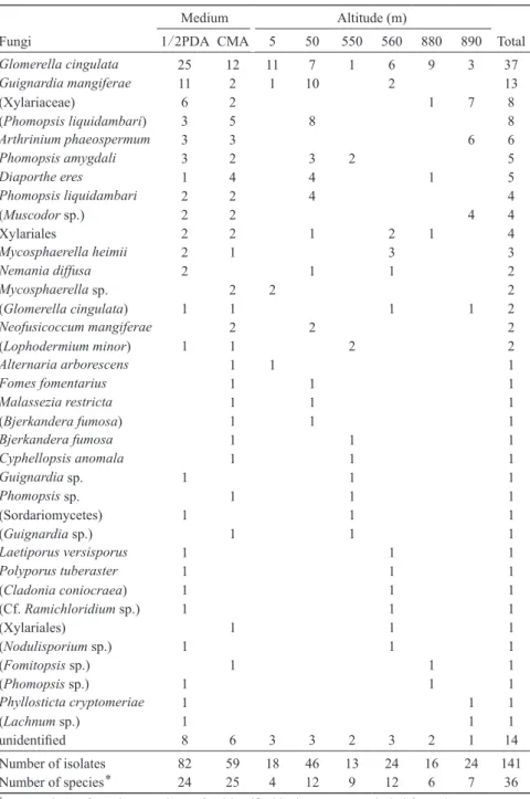
Table 1. Endophytic fungi isolated from leaves of Aucuba japonica identified by molecular phylogenetic analysis
FC Identification1) Family Order Phyllum2) Site Altitude
(m) Isolation
Medium Leaves3) Part14)
3037 (Bjerkandera fumosa) Meruliaceae Polyporaels B Shiroyama 50 CMA C M
3161 (Cf. Ramichloridium sp.) Mycosphaerellaceae Capnodiales A Hatajuku 560 1/2PDA C M 3048 (Cladonia coniocraea) Cladoniaceae Lecanorales A Hatajuku 560 1/2PDA A A 3114 (Glomerella cingulata) Glomerellaceae incertae sedis A Komagatake 890 CMA D M 3157 (Glomerella cingulata) Glomerellaceae incertae sedis A Hatajuku 560 1/2PDA B M
3170 (Fomitopsis sp.) Fomitopsidaceae Polyporaels B Ubako 880 CMA C M
3164 (Guignardia sp.) Botryosphaeriaceae Botryosphaeriales A Miyagami 550 CMA A M
3171 (Lachnum sp.) Lachnaceae Helotiales A Komagatake 890 1/2PDA A A
3068 (Lophodermium minor) Rhytismataceae Polyporaels A Miyagami 550 CMA A B
3073 (Lophodermium minor) Rhytismataceae Rhytismatales A Miyagami 550 1/2PDA B M
3107 (Muscodor sp.) Xylariaceae Xyalriales A Komagatake 890 CMA C B
3108 (Muscodor sp.) Xylariaceae Xyalriales A Komagatake 890 1/2PDA D B
3109 (Muscodor sp.) Xylariaceae Xyalriales A Komagatake 890 1/2PDA D B
3110 (Muscodor sp.) Xylariaceae Xyalriales A Komagatake 890 CMA D B
3053 (Nodulisporium sp.) Xylariaceae Xyalriales A Hatajuku 560 1/2PDA B A
3012 (Phomopsis liquidambari) Diaporthaceae Diaporthales A Miyashita 50 1/2PDA C A 3013 (Phomopsis liquidambari) Diaporthaceae Diaporthales A Miyashita 50 CMA C A 3010 (Phomopsis liquidambari) Diaporthaceae Diaporthales A Miyashita 50 CMA B M 3011 (Phomopsis liquidambari) Diaporthaceae Diaporthales A Miyashita 50 1/2PDA B M 3015 (Phomopsis liquidambari) Diaporthaceae Diaporthales A Miyashita 50 CMA C M 3017 (Phomopsis liquidambari) Diaporthaceae Diaporthales A Miyashita 50 CMA C M 3018 (Phomopsis liquidambari) Diaporthaceae Diaporthales A Miyashita 50 1/2PDA C M 3020 (Phomopsis liquidambari) Diaporthaceae Diaporthales A Miyashita 50 CMA D M
3047 (Phomopsis sp.) Diaporthaceae Diaporthales A Ubako 880 1/2PDA B M
3067 (Sordariomycetes) − − A Miyagami 550 1/2PDA A B
3129 (Xylariaceae) Xylariaceae Xyalriales A Ubako 880 1/2PDA A A
3096 (Xylariaceae) Xylariaceae Xyalriales A Komagatake 890 CMA B A
3104 (Xylariaceae) Xylariaceae Xyalriales A Komagatake 890 CMA C A
3097 (Xylariaceae) Xylariaceae Xyalriales A Komagatake 890 1/2PDA B B
3099 (Xylariaceae) Xylariaceae Xyalriales A Komagatake 890 1/2PDA B B
3094 (Xylariaceae) Xylariaceae Xyalriales A Komagatake 890 1/2PDA A M
3103 (Xylariaceae) Xylariaceae Xyalriales A Komagatake 890 1/2PDA B M
3141 (Xylariaceae) Xylariaceae Xyalriales A Komagatake 890 1/2PDA C M
3064 (Xylariales) − Xyalriales A Hatajuku 560 CMA D B
3089 Alternaria arborescens Pleospoaceae Pleosporales A Manazuru 5 CMA D B
3090 Arthrinium phaeospermum Apiosporaceae incertae sedis A Komagatake 890 CMA A A 3091 Arthrinium phaeospermum Apiosporaceae incertae sedis A Komagatake 890 CMA A B 3139 Arthrinium phaeospermum Apiosporaceae incertae sedis A Komagatake 890 1/2PDA A B 3140 Arthrinium phaeospermum Apiosporaceae incertae sedis A Komagatake 890 1/2PDA A B 3092 Arthrinium phaeospermum Apiosporaceae incertae sedis A Komagatake 890 CMA A M 3093 Arthrinium phaeospermum Apiosporaceae incertae sedis A Komagatake 890 1/2PDA A M
3070 Bjerkandera fumosa Meruliaceae Polyporaels B Miyagami 550 CMA B A
3069 Cyphellopsis anomala Cyphellaceae Agaricales A Miyagami 550 CMA A M
3001 Diaporthe eres Diaporthaceae Diaporthales A Miyashita 50 CMA A A
3002 Diaporthe eres Diaporthaceae Diaporthales A Miyashita 50 CMA A A
3117 Diaporthe eres Diaporthaceae Diaporthales A Ubako 880 CMA B B
3005 Diaporthe eres Diaporthaceae Diaporthales A Miyashita 50 1/2PDA A B
3009 Diaporthe eres Diaporthaceae Diaporthales A Miyashita 50 CMA B B
3045 Fomes fomentarius Polyporaceae Polyporaels B Shiroyama 50 CMA D M
3120 Glomerella cingulata Glomerellaceae incertae sedis A Ubako 880 CMA C A
1)For detail, see the text. 2) A: Ascomycota, B: Basidiomycota. 3) Four collected leaves were named as A-D for practi- cality (see text). 4) A: apical, B: basal, M: middle part of the leaves.
FC Identification1) Family Order Phyllum2) Site Altitude (m) Isolation
Medium Leaves3) Part14)
3121 Glomerella cingulata Glomerellaceae incertae sedis A Ubako 880 1/2PDA C A
3125 Glomerella cingulata Glomerellaceae incertae sedis A Ubako 880 1/2PDA D A
3031 Glomerella cingulata Glomerellaceae incertae sedis A Shiroyama 50 1/2PDA B A
3055 Glomerella cingulata Glomerellaceae incertae sedis A Hatajuku 560 CMA B A
3078 Glomerella cingulata Glomerellaceae incertae sedis A Manazuru 5 1/2PDA B A
3079 Glomerella cingulata Glomerellaceae incertae sedis A Manazuru 5 CMA B A
3149 Glomerella cingulata Glomerellaceae incertae sedis A Manazuru 5 1/2PDA A A
3122 Glomerella cingulata Glomerellaceae incertae sedis A Ubako 880 CMA C B
3042 Glomerella cingulata Glomerellaceae incertae sedis A Shiroyama 50 1/2PDA D B
3043 Glomerella cingulata Glomerellaceae incertae sedis A Shiroyama 50 1/2PDA D B
3144 Glomerella cingulata Glomerellaceae incertae sedis A Shiroyama 50 1/2PDA B B
3052 Glomerella cingulata Glomerellaceae incertae sedis A Hatajuku 560 CMA A B
3165 Glomerella cingulata Glomerellaceae incertae sedis A Hatajuku 560 CMA C B
3080 Glomerella cingulata Glomerellaceae incertae sedis A Manazuru 5 1/2PDA B B
3083 Glomerella cingulata Glomerellaceae incertae sedis A Manazuru 5 1/2PDA C B
3084 Glomerella cingulata Glomerellaceae incertae sedis A Manazuru 5 CMA C B
3169 Glomerella cingulata Glomerellaceae incertae sedis A Manazuru 5 1/2PDA D B
3135 Glomerella cingulata Glomerellaceae incertae sedis A Miyashita 50 CMA C B
3046 Glomerella cingulata Glomerellaceae incertae sedis A Ubako 880 1/2PDA B M
3115 Glomerella cingulata Glomerellaceae incertae sedis A Ubako 880 1/2PDA A M
3119 Glomerella cingulata Glomerellaceae incertae sedis A Ubako 880 1/2PDA B M
3124 Glomerella cingulata Glomerellaceae incertae sedis A Ubako 880 1/2PDA C M
3128 Glomerella cingulata Glomerellaceae incertae sedis A Ubako 880 1/2PDA D M
3112 Glomerella cingulata Glomerellaceae incertae sedis A Komagatake 890 CMA D M
3113 Glomerella cingulata Glomerellaceae incertae sedis A Komagatake 890 1/2PDA D M
3142 Glomerella cingulata Glomerellaceae incertae sedis A Komagatake 890 CMA A M
3057 Glomerella cingulata Glomerellaceae incertae sedis A Hatajuku 560 CMA B M
3058 Glomerella cingulata Glomerellaceae incertae sedis A Hatajuku 560 1/2PDA B M
3059 Glomerella cingulata Glomerellaceae incertae sedis A Hatajuku 560 1/2PDA B M
3076 Glomerella cingulata Glomerellaceae incertae sedis A Manazuru 5 1/2PDA A M
3077 Glomerella cingulata Glomerellaceae incertae sedis A Manazuru 5 1/2PDA A M
3085 Glomerella cingulata Glomerellaceae incertae sedis A Manazuru 5 1/2PDA C M
3150 Glomerella cingulata Glomerellaceae incertae sedis A Manazuru 5 1/2PDA B M
3155 Glomerella cingulata Glomerellaceae incertae sedis A Miyagami 550 1/2PDA A M
3021 Glomerella cingulata Glomerellaceae incertae sedis A Miyashita 50 1/2PDA D M
3022 Glomerella cingulata Glomerellaceae incertae sedis A Miyashita 50 CMA D M
3145 Guignardia mangiferae Botryosphaeriaceae Botryosphaeriales A Shiroyama 50 1/2PDA C A 3159 Guignardia mangiferae Botryosphaeriaceae Botryosphaeriales A Hatajuku 560 1/2PDA C A 3087 Guignardia mangiferae Botryosphaeriaceae Botryosphaeriales A Manazuru 5 1/2PDA D A 3132 Guignardia mangiferae Botryosphaeriaceae Botryosphaeriales A Miyashita 50 1/2PDA B A 3137 Guignardia mangiferae Botryosphaeriaceae Botryosphaeriales A Miyashita 50 1/2PDA D A
3056 Guignardia mangiferae Botryosphaeriaceae Botryosphaeriales A Hatajuku 560 CMA B B
3133 Guignardia mangiferae Botryosphaeriaceae Botryosphaeriales A Miyashita 50 1/2PDA B B 3136 Guignardia mangiferae Botryosphaeriaceae Botryosphaeriales A Miyashita 50 1/2PDA D B
3006 Guignardia mangiferae Botryosphaeriaceae Botryosphaeriales A Miyashita 50 CMA A M
3130 Guignardia mangiferae Botryosphaeriaceae Botryosphaeriales A Miyashita 50 1/2PDA A M 3131 Guignardia mangiferae Botryosphaeriaceae Botryosphaeriales A Miyashita 50 1/2PDA A M 3134 Guignardia mangiferae Botryosphaeriaceae Botryosphaeriales A Miyashita 50 1/2PDA B M 3138 Guignardia mangiferae Botryosphaeriaceae Botryosphaeriales A Miyashita 50 1/2PDA D M
3172 Guignardia sp. Botryosphaeriaceae Botryosphaeriales A Miyagami 550 1/2PDA A A
1)For detail, see the text. 2) A: Ascomycota, B: Basidiomycota. 3) Four collected leaves were named as A-D for practi- cality (see text). 4) A: apical, B: basal, M: middle part of the leaves.
Table 1. (Cont.)
FC Identification1) Family Order Phyllum2) Site Altitude (m) Isolation
Medium Leaves3) Part14)
3054 Laetiporus versisporus Fomitopsidaceae Polyporaels B Hatajuku 560 1/2PDA B A
3044 Malassezia restricta − Malasseziales B Shiroyama 50 CMA D M
3160 Mycosphaerella heimii Mycosphaerellaceae Capnodiales A Hatajuku 560 1/2PDA C B
3158 Mycosphaerella heimii Mycosphaerellaceae Capnodiales A Hatajuku 560 1/2PDA B B
3060 Mycosphaerella heimii Mycosphaerellaceae Capnodiales A Hatajuku 560 CMA B M
3151 Mycosphaerella sp. Mycosphaerellaceae Capnodiales A Manazuru 5 CMA C M
3152 Mycosphaerella sp. Mycosphaerellaceae Capnodiales A Manazuru 5 CMA C M
3049 Nemania diffusa Xylariaceae Xyalriales A Hatajuku 560 1/2PDA A B
3028 Nemania diffusa Xylariaceae Xyalriales A Shiroyama 50 1/2PDA A M
3007 Neofusicoccum mangiferae Botryosphaeriaceae Botryosphaeriales A Miyashita 50 CMA A M 3008 Neofusicoccum mangiferae Botryosphaeriaceae Botryosphaeriales A Miyashita 50 CMA A M
3035 Phomopsis amygdali Diaporthaceae Diaporthales A Shiroyama 50 CMA C A
3036 Phomopsis amygdali Diaporthaceae Diaporthales A Shiroyama 50 CMA C A
3003 Phomopsis amygdali Diaporthaceae Diaporthales A Miyashita 50 1/2PDA A A
3074 Phomopsis amygdali Diaporthaceae Diaporthales A Miyagami 550 1/2PDA C B
3075 Phomopsis amygdali Diaporthaceae Diaporthales A Miyagami 550 1/2PDA C B
3019 Phomopsis liquidambari Diaporthaceae Diaporthales A Miyashita 50 CMA D A
3004 Phomopsis liquidambari Diaporthaceae Diaporthales A Miyashita 50 CMA A B
3014 Phomopsis liquidambari Diaporthaceae Diaporthales A Miyashita 50 1/2PDA C B
3016 Phomopsis liquidambari Diaporthaceae Diaporthales A Miyashita 50 1/2PDA C M
3156 Phomopsis sp. Diaporthaceae Diaporthales A Miyagami 550 CMA C B
3101 Phyllosticta cryptomeriae Botryosphaeriaceae Botryosphaeriales A Komagatake 890 1/2PDA B M
3061 Polyporus tuberaster Polyporaceae Polyporaels B Hatajuku 560 1/2PDA D A
3071 unidentified A Miyagami 550 1/2PDA B A
3082 unidentified A Manazuru 5 1/2PDA C B
3072 unidentified A Miyagami 550 1/2PDA B B
3118 unidentified A Ubako 880 CMA B M
3126 unidentified A Ubako 880 1/2PDA D M
3102 unidentified A Komagatake 890 CMA B M
3032 unidentified A Shiroyama 50 CMA B M
3033 unidentified A Shiroyama 50 CMA B M
3038 unidentified A Shiroyama 50 1/2PDA C M
3066 unidentified A Hatajuku 560 CMA D M
3162 unidentified A Hatajuku 560 1/2PDA D M
3163 unidentified A Hatajuku 560 1/2PDA D M
3081 unidentified A Manazuru 5 1/2PDA B M
3154 unidentified A Manazuru 5 CMA D M
3063 Xylariales − Xyalriales A Hatajuku 560 CMA D A
3123 Xylariales − Xyalriales A Ubako 880 1/2PDA C B
3050 Xylariales − Xyalriales A Hatajuku 560 1/2PDA A B
3027 Xylariales − Xyalriales A Shiroyama 50 CMA A M
1)For detail, see the text. 2) A: Ascomycota, B: Basidiomycota. 3) Four collected leaves were named as A-D for practi- cality (see text). 4) A: apical, B: basal, M: middle part of the leaves.
Table 1. (Cont.)
Table 2. Number of isolates of the endophytic fungi from Aucuba japonica at various altitude with a reference to the difference of the medium for isolation.
Medium Altitude (m)
Fungi 1/2PDA CMA 5 50 550 560 880 890 Total
Glomerella cingulata 25 12 11 7 1 6 9 3 37
Guignardia mangiferae 11 2 1 10 2 13
(Xylariaceae) 6 2 1 7 8
(Phomopsis liquidambari) 3 5 8 8
Arthrinium phaeospermum 3 3 6 6
Phomopsis amygdali 3 2 3 2 5
Diaporthe eres 1 4 4 1 5
Phomopsis liquidambari 2 2 4 4
(Muscodor sp.) 2 2 4 4
Xylariales 2 2 1 2 1 4
Mycosphaerella heimii 2 1 3 3
Nemania diffusa 2 1 1 2
Mycosphaerella sp. 2 2 2
(Glomerella cingulata) 1 1 1 1 2
Neofusicoccum mangiferae 2 2 2
(Lophodermium minor) 1 1 2 2
Alternaria arborescens 1 1 1
Fomes fomentarius 1 1 1
Malassezia restricta 1 1 1
(Bjerkandera fumosa) 1 1 1
Bjerkandera fumosa 1 1 1
Cyphellopsis anomala 1 1 1
Guignardia sp. 1 1 1
Phomopsis sp. 1 1 1
(Sordariomycetes) 1 1 1
(Guignardia sp.) 1 1 1
Laetiporus versisporus 1 1 1
Polyporus tuberaster 1 1 1
(Cladonia coniocraea) 1 1 1
(Cf. Ramichloridium sp.) 1 1 1
(Xylariales) 1 1 1
(Nodulisporium sp.) 1 1 1
(Fomitopsis sp.) 1 1 1
(Phomopsis sp.) 1 1 1
Phyllosticta cryptomeriae 1 1 1
(Lachnum sp.) 1 1 1
unidentified 8 6 3 3 2 3 2 1 14
Number of isolates 82 59 18 46 13 24 16 24 141
Number of species* 24 25 4 12 9 12 6 7 36
*For numbers of species, numbers of unidentified isolates were excluded from the count.
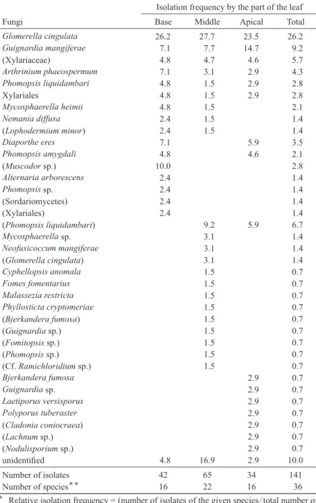
Table 3. Relative isolation frequency* and total number of isolates of endophytic fungi from various part of the leaves of Aucuba japonica
Isolation frequency by the part of the leaf
Fungi Base Middle Apical Total
Glomerella cingulata 26.2 27.7 23.5 26.2
Guignardia mangiferae 7.1 7.7 14.7 9.2
(Xylariaceae) 4.8 4.7 4.6 5.7
Arthrinium phaeospermum 7.1 3.1 2.9 4.3
Phomopsis liquidambari 4.8 1.5 2.9 2.8
Xylariales 4.8 1.5 2.9 2.8
Mycosphaerella heimii 4.8 1.5 2.1
Nemania diffusa 2.4 1.5 1.4
(Lophodermium minor) 2.4 1.5 1.4
Diaporthe eres 7.1 5.9 3.5
Phomopsis amygdali 4.8 4.6 2.1
(Muscodor sp.) 10.0 2.8
Alternaria arborescens 2.4 1.4
Phomopsis sp. 2.4 1.4
(Sordariomycetes) 2.4 1.4
(Xylariales) 2.4 1.4
(Phomopsis liquidambari) 9.2 5.9 6.7
Mycosphaerella sp. 3.1 1.4
Neofusicoccum mangiferae 3.1 1.4
(Glomerella cingulata) 3.1 1.4
Cyphellopsis anomala 1.5 0.7
Fomes fomentarius 1.5 0.7
Malassezia restricta 1.5 0.7
Phyllosticta cryptomeriae 1.5 0.7
(Bjerkandera fumosa) 1.5 0.7
(Guignardia sp.) 1.5 0.7
(Fomitopsis sp.) 1.5 0.7
(Phomopsis sp.) 1.5 0.7
(Cf. Ramichloridium sp.) 1.5 0.7
Bjerkandera fumosa 2.9 0.7
Guignardia sp. 2.9 0.7
Laetiporus versisporus 2.9 0.7
Polyporus tuberaster 2.9 0.7
(Cladonia coniocraea) 2.9 0.7
(Lachnum sp.) 2.9 0.7
(Nodulisporium sp.) 2.9 0.7
unidentified 4.8 16.9 2.9 10.0
Number of isolates 42 65 34 141
Number of species** 16 22 16 36
* Relative isolation frequency = (number of isolates of the given species/total number of the isolates in each part)×100.
** For numbers of species, numbers of unidentified isolates were excluded from the count.
motor road, respectively. The high altitude may also have affected the fungi, resulting the less di- verse endophytic mycobiota by low temperature.
Hashizume et al. (2008) compared the endo- phytes in Quercus acuta in different altitude, and showed that endophytic mycobiota changed by- altitude. They also suggest that the optimum tem- perature of the fungi may be the major reason for this difference. This explanation may be also adoptable to the present result.
Distribution of the endophytes in the leaves Because two pieces were inoculated for the middle part of the leaves while single piece was used for isolation from the basal and apical part, relative isolation frequency (isolates of the given species/total number of the isolates in each part
×100) was used for comparison (Table 3). Five members were isolated from all parts of the leaves: Glomerella cingulata, Guignardia mangiferae, Arthrinum phaeospermum, Phomop- sis liquidambari, and Xylariales including Xylari- aceae. These members distributed in all parts of the leaves, and the relative isolation frequency of them did not differ considerably by part of the leaves. The middle part of the leaf embraced the largest numbers of species, and fewer members were unique to the base or apical part. The pres- ent results are similar to those in Sahashi et al.
(1999) and Kaneko et al. (2003) who reported that isolation frequency of the major endophytes did not differ in part of the leaves in Fagus crena- ta. The endophytic mycobiota in leaves of A.
japonica were found to be homogeneous in the leaves.
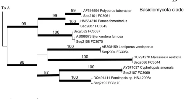
Phylogenetic aspects of the isolates
Two major clades, one for Ascomycota and the other for Basidiomycota were recognized in NJ tree (Figs. 3–6). The Ascomycota clade was sub- divided into several major clades, but they were not strongly supported. However, most of the ter- minal nodes, mostly representing the species or genera, were strongly supported. In some cases, groups higher than genera were strongly support- ed. They represented Sordariomycetes clade1
(mainly Xylariales), Sordariomycetes clade 2, Glomerella cingulata (anamorph: Glomerella cin- gulata), Diaporthe spp. (anamorph: Phomopsis spp.), Guignardia spp. (anamorph: Phyllosticta spp.), Mycosphaerellaceous clade (Mycosphaer- ella with Ramichloridium anamorph), and Lach- num spp. Among these, some unidentified endo- phytes with strong support were inserted.
Phylogenetic relationships of these unidentified endophytes and the aforementioned groups were not clarified.
The Basidiomycota clade was subdivided into two strongly supported clades. One included Bjerkandera, Fomes, and Polyporus. All these are known as white rot fungi, decomposing lignin in the tree. However, none of them are known as a a pathogen from A. japonica. Fomes fomentarius is a stem heart rot pathogen of Acer, Betula, Carpi- nus, Larix, Populus, Prunus, Quercus, Sorbus, Tilia, and Ulmus (Kobayashi, 2007) . It is particu- larly well known to occur on Fagus crenata (Ki- shi, 1998). Although F. crenata was associated with A. japonica in high altitude, the site where F.
fomentarius was isolated was about 50 m altitude where A. japonica was not associated with F. cre- nata. It is therefore suggested that some patho- genic fungi, having a selectivity to their host may survive as endophytes in other plant when they are away from their primary habitat.
Basidiomycetes are infrequently isolated as en- dophytes (Bills and Polishook, 1991), and claimed to be isolation bias (Stone et al., 2004).
However, Oses et al. (2006) and Arnold et al.
(2007) detected basidiomycetes from environ- mental DNA, suggesting that more numbers of basidiomycetous endophytes exist than expected.
Oses et al. (2006) elucidated the possible contri- bution of basidiomycetous endophytes to white rot decay of trees.
Xylariales is also known as white rot fungi.
More than several groups of lignin decomposing fungi were found to contribute to decompose A.
japonica leaves, together with brown rot fungi.
Xyalrialean isolates shear about 14% of the ob- tained isolates in the present study. Okane et al.
(1997) reported that xylariaceous fungi and Phyl-
losticta spp. comprised of about 50% of obtained isolates in evergreen plants in subtropical area.
Xylariales seems to be cosmopolitan in the ever- green broad-leaved trees in subtropical area.
The other clade in Basidiomycota is composed of brown rot fungi Fomitopsis and Laetiporus, and Cyphellopsis and Malassezia. Substrate se- lectivity of the latter two is not known.
Biodiversity of fungal endophytes in Aucuba japonicae in Sagami Sea and its adjacent areas
In the inventory of Japanese fungi (Katumoto, 2010), 26 species were listed from A. japonica.
Of these, Glomerella cingulata (anamorph: Col- letotrichum gloeosporioides) is known as a patho- gen of the anthracnose of A. japonica (Kishi, 1998). It is also known as a pathogen of various plants. Diaporthe aucubae and Phomopsis aucu- bae are also known as canker disease pathogens.
The sequences of these fungi were not registered in GenBank, and further study is required to iden- tify the isolates obtained in the present study with certainty. Phyllosticta harai is known for brown rot fungus, but the sequences of the isolates ob- tained in the present study was not identical with it.
Elucidation of more comprehensive endophytic mycobiota requires multiple media and large scale collecting because isolation of singletons may be ruled by chance. Due to the distribution of the host, fungi specific to A. japonica may con- tain east Asian elements, and extensive studies in other areas to compare the results with those in the present study will contribute to reveal the fun- gal diversity hot spot in Asia.
Some endophytic fungi are known to stay on the leaves after the leaves were fallen from the tree, and contribute in decomposition as sapro- phytes (Promputtha et al, 2007). Field mycologi-
Fig. 3. Overall topology of a neighbor-joining (NJ) tree of endophytic fungi from Aucuba japonica with ref- erence sequences from GenBank inferred from ITS- 5.8S rRNA sequences. A1 and A2 represent Ascomy- cota clade, B represents Basidiomycota clade. Details of each clade are shown in Figs 4, 5 and 6, respec- tively.
0.05
A1
A2
B
Fig. 4. Part of neighbor-joining (NJ) tree (clade A1 in Fig. 3) of endophytic fungi from Aucuba japonica with refer- ence sequences from GenBank inferred from ITS-5.8S rRNA sequences. Remarkable fungal groups are indicated.
Bootstrap values (BP) of 1000 replications are shown where BP exceeded over 80%. Caldes with BP>80% are shown with thickened branches.
Seq2091 FC3050 Seq2102 FC3063
GQ906959 Xylariales cf. JW38-4 EU680529 Sordariomycete sp. 7653 GU292817 Nemania diffusa Seq2090 FC3049 Seq2103 FC3064
GQ906961 Xylariales cf. JP89-2 Seq2075 FC3027
EU686201 Fungal endophyte sp. ECD-2008 isolate d12 Seq2135 FC3102
GQ906963 Nodulisporium sp. JW89-5 Seq2093 FC3053
AJ279456 Arthrinium phaeospermum Seq2170 FC3139
GQ848369 Muscodor sp. CMU-Cib 461 AM921731 Xylariaceae sp. IZ-1249 Seq2129 FC3094
Seq2172 FC3141 Seq2130 FC3096 Seq2160 FC3129
FJ799947 Sordariomycetes sp. SAB-2009a Seq2105 FC3067
Seq2092 FC3052 Seq2177 FC3150
HM131512 Colletotrichum sp. LC922 FJ456938 Colletotrichum gloeosporioides FJ940901 Colletotrichum gloeosporioides Seq2150 FC3120
Seq2145 FC3114 Seq2183 FC3157
Seq2104 FC3066 Seq2109 FC3071
EU686922 Fungal endophyte isolate 1657 FJ466716 Fungal endophyte sp. UFMGCB_537 Seq2148 FC3118
EU686888 Fungal endophyte isolate 1506 Seq2187 FC3162
Seq2188 FC3163 AB365875 Fungal sp. QA-b Seq2180 FC3154
Seq2068 FC3016 Seq2071 FC3019 Seq2066 FC3014 Seq2056 FC3004
HM537071 Fungal endophyte sp. g101 FJ478124 Phomopsis liquidambari xsd08082 AF103000 Phomopsis sp. FAU 1053 Seq2070 FC3018
Seq2053 FC3001 Seq2054 FC3002
FJ478132 Diaporthe eres strain xsd0809 AB107890 Phomopsis sp. MAFF 665006 GU133063 Phomopsis amygdali JX-Pa-1 Seq2112 FC3074
Seq2182 FC3156
GU462142 Phomopsis sp. NY302 EU571102 Phomopsis sp. VAAT- PZ15 HM537052 Fungal endophyte sp. g68
Seq2088 FC3047 Seq2118 FC3081
0.05 87
89 100 100 100 82 82
100 100
100
100
100 100
100 100
91
93 91
84
A1
Sordariomycetes clade 1 (mainly Xylariales)
Sordariomycetes clade 2
Glomerella cingulata (Colletotrichum gloeosporioides)
clade Ascomycota
clade
To A2
Diaporthe/Phomopsis
Fig. 5. Part of neighbor-joining (NJ) tree (clade A2 in Fig. 3) of endophytic fungi from Aucuba japonica with refer- ence sequences from GenBank inferred from ITS-5.8S rRNA sequences. Remarkable fungal groups are indicated.
Bootstrap values (BP) of 1000 replications are shown where BP exceeded over 80%. Caldes with BP>80% are shown with thickened branches.
Mycosphaerellaceae clade
DQ394378 Cladonia coniocraea Seq2089 FC3048
EU821360 Guignardia mangiferae Seq2175 FC3145
Seq2189 FC3164 Seq2194 FC3172
AB041242 Phyllosticta pyrolae AB454300 Phyllosticta cryptomeriae Seq2134 FC3101
HM021155 Neofusicoccum mangiferae Seq2059 FC3007
HM537022 Fungal endophyte sp. g6 Seq2078 FC3032
GQ852745 Mycosphaerella heimii Seq2100 FC3060
EU167579 Mycosphaerella crystallina FJ037771 Mycosphaerella sp. ZJ12-2A Seq2178 FC3151
GU214688 Cf. Ramichloridium sp. CP10672 Seq2186 FC3161
EF419954 Fungal endophyte isolate 9183 Seq2083 FC3038
Seq2110 FC3072
AY154706 Alternaria arborescens Seq2124 FC3089
AB481251 Lachnum asiaticum AB481275 Lachnum sp. FC-2076
Seq2193 FC3171
EF434059 Uncultured fungus clone TB18 OTU142 AJ877183 Uncultured fungus clone YJ4-33
Seq2158 FC3126
FJ861983 Lophodermium minor strain R83 Seq2106 FC3068
0.05
A2
100
100 97
98 98
83 100
100
100 99
87
100
99 100
100 87 94
93
80 100
100
Guignardia/Phyllostictaclade
Lachnumclade To A1
To B
Ascomycota clade
(cont’d from A1)
cal studies should also be carried out by collect- ing the fruiting body of the fungi on A. japonica in situ to give a reference to the identification of the isolates with only sterile hyphae.
Acknowledgements
The authors of the present paper wish to ex- press their gratitude to Dr. Izumi Okane, Biologi- cal Resource Center (NBRC), Department of Biotechnology, National Institute of Technology and Evaluation for carefully reading the manu- script.
References
Agrios, G.N. 2005. Plant pathology. 5th ed. pp.992. El- sevier Academic Press, Amsterdam.
Arnold A.E., Henk, D.A., Eels, R.L., Lutzoni, F. and R.
Vilgalys, 2007. Diversity and phylogenetic affinities of foliar fungal endophytes in loblolly pine inferred by
culturing and environmental PCR. Mycologia, 99: 185–
206.
Bills, G.F. and J.D. Polishook, 1991. Microfungi from Carpinus caroliana. Canadian Journal of Botany, 69:
1477–1482.
Degawa Y, Doi, Y. and T. Hosoya, 2006. Records of All- escheriella crocea (Basidiomycetous Anamorphic Fun- gus) from Sagami Bay Coast, Japan. Memoirs of the National Science Museum, 42: 233–238.
Hall, T.A., 1999. BioEdit: a user-friendly biological se- quence alignment editor and analysis program for Win- dows 95/98/NT. Nucleic Acids Symposium Series, 41:
95–98.
Hashizume, Y., Sahashi, N. and K. Fukuda, 2008. The in- fluence of altitude on endophytic microbiota in Quercus acuta leaves collected in two areas 1000 km apart. For- est Pathology, 31: 141–147.
Hata, K. and K. Futai, 1995. Endophytic fungi associated with healthy pine needles and needles infested by the pine needle gall midge, Thecodiplosis japonensis. Ca- nadian Journal of Botany, 73: 384–390.
Hawksworth, D.L., 1991. The fungal dimension of biodi- versity: magnitude, significance, and conservation. My- cological Research, 95: 641–655.
Fig. 6. Part of neighbor-joining (NJ) tree (clade B in Fig. 3) of endophytic fungi from Aucuba japonica with reference sequences from GenBank inferred from ITS-5.8S rRNA sequences. Remarkable fungal groups are indicated. Boot- strap values (BP) of 1000 replications are shown where BP exceeded over 80%. Caldes with BP>80% are shown with thickened branches.
AF516594 Polyporus tuberaster Seq2101 FC3061
HM584810 Fomes fomentarius Seq2087 FC3045
Seq2082 FC3037
AJ006673 Bjerkandera fumosa Seq2108 FC3070
AB308159 Laetiporus versisporus Seq2094 FC3054
GU291270 Malassezia restricta Seq2086 FC3044
AY571037 Cyphellopsis anomala Seq2107 FC3069
DQ491411 Fomitopsis sp. HSJ-2006a Seq2192 FC3170
0.05
100 99 99
99
100
100 100
100 100
87 98
B
Basidiomycota clade
To A