九州大学学術情報リポジトリ
Kyushu University Institutional Repository
ビスフェノールAアナログが人体に与える有害作用の 評価に関する研究
袈裟丸, 仁志
http://hdl.handle.net/2324/2236029
出版情報:Kyushu University, 2018, 博士(理学), 課程博士 バージョン:
権利関係:
Study on the assessment for the adverse effects of the bisphenol A analogs in the human body
Hitoshi Kesamaru
Laboratory of Biomolecular Chemistry Department of Chemistry
Graduate School of Science Kyushu University
March 2019
CONTENTS
Page Study on the assessment for the adverse effects of the bisphenol A analogs
in the human body
CHAPTER 1
Influence of receptor conformations in docking calculation based endocrine disruptor risk assessment: evaluation of binding affinity and transcriptional activity of estrogen receptor binding chemicals.
1. Introduction 2
2. Materials and Methods 6
3. Results and Discussion 8
4. Conclusion 14
5. References 15
CHAPTER 2
Evaluation of the influence of halogenation on the binding affinity and transcriptional activity of bisphenol A to the estrogen receptors.
1. Introduction 54
2. Materials and Methods 56
3. Results and Discussion 67
4. Conclusion 72
5. References 73
CHAPTER 3
Effects of halogenation of bisphenol A analogs on cytotoxicity for HeLa cells: di- halogenated BPA analogs inducing different types of cell death depending on halogen atom species.
1. Introduction 91
2. Materials and Methods 94
3. Results and Discussion 96
4. Conclusion 99
5. References 102
Acknowledgements 120
ABBREVIAIONS
The abbreviations according to biochemical nomenclature by IUPAC-IUB Joint Commission, Eur. J. Biochem., 138, 9-37 (1984), are used throughout. Unless otherwise specified, the amino acids are L-stereoisomers. Additional abbreviations are follows, but generally accepted abbreviations and symbols were used without definition.
(Z)-4-OHT (Z)-4-hydoroxytamoxifen ABC agonist-bound conformations AF-1/2 activation function 1/2
BPX bisphenol X (where X is any of the following A, AF, C, and E) cAMP cyclic adenosine monophosphate
DCC dextran coated charcoal
DES diethylstilbestrol
DMF dimethylformamide
DMSO dimethylsulfoxide
DTT dithiothreitol
E2 17β-estradiol
EDCs endocrine disrupting chemicals EMEM Eagle's minimim essential medium
ER estrogen receptor
ERK extracellular signal-regulated kinase ERRγ estrogen related receptor γ
FBS fatal bovine serum
GPR30 G protein-coupled receptor 30 GST glutathione S-transferase
H11 α-helix numbered 11
H12 α-helix numbered 12
HPTE 2,2-bis(p-hydroxy phenyl)-1,1,1-trichloroethane IC50 50% inhibition concentration
LBD ligand binding domain
LBP ligand binding pocket
LDH lactate dehydrogenase
MAPK mitogen-activated protein kinase
MOM methoxymethyl
MTT methylthiazole tetrazolium
NABC non-agonist-bound conformations
NMR nuclear magnetic resonance spectroscopy
NR nuclear receptor
PBS phosphate buffered saline
PDB protein data bank
PFCs perfluorinated compounds
PKA protein kinase A
PPAR peroxisome proliferator-activated receptor
RAL raloxifene
RMSD root mean square deviation
SDS-PAGE sodium dodecyl sulfate-poly-acrylamide gel electrophoresis
S.E. standard error
T3 3,3',5-Triiodo-L-thyronine TBBPA tetrabromobisphenol A TCBPA tetrachlorobisphenol A
TCDD 2,3,7,8-tetrachlorodibenzo-p-dioxin
TMS tetramethylsilane
1
CHAPTER 1
Importance of receptor conformations in docking calculation based risk assessment for endocrine disruptor: evaluation of binding potency and transcriptional activity of estrogen receptor α binding chemicals
Abstract
Risk assessment of chemical substances in environment is an important issue for the development and safe use of chemicals. To evaluate adverse effects of chemicals for living bodies in a high accuracy, it is necessary to investigate physiological activities from broad perspectives by using various kinds of assay methods. Among them, in silico docking calculation is one of the first-line screening methods to assess the risk of environmental chemicals, which can evaluate the binding affinity between a chemical and a receptor protein.
For using in silico docking calculations, employment of suitable receptor conformations as templates is essential for appropriate identification of the latent receptor-binding ability of chemicals. In this study, we performed docking calculations using a number of agonist- and antagonist-bound conformations of estrogen receptor α-ligand binding domains as templates to clarify the type of receptor conformations required for reasonable identification of endocrine disruptors. In the calculation results, ERα agonists bound selectively to the agonist conformations, whereas antagonists bound selectively to the non-antagonist conformations.
Furthermore, structural analyses of the ligand-binding domains and docking calculation utilizing C-terminal truncated receptors indicated that the conformation of C-terminal region of the ligand-binding domain, which altered its conformation by ligand binding, distinguished agonists from non-agonists. These results suggest that employment of appropriate receptor conformations as docking templates are necessary to predict
2
appropriately the receptor binding affinity and the transcriptional activity of chemicals using docking calculation-based risk assessment.
1. Introduction
At present, a number of chemical compounds exceed 100 million have been developed and reported in Chemical Abstracts Service (CAS) and many of them have been utilized for the chemical industries. However, it has been reported that chemical substances leaked from diverse consumer products and spread in the air, water, soil, and food. These chemicals, called environmental chemicals, have been concerned to exhibit various adverse effects on the living organisms. For example, early exposure of 2,3,7,8-tetrachlorodibenzo- p-dioxin (TCDD) caused decrease of sperm counts and motility in human or increase of cancers in children and young adults by endocrine disruption via receptor proteins1,2. As described in these reports, receptor-mediated endocrine disruption of environmental chemicals has become a concern for adverse effects on developmental processes, especially in fetuses and infants. Therefore, it is necessary to assess risk of these chemical substances promptly including newly developed chemicals. However, a large number of chemical substances are still unevaluated their activity by conventional toxicity tests, even though many of them are expected to exhibit various biological activities. Therefore, it is necessary to develop evaluation methods to assess the risk of chemicals efficiently and reasonably.
Since receptors are thought to be a first access point of environmental chemicals, the interaction between chemicals and receptors should be evaluated rapidly. Among the receptor proteins, nuclear receptors (NRs) are receptor proteins involved in regulating a wide range of physiological functions. NRs represent a protein superfamily comprising 48 different proteins in humans3. The ligand-binding domains (LBDs) of NRs share a highly homologous structure, including 12 α-helices (H1 to H12) and one or more underlying β-sheets. The conformation of NR protein is altered by the positioning of the H12 helix, which depends on the biological characteristics of the bound-ligand (agonist or antagonist). Following the agonist-ligand binding, NRs change their conformation from the apo-form to the activated form, while the antagonist-ligand binding results in achievement of an inactive conformation
3 of H12 (Figure 1). Compared with the change in conformation of H12, H11 shows relatively little but apparent position changes according to agonist or antagonist binding. Such conformational changes allow NRs to differentiate chemical agonists and antagonists3–6.
(Figure 1)
Under physiological conditions, NRs are susceptible to endocrine disruption by environmental chemicals7, including so-called endocrine-disrupting chemicals (EDCs).8 EDCs can bind to human NRs, disrupt gene transcription, and cause adverse effects on development, reproduction, cell differentiation, nervous system function, and immune responses in mammals and especially in fetuses and infants9–11. A representative example of an EDC is bisphenol A (BPA), which is used to manufacture polycarbonate plastics, epoxy resins, and other products. Exposure to BPA in mice and rats leads to early onset of sexual maturation in females12–14, increased prostate size in male offspring15–17, and altered immune function18–20. Therefore, to avoid health damage by BPA, several bisphenol analogs known as “next-generation bisphenols” have been developed and used as alternatives to BPA21. However, some BPA analogs, such as bisphenol AF (BPAF20) have been receiving much attention, since it was revealed that these analogs also bind to NRs and could induce endocrine disruption similar to BPA. For example, it was reported that bisphenol AF (BPAF), which is a fluorinated analog of BPA, strongly binds to estrogen receptors. Surprisingly, it was also revealed that BPAF acted as a full agonist for estrogen receptor α, while exhibiting strong antagonist activity for estrogen receptor β22. These results clearly indicate that some BPA analogs act as ligands with variety of activities via different NRs. Furthermore, it is highly likely that latent chemicals will emerge as EDCs similar to BPA; therefore, all NRs are regarded as candidate targets of these yet-undetermined EDCs23. Based on this likelihood, it is important to develop reliable assays capable of predicting the risk of NR-related endocrine disruption associated with various chemicals.
The recent emphasis on implementing strict ethical standards in research has increased the demand to switch from in vivo to in vitro or in silico methods of risk assessment of chemical substances. For example, according to the OECD guidelines, in vitro assays such as receptor binding assay and transcriptional activation assay to NRs are conducted as level
4
2 assessment of EDCs, which is early level for quick evaluation of a large number of substances, whereas in vivo evaluation is used for subsequent assessment for refined target substance24. Since in vitro and in silico assays can be carried out quickly and readily compared to in vivo methods, they are applied early stage of the assessment for a number of compounds as shown in the OECD guidelines. In addition, environmental chemicals exhibit adverse effects for living organisms not only by endocrine disruption via binding to nuclear receptors but also inducing several other types of biological toxicity including induction of mitochondrial injury and apoptotic death in several types of cells. Therefore, it is necessary to evaluate the risk of chemical substances from broad perspectives by using various in vitro and in silico methods. Regarding the in silico techniques, diverse computational method including quantitative structure-activity relationship models25, pharmacophore models26, docking calculations27,28, and molecular dynamics simulations29, have been frequently used as appropriate methods for assessment of the physiological impacts induced by a number of chemicals via various kinds of receptors. Among these techniques, docking calculations represent a possible first-line screening method for EDCs. To implement docking calculations, it is essential to utilize a receptor’s 3D-structure as a template for the calculations. To date, 3D-structures of the LBDs determined by X-ray crystallography are necessary to perform docking calculations. However, the quality and choice of the structures greatly affecting the results. In most cases, the receptor structure has been treated as rigid (i.e.
all atoms of the protein structure are fixed) during docking calculation using common docking programs such as Autodock, DOCK, and Autodock Vina30. Under the rigid conditions, if the receptor structure is not appropriately selected, binding affinity of the chemical will not be estimated reasonably, since the conformations of each residue on the ligand-binding pocket (LBP) are incapable of adapting to all structure of ligands possessing binding activity. For example, accurate antagonist-binding affinity cannot be calculated from a structure of an activated (agonist-bound) form of an NR, as H12 and H11 positioning in the activated form of the NR hinders antagonist binding (Figure 2). This renders an antagonist incapable of altering the active conformation of NR structures that obtained in the presence of an agonist by crystallography, without extensive structure optimization of the whole
5 antagonist-bound complex along with docking calculations. Therefore, it is necessary to select suitable NR-template structures, including agonist-bound conformations (ABCs) and non-agonist-bound conformations (NABCs), to enable accurate evaluation of binding affinities between NRs and ligands via docking calculations28,31. Nevertheless, it is not always possible to select an appropriate receptor structure for in silico studies. Thus, docking calculation studies using only one receptor structure may not be appropriate to evaluate the binding affinity of various chemicals. In this context, statistical analyses of the binding energy values obtained by docking calculations using multiple receptor structures might be an effective method to reduce prediction errors.
(Figure 2)
In this study, we performed docking calculations using estrogen receptor alpha (ERα) to examine whether the physiological activity of ER ligands could be predicted with high accuracy by statistical analyses of the obtained binding energy values. ERα is a member of the steroid-receptor family and a representative target of EDCs5. Since numerous agonist- bound and non-agonist-bound structures of ERα/ligand complexes have been reported in the Protein Data Bank (PDB; www.rcsb.org), ERα is suitable to use for statistical analysis of EDC binding. Therefore, some in silico docking studies using different conformations of ERα have been reported previously. For example, Nose et al. reported on an agonist/antagonist differential-docking screening method using a total of 4 ERα-LBDs, including 2 agonist- bound conformations and 2 antagonist-bound conformations25. Celik et al. also reported in silico study using 3 newly found quasi-stable structures of the ERα and LBD complexes and 3 crystallographic conformations of the protein32. In order to further advance these studies using multiple receptor structures and to clarify the optimal method for selecting the receptor structures in docking calculation, we tested eight known ERα agonists and antagonists against crystal structures representing 83 human ERα-LBDs in the present study. By statistically comparing the calculations results, we investigated whether docking calculations were capable of accurately predicting ERα chemical-binding activity. Moreover, using modified receptor structures in which H11 and H12 were truncated, we also investigated how H11 and H12 positioning affect the docking calculation.
6
2. Material and Methods
2.1. Preparation of receptor structures for docking calculations
As templates for docking calculations, 83 ERα-LBD structures reported by 2015 were prepared using crystal structures deposited in the PDB (Table 1). Preparation of the three- dimensional structures as docking templates was performed using Discovery Studio software (v4.0; Accelrys, San Diego, CA, USA). All calculations were performed using a Dell Precision T3610 workstation (Dell, Round Rock, TX, USA). In all cases, co-crystallized ligands and other small molecules except for water molecules were removed. The size of the LBP in ERα-LBDs and root mean square deviation (RMSD) between receptor structures were evaluated using Discovery Studio software. Receptor structures were classified as ABC or NABC (including both the antagonist-bound and apo conformations). Receptor structures that belong to ABC were categorized on the condition of the distance between the amide nitrogen of Leu540 and the oxygen atom on Asp351 side chain in wild type ERα, or alternatively Ser357 and Asp351 in Y537S mutant, were within hydrogen bond distance (~3.5 Å). On the other hand, NABC was composed of receptor structures other than that classified as ABC.
(Table 1)
2.2. In silico preparation of C-terminal-truncated ERα-LBDs
To assess the effects of deletions of the C-terminal regions of LBDs on ligand selectivity, C-terminal-truncated ERα-LBDs were prepared using Discovery Studio software.
A series of ΔK529C mutations, resulting in deletion of H12 (from K529 to the C-terminus), were computationally prepared for 83 ERα-LBD structures. Each prepared structure was energy minimized by Discovery Studio software. A series of ΔN519C mutants that deleted both H11 and H12 (from N519 to the C-terminus), were prepared in the same manner.
7 2.3. In silico ligand preparation for docking calculation
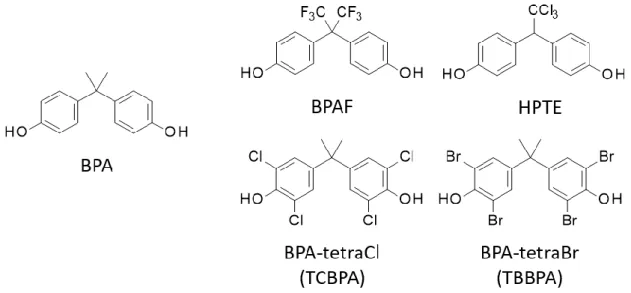
The ligands tested were as follows: 17β-estradiol (E2) and diethylstilbestrol (DES) as agonists for ERα, (Z)-4-hydroxytamoxifen [(Z)-4-OHT] and raloxifene (RAL) as antagonists for ERα, BPA and bisphenol C (BPC) as partial agonists for ERα, and bisphenol AF (BPAF) and 2,2-bis(p-hydroxyphenyl)-1,1,1-trichloroethane (HPTE) as EDCs act as agonists for ERα.
All chemical structures of the ligands were constructed using Accelrys Draw (v4.0; Accelrys) and energy minimized using the minimization protocol of Discovery Studio software.
Volume of ligands was evaluated using the analytical tools included in Discovery Studio software. The list of chemical structures used in this study is provided in Table 233–36.
(Table 2)
2.4. Docking calculations
The binding affinities of ligands were evaluated by docking calculations using Autodock version 4.2 (Scripps Research Institute, San Diego, CA, USA) on a Dell Precision T3500 workstation (Dell). The grid maps representing the receptor molecule were generated by AutoGrid (Scripps Research Institute), with each grid centered at the LBP. The docking- calculation area was set within a 126 x 126 x 126 grids at 0.197 Å per grid cube to include the entire LBP. Docking calculations were performed 100 times using the algorithm Lamarkian GA37. The run parameters used in this study were as follows: the number of GA runs was 100, the maximum number of energy evaluations was 2.5 x 10⁷, and the maximum number of generations was 1.0 x 10⁶. Other parameters were represented by default values implemented by the program. In the calculation, the free energy change (ΔG) associated with each conformation was defined as the sum of the free energy changes, van der Waals forces, electrostatic interactions, hydrogen-bonding events, desolvation activity, and torsion energetics as designed by the developer. Dissociation constant (Kd) was calculated from ΔG using a theoretical formula (i).
ΔG = RTlnKd (i)
The structure resulting from 100 rounds of free energy minimization was adopted as the calculation result. The lowest binding energy value among the calculation results from
8
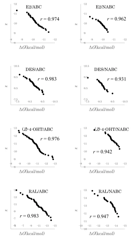
the 100 runs was used for the analysis. A total of 83 ERα structures (Table 1), and 8 ligands (Table 1) were used in this study. The calculation results were analyzed and compared according to their ΔG values between ABCs and NABCs. Data normalization was determined using a normal quantile-quantile plot (Figure 3)38. Subsequently, comparison of the means and standard errors of the mean was performed using Welch's t-test39.
(Figure 3)
3. Results & Discussion
3.1. Analysis and classification of receptor structures
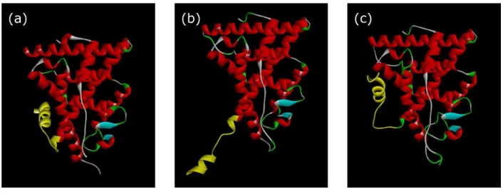
First of all, crystal structures of ERα were classified by the positioning of the H12 helix. The LBD of NRs share a highly homologous structure, including 12 α-helices (H1 to H12) and one or more underlying β-sheets. Within the LBD, an LBP mainly composed of multiple hydrophobic residues and fewer hydrophilic residues is responsible for ligand binding. Upon ligand binding, the LBD attains an altered conformation of H12 depending upon the structure of the ligand (Figure 4a). The conformation of H12 was primarily categorized as representing either an activated conformation (ABC) or a non-activated conformation (NABC). When an agonist binds to wild type ERα-LBD, the amide hydrogen atom of Leu540 on H12 forms a hydrogen bond with the oxygen atom of Asp351 side chain on H340. Additionally, in case of the Y537S mutant of ERα, it was reported that Asp351 formed a hydrogen bond with Ser537, forcing it to form an activation conformation41. Therefore, ABC and NABC were classified by whether the distance between Leu540 and Asp351 for the wild type ERα and Ser537 and Asp351 for the Y537S mutant ERα were within a typical hydrogen bond distance of ~3.5 Å or not as indexes (Figures 4b and 4c). By applying these criteria to 83 ERα-LBD (Table 1), we assigned 56 ERα-LBDs to the ABC group and 27 ERα-LBDs to the NABC group.
(Figure 4)
The ratio of ERα-LBD structures in the ABC group to those in the NABC group was almost 2:1. Therefore, we assumed that docking calculations using all LBDs would result in
9 a higher identification rate of chemicals that shows high-affinity to ABC as compared with chemicals lacking affinity to ABC. Thus, it was concerned that calculation results by using ABC and NABC should be evaluated separately.
We also calculated the volume of each LBP on LBD structure, as it was reported that differences in LBP volume could affect ligand selectivity in vitro2, 42,43. It was estimated that the average LBP volume in structures from the ABC (55 structures) and NABC (10 structures) groups were 632 ± 67 Å3 and 707 ± 89 Å3, respectively. Thus, structures from the NABC group had larger LBPs than those from the ABC group. This difference in LBP volume might provide a reason for differences in ligand selectivity observed in silico docking calculations.
3.2. Effects of water molecules on LBP/ligand-docking calculations
To investigate the effects of water molecules observed in the crystal structures, docking calculations were performed using the same LBDs in the presence or absence of water molecules, as those water molecules might serve as spacers or mediators of hydrogen bonds between a ligand and residues of the LBP. Ratios of the two calculated Kd values [Kdw
(water preserved) and Kdnw (water removed)] are shown in Table 3, and in the calculations, E2 and (Z)-4-OHT were used as one of the representative agonists and antagonists, respectively. For structures in the ABC group, the Kdw: Kdnw ratio was 0.96 in the presence of E2, and 1.5 in the presence of (Z)-4-OHT. For structures in the NABC group, the Kdw: Kdnw ratio was 0.97 for E2, and 1.5 for (Z)-4-OHT, indicating highly similar ratios between the two groups. E2 showed relatively high affinity for structures in the ABC group, whereas (Z)-4-OHT showed high affinity for structures in the NABC group. These results suggested that the presence of water molecules in and around the LBD did not significantly influence the results of docking calculations performed on structures in either the ABC or NABC group.
Thus, water molecules in the LBP crystal structures were preserved in the subsequent analysis.
(Table 3)
3.3. Comparison of ligand-binding energy between ABC and NABC of ERα-LBD
10
structures
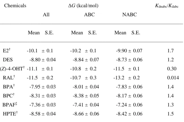
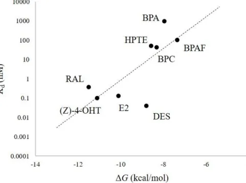
The eight chemicals included agonists, antagonists, and partial agonists (Table 4) were investigated their binding affinity to ERα-LBD structures by docking calculation using the classified structures as mentioned above. Using the average of calculated ΔG values against each test chemical as listed in Table 4, we obtained the dissociation constant (Kd) values associated with their receptor-binding activity against the target structures. To confirm the reproducibility of the docking calculation, the calculated average ΔG values were plotted against log Kd values previously determined using wet assays, and Pearson's product-moment correlation (r) was calculated44. As a result, strong correlation (r = 0.737) was observed between the calculated average ΔG values and Kd values determined by wet assays (Figure 5).
(Figure 5 and Table 4)
The statistical difference between ΔG values from the ABC group and those from the NABC group was determined by Welch's t-test. It was revealed that the ΔG values of agonist- ligand that bind to ABC group was statistically smaller than that observed in the NABC group, whereas the ΔG values of antagonist binding to NABC group was smaller than that observed in the ABC group. Based on these values, Kd ratios, i.e. the Kd(nabc)/Kd(abc), were calculated for each chemical to investigate the selectivity of the conformations that indicated which receptor group preferentially bound to the chemical (Table 4). Usually, E2 (a potent mammalian estrogenic steroid hormone) binds as an agonist to induce an activated conformation in ERα. Our results showed that E2 bound to ERα structures in the ABC group with 1.7-fold greater affinity as compared with structures in the NABC group on an average.
Although E2 and DES share similar molecular features, including molecular volumes (E2 = 234.51 Å3; DES = 245.02 Å3), two characteristic hydroxyl groups, and ERα agonistic activity, DES exhibited only 1.2-fold greater affinity for the ABC group as compared with those for the NABC group. In contrast with the antagonists, we observed no significant differences in agonist binding between the ABC and NABC groups (Table 4). Additionally, the calculated binding affinity of DES for structures in the ABC group (ΔG = −8.84 kcal/mol) was weaker than that of E2 (ΔG = −10.2 kcal/mol), despite a similar 50% effective concentrations (EC50;
11 E2 = 32 pM; DES = 13 pM)45. To discuss the relatively low selectivity of DES, we analyzed DES/ERα-LBD complex structures determined by X-ray crystallography and docking calculations performed in this study. The complex structure that was determined by X-ray crystallography of DES and ERα-LBD (PDB: 3ERD) was clearly different from the calculated complexes using ABC in this study. Among the amino acid residues of ERα-LBDs, Met421 and His524 of 3ERD changed their conformation to make preferable interaction with DES (Figure 6). Thus, this discrepancy in the calculated binding affinity may be caused by the lack of the specific DES-accepting conformation of ERα-LBDs except for 3ERD. It was considered that ERα-LBD can induce such a unique conformation only as DES binding occurred.
(Figure 6)
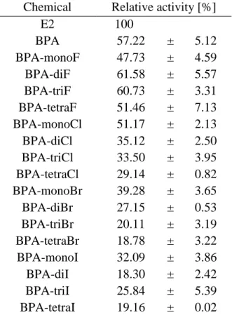
BPA is a known ERα ligand; however, its binding affinity is 500- to 15,000-fold lower than that of natural ligand E216,46–48, and it also shows 160,000-fold lower induction of in vivo estrogenic activity against ethynyl-estradiol following oral administration49. In this study, we estimated that the BPA-binding affinity for all ERα-LBDs were 39-fold lower than that of E2. This estimated weak binding affinity agreed with the previous results cited above and also suggest that BPA is a very weak or partial ERα-agonist. According to Kd-ratio analysis, BPA exhibited a 1.4-fold higher affinity for structures in the ABC group relative to those in the NABC group (Table 4), indicating that BPA may act as an ERα-agonist. Other bisphenol analogs, BPC, BPAF, and HPTE, bound to structures in the ABC group with 1.4-, 1.3-, and 1.5-fold higher affinity relative to their binding to LBDs in the NABC group, respectively (Table 4). These results confirmed that the BPA analogs may act as ERα- agonists, in agreement with previous in vitro results22,34,35.
In this study, we used RAL and (Z)-4-OHT as representative antagonists among the chemicals tested in this in silico calculation study. RAL bound to ERα-LBDs in the NABC group with 70-fold higher affinity relative to its binding to LBDs in the ABC group. We also observed that the antagonist (Z)-4-OHT bound to ERα-LBDs in the NABC group with 3.3- fold higher affinity relative to its binding to LBDs in the ABC group. The observed NABC
12
selectivity of antagonists may be due to the large LBP volumes of the LBDs in NABC, which is able to properly accept relatively large antagonist ligands.
Overall, our docking calculation studies predicted that ERα agonists preferentially bound to ABC group of LBD structure, whereas antagonist ligands showed stronger binding affinity to NABC than that of ABC. Since these results were consistent with previous reports28,31, the necessity of proper selection of ABC/NABC for docking calculation was appropriately confirmed by the results of this study. In addition, we suggest that the possible transcriptional activity of chemicals could be predicted by investigating the selectivity for ABC or NABC using the docking calculation described in this study.
3.4. Importance of C-terminal moiety on ERα-LBD for docking calculation
The C-terminal region of ERα-LBDs demonstrated an apparent variety of conformations between the structures determined as ligand/receptor complexes (Figure 1).
To investigate the effects of conformational variance on the in silico docking calculations, we compared the RMSD values of all full-length ERα-LBD structures (Table 5). The mean RMSD for all full-length ERα-LBD structures was 2.55 ± 0.03 Å, whereas the values for structures within the ABC and NABC groups were 1.22 ± 0.01 and 1.40 ± 0.02 Å, respectively. As expected, a large RMSD value (4.09 ± 0.09 Å) was derived from comparisons between the ABC and NABC groups.
(Table 5)
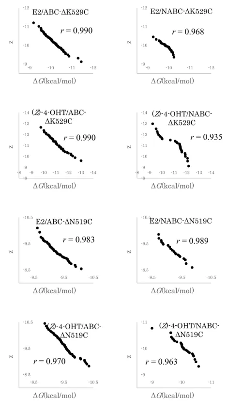
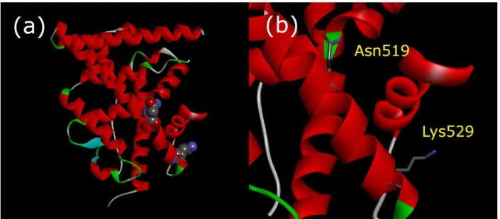
Furthermore, to consider the importance of the C-terminal region in the docking calculation, we prepared two in silico C-terminal-curtailing mutants involving truncation at K529 (ΔK529C) and at N519 (ΔN519C). As shown in Figure 7, Asn519 is located in the middle of H11, and K529 is located at the end of H11 in close proximity to the loop between H11 and H12 (Figure 7). A series of ΔK529C (lacking the H12 moiety) and ΔN519C (lacking the H11 and H12 moiety) mutants was prepared for all 83 ERα-LBDs that were categorized in the ABC and NABC groups, respectively. To evaluate the effects of the C-terminal region of the LBDs in regard to ligand binding, docking calculations of E2, BPA, (Z)-4-OHT, and RAL were performed using ΔK529C series as templates. Using the obtained ΔG values, ΔΔG
13 was defined as the difference in ΔG values between the ABC and the NABC groups (Figure 8). When the ΔK529C variants were used as templates for the docking calculation of agonist ligands (E2 and BPA), ΔΔG values were slightly increased compared to those with full-length structures (Figure 8). On the other hand, in the presence of antagonist ligands ((Z)-4-OHT and RAL), ΔΔG with the ΔK529C variants significantly decreased compared to those with the full-length structures. Decreasing of ΔΔG values could indicate that the selectivity of antagonist ligands for the ΔK529C variants was lower than that for the full-length LBDs.
Thus, H12 seems to be important for ligand selectivity particularly in case of antagonists in receptor binding. Here, we defined “ligand selectivity” as the ratio selectivity of agonist to that of antagonist in ligand binding to LBP. Additional docking calculations were performed using the ΔK519C series to determine the importance of H11, in addition to that of H12. We observed that ΔΔG with the ΔN519C variants from both the ABC and NABC groups showed low ligand selectivity for E2 and BPA (ΔΔG = 0.02 kcal/mol and 0.10 kcal/mol, respectively).
Therefore, in the case of agonists, H11 was judged to be important for ligand selectivity. By contrast, in the presence of (Z)-4-OHT and RAL, the ligand selectivity was lower in both ΔK529C and ΔN519C than in the full-length LBD (Figure 8). The relative decrease in the ABC/NABC selectivity of ligands using the C-terminal-truncated LBDs in the docking calculation indicate that ligand selectivity is chiefly determined by the C-terminal region of ERα-LBD (Figure 8). Therefore, the docking calculations were unable to estimate ligand selectivity in the absence of the region that included H11 and H12 of ERα-LBD (from N519 to the C-terminus).
(Figures 7 and 8)
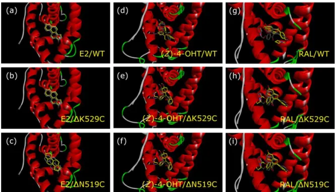
For further evaluation of the importance of H12 and H11 on ligand orientation in the docking calculation, docked conformation of E2, (Z)-4-OHT, and RAL were analyzed. For the analysis, the complex structures consisting of ligands and full-length-, ΔK529C-, or ΔN519C-LBDs that were constructed from 1GWR (ABC), and 3ERT and 1ERR (NABC) were utilized (Figure 9). By the deletion of both H11 and H12 of 1GWR, binding conformation of E2 was changed. In contrast, when the H11 was preserved, conformation of
14
E2 was almost identical to that observed in the full-length LBD. Therefore, the binding conformation and the ligand orientation of E2 may be mainly affected by H11. On the other hand, conformations of antagonists, (Z)-4-OHT and RAL, were quite different between full- length-1GWR and the truncated LBDs, especially between full-length-1GWR and 1GWR- ΔK529C (Figure 9). Furthermore, binding conformation of (Z)-4-OHT and RAL in the truncated LBDs belonging to ABC, 1GWRΔK529C and 1GWRΔN519C, were almost similar to those in the NABC group. In contrast, conformations of antagonists, (Z)-4-OHT and RAL, exhibited only little change in the NABC regardless of existence of H11 and H12 (Figure 10). Thus, the conformation of antagonist ligands in the LBPs is affected depending on the orientation or existence of H12.
(Figures 9 and 10)
4. Conclusion
In this study, we performed in silico ligand-docking studies using multiple ERα- LBDs as templates to assess whether the physiological activity of ER ligands could be predicted with high accuracy by statistical analyses of the calculated binding energy values using ABC and NABC of LBDs. In particular, we focused on and tested bisphenol analogs in the docking calculation and found weak agonistic activity of the bisphenol analogs via ERα. Our results suggested that receptor-binding affinity and potential transcriptional activity for various bisphenol analogs could be estimated properly by the calculation.
Moreover, we revealed that the C-terminal regions of ERα-LBDs involving H11, H12, and an intermediate loop structure were important for estimation of ligand binding affinity by the docking calculation. Our study highlights that for accurate discrimination of EDCs that exhibit receptor-binding properties and presumed biological activity, it is necessary to analyze statistically results of the docking calculation equipped with different receptor conformations. Acquisition of both ABC and NABC structures of NRs will enable the further development of in silico docking-calculation-based risk assessments capable of identification of EDCs.
15 References
1. Birnbaum, L. S. & Fenton, S. E. Cancer and developmental exposure to endocrine disruptors. Environ. Health Perspect. 111, 389–394 (2003).
2. Mocarelli, P., Gerthoux, P. M., Patterson, D. G., Milani, S., Limonta, G., Bertona, M., Signorini, S., Tramacere, P., Colombo, L., Crespi, C., Brambilla, P., Sarto, C., Carreri, V., Sampson, E. J., Turner, W. E. & Needham, L. L. Dioxin exposure, from infancy through puberty, produces endocrine disruption and affects human semen quality.
Environ. Health Perspect. 116, 70–77 (2008).
3. Heldring, N., Pike, A., Andersson, S., Matthews, J., Cheng, G., Treuter, E., Warner, M., Hartman, J., Tujague, M. & Stro, A. Estrogen Receptors: How Do They Signal and What Are Their Targets. Physiol Rev 87, 905–931 (2007).
4. Brzozowski, A. M., Pike, A. C. W., Dauter, Z., Hubbard, R. E., Bonn, T., Engström, O., Öhman, L., Greene, G. L., Gustafsson, J. Å. & Carlquist, M. Molecular basis of agonism and antagonism in the oestrogen receptor. Nature 389, 753–758 (1997).
5. Germain, P., Staels, B., Dacquet, C., Spedding, M. & Laudet, V. Overview of Nomenclature of Nuclear Receptors. Pharmacol. Rev. 58, 685–704 (2006).
6. Olefsky, J. M. Nuclear Receptor Minireview Series. J. Biol. Chem. 276, 36863–36864 (2001).
7. Rouiller-Fabre, V., Guerquin, M. J., N’Tumba-Byn, T., Muczynski, V., Moison, D., Tourpin, S., Messiaen, S., Habert, R. & Livera, G. Nuclear receptors and endocrine disruptors in fetal and neonatal testes: A gapped landscape. Frontiers in Endocrinology 6, 58 (2015).
8. Janošek, J., Hilscherová, K., Bláha, L. & Holoubek, I. Environmental xenobiotics and nuclear receptors - Interactions, effects and in vitro assessment. Toxicol. Vitr. 20, 18–
37 (2006).
16
9. Bergman, Å., Heindel, J., Jobling, S., Kidd, K. & Zoeller, R. T. State-of-the-science of endocrine disrupting chemicals, 2012. WHO Press 1–260 (2013).
10. Vom Saal, F. S., Cooke, P. S., Buchanan, D. L., Palanza, P., Thayer, K. A., Nagel, S.
C., Parmigiani, S. & Welshons, W. V. A Physiologically Based Approach To the Study of Bisphenol a and Other Estrogenic Chemicals On the Size of Reproductive Organs, Daily Sperm Production, and Behavior. Toxicol. Ind. Health 14, 239–260 (1998).
11. Markey, C. M., Luque, E. H., De Toro, M. M., Sonnenschein, C. & Soto, A. M. In utero exposure to bisphenol A alters the development and tissue organization of the mouse mammary gland. Biol. Reprod. 65, 1215–1223 (2001).
12. Honma, S., Suzuki, A., Buchanan, D. L., Katsu, Y., Watanabe, H. & Iguchi, T. Low dose effect of in utero exposure to bisphenol A and diethylstilbestrol on female mouse reproduction. Reprod. Toxicol. 16, 117–122 (2002).
13. Howdeshell, Kembra L, Hotchkiss, Andrew K, Thayer, Kristina A, Vandenbergh, John G, vom Saal, F. S. Exposure to bisphenol Advances puberty. Nature 401, 763–
764 (1999).
14. Nikaido, Y., Yoshizawa, K., Danbara, N., Tsujita-Kyutoku, M., Yuri, T., Uehara, N.
& Tsubura, A. Effects of maternal xenoestrogen exposure on development of the reproductive tract and mammary gland in female CD-1 mouse offspring. Reprod.
Toxicol. 18, 803–811 (2004).
15. Gupta, C. Reproductive malformation of the male offspring following maternal exposure to estrogenic chemicals. Proc. Soc. Exp. Biol. Med. 224, 61–68 (2000).
16. Nagel, S. C., Vom Saal, F. S., Thayer, K. A., Dhar, M. G., Boechler, M. & Welshons, W. V. Relative binding affinity-serum modified access (RBA-SMA) assay predicts the relative in vivo bioactivity of the xenoestrogens bisphenol A and octylphenol.
Environ. Health Perspect. 105, 70–76 (1997).
17 17. Timms, B. G., Howdeshell, K. L., Barton, L., Bradley, S., Richter, C. A. & vom Saal, F. S. Estrogenic chemicals in plastic and oral contraceptives disrupt development of the fetal mouse prostate and urethra. Proc. Natl. Acad. Sci. 102, 7014–7019 (2005).
18. Sawai, C., Anderson, K. & Walser-Kuntz, D. Effect of bisphenol A on murine immune function: Modulation of interferon-γ, IgG2a, and disease symptoms in NZB × NZW F1 mice. Environ. Health Perspect. 11, 1883–1887 (2003).
19. Yoshino, S., Yamaki, K., Yanagisawa, R., Takano, H., Hayashi, H. & Mori, Y. Effects of bisphenol A on antigen-specific antibody production, proliferative responses of lymphoid cells, and TH1 and TH2 immune responses in mice. Br. J. Pharmacol. 138, 1271–1276 (2003).
20. Yoshino, S., Yamaki, K., Li, X., Sai, T., Yanagisawa, R., Takano, H., Taneda, S., Hayashi, H. & Mori, Y. Prenatal exposure to bisphenol A up-regulates immune responses, including T helper 1 and T helper 2 responses, in mice. Immunology 112, 489–495 (2004).
21. Sartain, C. V & Hunt, P. A. An old culprit but a new story: bisphenol A and “NextGen”
bisphenols. Fertil. Steril. 106, 820–826 (2016).
22. Matsushima, A., Liu, X., Okada, H., Shimohigashi, M. & Shimohigashi, Y. Bisphenol AF is a full agonist for the estrogen receptor ERα but a highly specific antagonist for ERβ. Environ. Health Perspect. 118, 1267–1272 (2010).
23. Tabb, M. M. & Blumberg, B. New Modes of Action for Endocrine-Disrupting Chemicals. Mol. Endocrinol. 20, 475–482 (2006).
24. Gelbke, H. P., Kayser, M. & Poole, A. OECD test strategies and methods for endocrine disruptors. Toxicology 205, 17–25 (2004).
25. Gao, H., Katzenellenbogen, J. A., Garg, R. & Hansch, C. Comparative QSAR Analysis of Estrogen Receptor Ligands. Chem. Rev. 99, 723–744 (1999).
18
26. Yang, S. Y. Pharmacophore modeling and applications in drug discovery: Challenges and recent advances. Drug Discov. Today 15, 444–450 (2010).
27. Luty, B. A., Wasserman, Z. R., Stouten, P. F. W., Hodge, C. N., Zacharias, M. &
McCammon, J. A. A molecular mechanics/grid method for evaluation of ligand - receptor interactions. J. Comput. Chem. 16, 454–464 (1995).
28. Nose, T., Tokunaga, T. & Shimohigashi, Y. Exploration of endocrine-disrupting chemicals on estrogen receptor α by the agonist/antagonist differential-docking screening (AADS) method: 4-(1-Adamantyl)phenol as a potent endocrine disruptor candidate. Toxicol. Lett. 191, 33–39 (2009).
29. Humphrey, W., Dalke, A. & Schulten, K. VMD: Visual molecular dynamics. J. Mol.
Graph. 14, 33–38 (1996).
30. Pagadala, N. S., Syed, K. & Tuszynski, J. Software for molecular docking: a review.
Biophys. Rev. 9, 91–102 (2017).
31. Ng, H. W., Zhang, W., Shu, M., Luo, H., Ge, W., Perkins, R., Tong, W. & Hong, H.
Competitive molecular docking approach for predicting estrogen receptor subtype α agonists and antagonists. BMC Bioinformatics 15, (2014).
32. Celik, L., Lund, J. D. D. & Schiøtt, B. Exploring interactions of endocrine-disrupting compounds with different conformations of the human estrogen receptor α ligand binding domain: A molecular docking study. Chem. Res. Toxicol. 21, 2195–2206 (2008).
33. Kuiper, G. G. J. M., Carlsson, B., Grandien, K., Enmark, E., Häggblad, J., Nilsson, S.
& Gustafsson, J. Å. Comparison of the ligand binding specificity and transcript tissue distribution of estrogen receptors and α and β. Endocrinology 138, 863–870 (1997).
19 34. Sun, J., Huang, Y. R., Harrington, W. R., Sheng, S., Katzenellenbogen, J. A. &
Katzenellenbogen, B. S. Antagonists selective for estrogen receptor α. Endocrinology 143, 941–947 (2002).
35. Delfosse, V., Grimaldi, M., Pons, J.-L., Boulahtouf, A., le Maire, A., Cavailles, V., Labesse, G., Bourguet, W. & Balaguer, P. Structural and mechanistic insights into bisphenols action provide guidelines for risk assessment and discovery of bisphenol A substitutes. Proc. Natl. Acad. Sci. 109, 14930–14935 (2012).
36. Gaido, K. W., Leonard, L. S., Maness, S. C., Hall, J. M., McDonnell, D. P., Saville, B. & Safe, S. Differential interaction of the methoxychlor metabolite 2,2-bis-(p- hydroxyphenyl)-1,1,1-trichloroethane with estrogen receptors α and β. Endocrinology 140, 5746–5753 (1999).
37. Morris, G. M., Goodsell, D. S., Halliday, R. S., Huey, R., Hart, W. E., Belew, R. K. &
Olson, A. J. Automated docking using a Lamarckian genetic algorithm and an empirical binding free energy function. J. Comput. Chem. 19, 1639–1662 (1998).
38. Wilk, M. B. & Gnanadesikan, R. Probability Plotting Methods for the Analysis of Data.
Biometrika 55, 1–17 (1968).
39. Welch, A. B. L. The Generalization of ` Student ’ s ’ Problem when Several Different Population Variances are Involved Published by : Biometrika Trust Stable URL : http://www.jstor.org/stable/2332510. Biometrika 34, 28–35 (1947).
40. Kim, J. H., Lee, M. H., Kim, B. J., Kim, J. H., Han, S. J., Kim, H. Y. & Stallcup, M.
R. Role of aspartate 351 in transactivation and active conformation of estrogen receptor α. J. Mol. Endocrinol. 35, 449–464 (2005).
41. Nettles, K. W., Bruning, J. B., Gil, G., Nowak, J., Sharma, S. K., Hahm, J. B., Kulp, K., Hochberg, R. B., Zhou, H., Katzenellenbogen, J. A., Katzenellenbogen, B. S., Kim, Y., Joachmiak, A. & Greene, G. L. NFκB selectivity of estrogen receptor ligands
20
revealed by comparative crystallographic analyses. Nat. Chem. Biol. 4, 241–247 (2008).
42. Pike, A. C. W., Brzozowski, A. M., Hubbard, R. E., Bonn, T., Thorsell, A. G., Engström, O., Ljunggren, J., Gustafsson, J. Å. & Carlquist, M. Structure of the ligand- binding domain of oestrogen receptor beta in the presence of a partial agonist and a full antagonist. EMBO J. 18, 4608–4618 (1999).
43. Shiau, A. K., Barstad, D., Loria, P. M., Cheng, L., Kushner, P. J., Agard, D. A. &
Greene, G. L. The structural basis of estrogen receptor/coactivator recognition and the antagonism of this interaction by tamoxifen. Cell 95, 927–937 (1998).
44. Hazra, A. & Gogtay, N. Biostatistics series module 6: Correlation and linear regression.
Indian J. Dermatol. 61, 593–601 (2016).
45. Folmar, L. C., Hemmer, M. J., Denslow, N. D., Kroll, K., Chen, J., Cheek, A., Richman, H., Meredith, H. & Grau, E. G. A comparison of the estrogenic potencies of estradiol, ethynylestradiol, diethylstilbestrol, nonylphenol and methoxychlor in vivo and in vitro. Aquat. Toxicol. 60, 101–110 (2002).
46. Blair, R. M., Fang, H., Branham, W. S., Hass, B. S., Dial, S. L., Moland, C. L., Tong, W., Shi, L., Perkins, R. & Sheehan, D. M. The Estrogen Receptor Relative Binding Affinities of 188 Natural and Xenochemicals : Structural Diversity of Ligands. Toxicol.
Sci. 54, 138–153 (2000).
47. Sheeler, C. Q., Dudley, M. W. & Khan, S. A. Environmental estrogens induce transcriptionally active estrogen receptor dimers in yeast: Activity potentiated by the coactivator RIP140. Environ. Health Perspect. 108, 97–103 (2000).
48. CERI. Report on evaluation and method development for hormone-like effects of exogenous substances. (2000).
21 49. OECD. Second Meeting of the Validation Management Group on Screening and
Testing for Endocrine Disrupters (Mammalian Effects). 20–21 January (2000).
50. Tanenbaum, D. M., Wang, Y., Williams, S. P. & Sigler, P. B. Crystallographic comparison of the estrogen and progesterone receptor’s ligand binding domains. Natl.
Acad. Sci. 95, 5998–6003 (1998).
51. Eiler, S., Gangloff, M., Duclaud, S., Moras, D. & Ruff, M. Overexpression, purification, and crystal structure of native erα lbd. Protein Expr. Purif. 22, 165–173 (2001).
52. Wärnmark, A., Treuter, E., Gustafsson, J. Å., Hubbard, R. E., Brzozowski, A. M. &
Pike, A. C. W. Interaction of transcriptional intermediary factor 2 nuclear receptor box peptides with the coactivator binding site of estrogen receptor α. J. Biol. Chem. 277, 21862–21868 (2002).
53. Shiau, A. K., Barstad, D., Radek, J. T., Meyers, M. J., Nettles, K. W., Katzenellenbogen, B. S., Katzenellenbogen, J. A., Agard, D. A. & Greene, G. L.
Structural characterization of a subtype-selective ligand reveals a novel mode of estrogen receptor antagonism. Nat. Struct. Biol. 9, 359–364 (2002).
54. Wu, Y. L., Yang, X., Ren, Z., McDonnell, D. P., Norris, J. D., Willson, T. M. & Greene, G. L. Structural basis for an unexpected mode of SERM-Mediated ER antagonism.
Mol. Cell 18, 413–424 (2005).
55. Kim, S., Wu, J. Y., Birzin, E. T., Frisch, K., Chan, W., Pai, L. Y., Yang, Y. T., Mosley, R. T., Fitzgerald, P. M. D., Sharma, N., Dahllund, J., Thorsell, A. G., DiNinno, F., Rohrer, S. P., Schaeffer, J. M. & Hammond, M. L. Estrogen Receptor Ligands. II.
Discovery of Benzoxathiins as Potent, Selective Estrogen Receptor α Modulators. J.
Med. Chem. 47, 2171–2175 (2004).
56. Renaud, J., Bischoff, S. F., Buhl, T., Floersheim, P., Fournier, B., Halleux, C., Kallen, J., Keller, H., Schlaeppi, J. M. & Stark, W. Estrogen receptor modulators:
22
Identification and structure-activity relationships of potent ERα-selective tetrahydroisoquinoline ligands. J. Med. Chem. 46, 2945–2957 (2003).
57. Manas, E. S., Unwalla, R. J., Xu, Z. B., Malamas, M. S., Miller, C. P., Harris, H. A., Hsiao, C., Akopian, T., Hum, W. T., Malakian, K., Wolfrom, S., Bapat, A., Bhat, R.
A., Stahl, M. L., Somers, W. S. & Alvarez, J. C. Structure-based design of estrogen receptor-β selective ligands. J. Am. Chem. Soc. 126, 15106–15119 (2004).
58. Manas, E. S., Xu, Z. B., Unwalla, R. J. & Somers, W. S. Understanding the selectivity of genistein for human estrogen receptor-β using X-ray crystallography and computational methods. Structure 12, 2197–2207 (2004).
59. Blizzard, T.A., Dininno, F., Morgan, J.D., Chen, H.Y., Wu, J.Y., Kim, S., Chan, W., Birzin, E.T., Yang, Y.T., Pai, L.Y., Fitzgerald, P.M., Sharma, N., Li, Y., Zhang, Z., Hayes, E.C., Dasilva, C.A., Tang, W., Rohrer, S.P., Schaeffer, J.M. & Hammond, M.L.
Estrogen receptor ligands. Part 9: Dihydrobenzoxathiin SERAMs with alkyl substituted pyrrolidine side chains and linkers. Bioorganic Med. Chem. Lett. 15, 107–
113 (2005).
60. Renaud, J., Bischoff, S. F., Buhl, T., Floersheim, P., Fournier, B., Geiser, M., Halleux, C., Kallen, J., Keller, H. & Ramage, P. Selective estrogen receptor modulators with conformationally restricted side chains. Synthesis and structure-activity relationship of ERα-selective tetrahydroisoquinoline ligands. J. Med. Chem. 48, 364–379 (2005).
61. Tan, Q., Blizzard, T.A., Morgan, J.D., Birzin, E.T., Chan, W., Yang, Y.T., Pai, L.Y., Hayes, E.C., DaSilva, C.A., Warrier, S., Yudkovitz, J., Wilkinson, H.A., Sharma, N., Fitzgerald, P.M., Li, S., Colwell, L., Fisher, J.E., Adamski, S., Reszka, A.A., Kimmel, D., DiNinno, F., Rohrer, S.P., Freedman, L.P., Schaeffer, J.M. & Hammond, M.L.
Estrogen receptor ligands. Part 10: Chromanes: Old scaffolds for new SERAMs.
Bioorganic Med. Chem. Lett. 15, 1675–1681 (2005).
23 62. Hsieh, R. W., Rajan, S. S., Sharma, S. K., Guo, Y., DeSombre, E. R., Mrksich, M. &
Greene, G. L. Identification of ligands with bicyclic scaffolds provides insights into mechanisms of estrogen receptor subtype selectivity. J. Biol. Chem. 281, 17909–
17919 (2006).
63. Hsieh, R. W., Rajan, S. S., Sharma, S. K. & Greene, G. L. Molecular characterization of a B-ring unsaturated estrogen: Implications for conjugated equine estrogen components of Premarin. Steroids 73, 59–68 (2008).
64. Dykstra, K. D., Guo, L., Birzin, E. T., Chan, W., Yang, Y. T., Hayes, E. C., DaSilva, C. A., Pai, L. Y., Mosley, R. T., Kraker, B., Fitzgerald, P. M. D., DiNinno, F., Rohrer, S. P., Schaeffer, J. M. & Hammond, M. L. Estrogen receptor ligands. Part 16: 2-Aryl indoles as highly subtype selective ligands for ERα. Bioorganic Med. Chem. Lett. 17, 2322–2328 (2007).
65. Heldring, N., Pawson, T., McDonnell, D., Treuter, E., Gustafsson, J. Å. & Pike, A. C.
W. Structural insights into corepressor recognition by antagonist-bound estrogen receptors. J. Biol. Chem. 282, 10449 (2007).
66. Koide, A., Abbatiello, S., Rothgery, L. & Koide, S. Probing protein conformational changes in living cells by using designer binding proteins: application to the estrogen receptor. Proc Natl Acad Sci USA 99, 1253–1258 (2002).
67. Vajdos, F. F., Hoth, L. R., Geoghegan, K. F., Simons, S. P., LeMotte, P. K., Danley, D. E., Ammirati, M. J. & Pandit, J. The 2.0 Å crystal structure of the ERα ligand- binding domain complexed with lasofoxifene. Protein Sci. 16, 897–905 (2007).
68. Nettles, K. W., Bruning, J. B., Gil, G., O’Neill, E. E., Nowak, J., Hughs, A., Kim, Y., DeSombre, E. R., Dilis, R., Hanson, R. N., Joachimiak, A. & Greene, G. L. Structural plasticity in the oestrogen receptor ligand-binding domain. EMBO Rep. 8, 563–568 (2007).
24
69. Richardson, T. I., Dodge, J. A., Durst, G. L., Pfeifer, L. A., Shah, J., Wang, Y., Durbin, J. D., Krishnan, V. & Norman, B. H. Benzopyrans as selective estrogen receptor β agonists (SERBAs). Part 3: Synthesis of cyclopentanone and cyclohexanone intermediates for C-ring modification. Bioorganic Med. Chem. Lett. 17, 4824–4828 (2007).
70. Norman, B. H., Richardson, T. I., Dodge, J. A., Pfeifer, L. A., Durst, G. L., Wang, Y., Durbin, J. D., Krishnan, V., Dinn, S. R., Liu, S., Reilly, J. E. & Ryter, K. T.
Benzopyrans as selective estrogen receptor β agonists (SERBAs). Part 4:
Functionalization of the benzopyran A-ring. Bioorganic Med. Chem. Lett. 17, 5082–
5085 (2007).
71. Bruning, J. B., Parent, A. A., Gil, G., Zhao, M., Nowak, J., Pace, M. C., Smith, C. L., Afonine, P. V., Adams, P. D., Katzenellenbogen, J. A. & Nettles, K. W. Coupling of receptor conformation and ligand orientation determine graded activity. Nat. Chem.
Biol. 6, 837–843 (2010).
72. Dai, S. Y., Chalmers, M. J., Bruning, J., Bramlett, K. S., Osborne, H. E., Montrose- Rafizadeh, C., Barr, R. J., Wang, Y., Wang, M., Burris, T. P., Dodge, J. A. & Griffin, P. R. Prediction of the tissue-specificity of selective estrogen receptor modulators by using a single biochemical method. Proc. Natl. Acad. Sci. 105, 7171–7176 (2008).
73. Li, M. J., Greenblatt, H. M., Dym, O., Albeck, S., Pais, A., Gunanathan, C., Milstein, D., Degani, H. & Sussman, J. L. Structure of estradiol metal chelate and estrogen receptor complex: The basis for designing a new class of selective estrogen receptor modulators. J. Med. Chem. 54, 3575–3580 (2011).
74. Phillips, C., Roberts, L. R., Schade, M., Bazin, R., Bent, A., Davies, N. L., Moore, R., Pannifer, A. D., Pickford, A. R., Prior, S. H., Read, C. M., Scott, A., Brown, D. G., Xu, B. & Irving, S. L. Design and structure of stapled peptides binding to estrogen receptors. J. Am. Chem. Soc. 133, 9696–9699 (2011).
25 75. Fang, J., Akwabi-Ameyaw, A., Britton, J. E., Katamreddy, S. R., Navas, F., Miller, A.
B., Williams, S. P., Gray, D. W., Orband-Miller, L. A., Shearin, J. & Heyer, D.
Synthesis of 3-alkyl naphthalenes as novel estrogen receptor ligands. Bioorganic Med.
Chem. Lett. 18, 5075–5077 (2008).
76. Osz, J., Brelivet, Y., Peluso-Iltis, C., Cura, V., Eiler, S., Ruff, M., Bourguet, W., Rochel, N. & Moras, D. Structural basis for a molecular allosteric control mechanism of cofactor binding to nuclear receptors. Proc. Natl. Acad. Sci. 109, E588–E594 (2012).
77. Srinivasan, S., Nwachukwu, J. C., Parent, A. A., Cavett, V., Nowak, J., Hughes, T. S., Kojetin, D. J., Katzenellenbogen, J. A. & Nettles, K. W. Ligand-binding dynamics rewire cellular signaling via estrogen receptor-α. Nat. Chem. Biol. 9, 326–332 (2013).
78. Delfosse, V., Grimaldi, M., Cavaillès, V., Balaguer, P. & Bourguet, W. Structural and functional profiling of environmental ligands for estrogen receptors. Environ. Health Perspect. 122, 1306–1313 (2015).
79. Nwachukwu, J. C., Srinivasan, S., Bruno, N. E., Parent, A. A., Hughes, T. S., Pollock, J. A., Gjyshi, O., Cavett, V., Nowak, J., Garcia-Ordonez, R. D., Houtman, R., Griffin, P. R., Kojetin, D. J., Katzenellenbogen, J. A., Conkright, M. D. & Nettles, K. W.
Resveratrol modulates the inflammatory response via an estrogen receptor-signal integration network. Elife 3, e02057–e02057 (2014).
80. Fanning, S. W., Mayne, C. G., Dharmarajan, V., Carlson, K. E., Martin, T. A., Novick, S. J., Toy, W., Green, B., Panchamukhi, S., Katzenellenbogen, B. S., Tajkhorshid, E., Griffin, P. R., Shen, Y., Chandarlapaty, S., Katzenellenbogen, J. A. & Greene, G. L.
Estrogen receptor alpha somatic mutations Y537S and D538G confer breast cancer endocrine resistance by stabilizing the activating function-2 binding conformation.
Elife 5, 1–25 (2016).
26
81. Delfosse, V., Maire, A. Le, Balaguer, P. & Bourguet, W. A structural perspective on nuclear receptors as targets of environmental compounds. Acta Pharmacol. Sin. 36, 88–101 (2014).
82. Zheng, Y., Zhu, M., Srinivasan, S., Nwachukwu, J. C., Cavett, V., Min, J., Carlson, K.
E., Wang, P., Dong, C., Katzenellenbogen, J. A., Nettles, K. W. & Zhou, H. B.
Development of Selective Estrogen Receptor Modulator (SERM)-Like Activity Through an Indirect Mechanism of Estrogen Receptor Antagonism: Defining the Binding Mode of 7-Oxabicyclo[2.2.1]hept-5-ene Scaffold Core Ligands.
ChemMedChem 7, 1094–1100 (2012).
27 Table 1. 3D-structures of ERα used in this study.
PDB
ID Ligand Mutation Resolution
(Å) Chain Reference
1A52 estradiol Q502E 2.8 A 50
1ERE estradiol 3.1 A 4
1ERR raloxifene 2.6 A 4
1G50 estradiol 2.9 A 51
1GWQ raloxifene core 2.45 A 52
1GWR estradiol 2.4 A 52
1L2I
(R,R)-5,11-cis-diethyl- 5,6,11,12-
tetrahydrochrysene- 2,8-diol
C417X 1.95 A 53
1R5K
(2E)-3-{4-[(1E)-1,2- diphenylbut-1-enyl]
phenyl} acrylic acid
2.7 A 54
1SJ0
(2S,3R)-2-(4-(2- (piperidin-1-
yl)ethoxy)phenyl)- 2,3- dihydro-3-(4-
hydroxyphenyl) benzo[b][1,4]oxathiin- 6-ol
1.9 A 55
1UOM
2-phenyl-1-[4-(2- piperidin-1-yl-ethoxy)- phenyl]- 1,2,3,4-tetra hydro- isoquinolin-6-ol
C381S, C417S, C530S
2.28 A 56
28
Continued Table 1
1X7E
[5-hydroxy-2-(4- hydroxyphenyl)-1- benzofuran- 7- yl]acetonitrile
2.8 A 57
1X7R genistein 2 A 58
1XP1
(2S,3R)-2-(4-{2- [(3R,4R)-3,4-
dimethylpyrrolidin- 1- yl]ethoxy}phenyl)-3-(4- hydroxyphenyl)-2,3- dihydro-1,4-
benzoxathiin-6-ol
1.8 A 59
1XP6
(2S,3R)-2-(4-{2- [(3S,4S)-3,4-dimethyl pyrrolidin- 1-yl]ethoxy}
phenyl)-3-(4-hydroxy phenyl)-2,3- dihydro- 1,4-benzoxathiin-6-ol
1.7 A 59
1XP9
(2S,3R)-3-(4-
hydroxyphenyl)-2-(4- {[(2S)-2- pyrrolidin-1- ylpropyl]oxy}phenyl)- 2,3-dihydro- 1,4- benzoxathiin-6-ol
1.8 A 59
1XPC
(2S,3R)-3-(4-
hydroxyphenyl)-2-(4- {[(2R)-2- pyrrolidin-1- ylpropyl]oxy}phenyl)- 2,3-dihydro- 1,4- benzoxathiin-6-ol
1.6 A 59
29 Continued Table 1
1XQC
(1S)-1-{4-[(9aR)- octahydro-2h-pyrido [1,2- a]pyrazin-2-yl]
phenyl}-2-phenyl-1,2,3, 4-tetrahydroisoquinolin- 6-ol1-[4-(octahydro- pyrido[1,2-a]pyrazin-2- yl)- phenyl]-2-phenyl- 1,2,3,4-tetrahydro- isoquinolin- 6-ol
C381S, C417S, C530S
2.05 A 60
1YIM
(2R,3R,4S)-3-(4- hydroxyphenyl)-4- methyl-2- [4-(2- pyrrolidin-1-
ylethoxy)phenyl]chrom an- 6-ol
1.9 A 61
1YIN
(2R,3R,4S)-5-fluoro-3- (4-hydroxyphenyl)-4- methyl-2-[4-(2- piperidin-1-ylethoxy) phenyl]chroman- 6-ol
2.2 A 61
1ZKY
4-[(1S,2S,5S)-5-
(hydroxymethyl)-6,8,9- trimethyl- 3-oxabicyclo [3.3.1]non-7-en-2- yl]phenol
C381X, C417X, C530X, Y537S
2.25 A 62
2B1V
4-[(1S,2S,5S)-5- (hydroxymethyl)-8- methyl- 3-oxabicyclo [3.3.1]non-7-en-2- yl]phenol
C381X, C417X, C530X
1.8 A 62
30
Continued Table 1
2B1Z 17-methyl-17-alpha- dihydroequilenin
C381X, C417X, C530X, Y537S
1.78 A 63
2B23 none
C381X, C417X, C530X, Y537S
2.1 A 41
2FAI
4-[(1S,2S,5S,9R)-5- (hydroxymethyl)-8,9- dimethyl- 3-
oxabicyclo[3.3.1]non-7- en-2-yl]phenol
C381X, C417X, C530X, Y537S
2.1 A 62
2G44
4-[(1S,2R,5S)-4,4,8- trimethyl-3-
oxabicyclo[3.3.1]non- 7-en-2-yl]phenol
C381X, C417X, C530X, Y537S
2.65 A
Unpublishe d data (Thesis)
2G5O
(9alpha,13beta,17beta)- 2-[(1Z)-but-1-en-1- yl]estra-1,3,5(10)- triene-3,17-diol
C381X, C417X, C530X, Y537S
1.65 A To be
published
2IOG
n-[(1R)-3-(4- hydroxyphenyl)-1- methylpropyl]- 2-[2- phenyl-6-(2-piperidin- 1-ylethoxy)-1h- indol-3- yl]acetamide
1.6 A 64
2IOK
n-[(1R)-3-(4- hydroxyphenyl)-1- methylpropyl]- 2-(2- phenyl-1h-indol-3- yl)acetamide
2.4 A 64
31 Continued Table 1
2JF9 4-hydroxytamoxifen 2.1 A 65
2JFA raloxifene 2.55 A 65
2OCF estradiol
C381X, C417X, Y537S
2.95 A 66
2OUZ
(5R,6S)-6-phenyl-5-[4- (2-pyrrolidin-1-
ylethoxy)phenyl]- 5,6,7,8-
tetrahydronaphthalen-2- ol
2 A 67
2P15
(17beta)-17-{(E)-2-[2- (trifluoromethyl)phenyl ]vinyl}estra- 1(10),2,4- triene-3,17-diol
Y537S 1.94 A 68
2Q70
(3aS,4R,9bR)-2,2- difluoro-4-(4- hydroxyphenyl)- 1,2,3,3a,4,9b-
hexahydrocyclopenta[c]
chromen- 8-ol
C381S, C417S, C530S
1.95 A 69
2QE4
(3aS,4R,9bR)-4-(4- hydroxyphenyl) -6- (methoxymethyl)- 1,2,3,3a,4,9b-
hexahydrocyclopenta[c]
chromen- 8-ol
C381S, C417S, C530S
2.4 A 70
2QZO
4-[1-allyl-7-
(trifluoromethyl)-1h- indazol- 3-yl]benzene- 1,3-diol
Y537S 1.72 A 71
32
Continued Table 1
2R6W
[6-hydroxy-2-(4- hydroxyphenyl)-1- benzothien- 3-yl]{4-[2- (4-methylpiperidin-1- yl)ethoxy]phenyl}meth anone
C381S, C417S, C530S
2 A 72
2R6Y
[6-hydroxy-2-(4- hydroxyphenyl)-1- benzothien- 3-yl][4-(2- pyrrolidin-1-
ylethoxy)phenyl]metha none
C381S, C417S, C530S
2 A 72
2YAT estradiol-pyridinium tetraacetic acid
C381S, C417S, C530S
2.6 A 73
2YJA estradiol 1.82 B 74
3DT3
5-(4-hydroxyphenoxy)- 6-(3-hydroxyphenyl)- 7- methylnaphthalen-2-ol
2.4 A 75
3ERT 4-hydroxytamoxifen 1.9 A 43
3HLV
(9beta,13alpha,16beta)- 3,16-dihydroxyestra- 1,3,5(10)-trien-17-one
Y537S 3 A To be
published
3HM1
(9beta,13alpha)-3- hydroxyestra-1,3,5(10)- trien-17-one
Y537S 2.33 A To be
published
3L03
(14beta,15alpha,16alph a,17alpha)-estra- 1,3,5(10)- triene- 3,15,16,17-tetrol
Y537S 1.9 A To be
published
33 Continued Table 1
3OS8
4-[1-benzyl-7- (trifluoromethyl)-1h- indazol- 3-yl]benzene- 1,3-diol
L372R,
L536S 2.03 A 35
3OS9
4-[1-allyl-7-
(trifluoromethyl)-1h- indazol- 3-yl]benzene- 1,3-diol
L372R,
L536S, 2.3 A 35
3OSA
4-[1-(3-methylbut-2-en- 1-yl)-7-
(trifluoromethyl)- 1h- indazol-3-yl]benzene- 1,3-diol
L372R,
L536S 2.3 A 35
3Q95 estriol Y537S 2.05 A To be
published 3Q97
4,4'-[2-(4-
ethoxyphenyl)but-1- ene-1,1-diyl]diphenol
Y537S 2.1 A To be
published
3UU7 bisphenol A Y537S 2.2 A 76
3UUA bisphenol AF Y537S 2.05 A 76
3UUC
4,4'-(2,2-
dichloroethene-1,1- diyl)diphenol
2.1 A 76
3UUD estradiol Y537S 1.6 A 76
4DMA
2'-bromo-6'-(furan-3- yl)-4'-
(hydroxymethyl)biphen yl- 4-ol
C530A 2.3 A 77
4IU7
4-[2-ethyl-7-
(trifluoromethyl)-2h- indazol- 3-yl]benzene- 1,3-diol
Y537S 2.29 A 78
34
Continued Table 1
4IUI
4-[1-butyl-7-
(trifluoromethyl)-1h- indazol- 3-yl]benzene- 1,3-diol
Y537S 2.3 A 78
4IV2
4-[1-(2-methylpropyl)- 7-(trifluoromethyl)- 1h- indazol-3-yl]benzene- 1,3-diol
Y537S 2.14 A 78
4IV4
4-[2-(2-methylpropyl)- 7-(trifluoromethyl)- 2h- indazol-3-yl]benzene- 1,3-diol
Y537S 2.3 A 78
4IVW
4-[2-benzyl-7- (trifluoromethyl)-2h- indazol- 3-yl]benzene- 1,3-diol
Y537S 2.06 A 78
4IVY
4-[1-(but-3-en-1-yl)-7- (trifluoromethyl)- 1h- indazol-3-yl]benzene- 1,3-diol
Y537S 1.95 A 78
4IW6
4-[2-(but-3-en-1-yl)-7- (trifluoromethyl)- 2h- indazol-3-yl]benzene- 1,3-diol
Y537S 1.98 A 78
4IW8
4-[1-(3-methylbut-2-en- 1-yl)-7-
(trifluoromethyl)-1h- indazol-3-yl]benzene- 1,3-diol
Y537S 2.04 A 78
4IWC 4,4'-thiene-2,5-
diylbis(3-methylphenol) Y537S 2.24 A 78
4IWF
2-chloro-3'-fluoro-3- [(E)-
(hydroxyimino)methyl]
biphenyl- 4,4'-diol
1.93 A 78
35 Continued Table 1
4MG5 chlordecone Y537S 2.05 A 79
4MG6 benzyl butyl benzene-
1,2-dicarboxylate Y537S 2.1 A 79
4MG7 ferutinine Y537S 2.15 A 79
4MG8 alpha-zearalanol Y537S 1.85 A 79
4MG9 butyl 4-
hydroxybenzoate Y537S 2.0 A 79
4MGA 4-(2,4,4-trimethyl
pentan-2-yl)phenol Y537S 1.8 A 79
4MGB
4,4'-propane-2,2- diylbis(2,6- dichlorophenol)
Y537S 1.85 A 79
4MGC
bis(2,4-
dihydroxyphenyl) methanone
Y537S 2.15 A 79
4MGD HPTE Y537S 1.9 A 79
4PP6 resveratrol Y537S 2.2 A 80
4PPP
5-[(E)-2-(3-fluoro-4- hydroxyphenyl)ethenyl]
benzene- 1,3-
diol(fluoro-resveratrol )
Y537S 2.69 A 80
4PPS
(1S,3aR,5R,7aS)-5-(4- hydroxyphenyl)-7a- methyloctahydro- 1h- inden-1-ol
Y537S 1.93 A 80
4PXM estradiol D538G 1.9 A 81
4Q13 none D538G 2.24 B 81
4Q50 4-hydroxytamoxifen D538G 3.07 A 81
36
Continued Table 1
4TUZ
(3S,7R,11E)-7,14,16- trihydroxy-3-methyl- 3,4,5,6,7,8,9,10- octahydro-1h-2-
benzoxacyclotetradecin- 1-one
Y537S 1.9 A 82
4TV1 propyl 4-
hydroxybenzoate Y537S 1.85 A 82
4ZN9
cyclohexa-2,5-dien-1-yl (1S,2R,4S)-5,6-bis(4- hydroxyphenyl)-7- oxabicyclo[2.2.1]hept- 5- ene-2-sulfonate
Y537S 2.21 A 43
37 Table 2. Chemical structure of compounds utilized in this study.
Structure
Name IUPAC name
Kd reported in the literatures 17β-estradiol (E2)
(8R,9S,13S,14S,17S)-13-methyl- 6,7,8,9,11,12,14,15,16,17-
decahydrocyclopenta[a]phenanthrene -3,17-diol
0.13 nM31
diethylstilbestrol (DES)
4,4'-(3E)-hex-3-ene-3,4-diyldiphenol
0.04 nM31
4-hydroxytamoxifen (4-(Z)-OHT) 4-[(Z)-1-[4-[2-
(dimethylamino)ethoxy]phenyl]-2- phenylbut-1-enyl]phenol
0.1 nM31