Precise determination of the scattered X-ray contamination
rate using diagnostic X-ray equipment for the construction
of the secondary X-ray field.
Poster No.: C-0007
Congress: ECR 2016
Type: Scientific Exhibit
Authors: I. Maehata, H. Hayashi, N. Kimoto, H. Okino, K. Takegami, Y. Kanazawa; Tokushima/JP
Keywords: Dosimetric comparison, Radiation safety, Physics, Dosimetry, Experimental, Digital radiography, Conventional radiography, Radioprotection / Radiation dose, Radiation physics
DOI: 10.1594/ecr2016/C-0007
Any information contained in this pdf file is automatically generated from digital material submitted to EPOS by third parties in the form of scientific presentations. References to any names, marks, products, or services of third parties or hypertext links to third-party sites or information are provided solely as a convenience to you and do not in any way constitute or imply ECR's endorsement, sponsorship or recommendation of the third party, information, product or service. ECR is not responsible for the content of these pages and does not make any representations regarding the content or accuracy of material in this file.
As per copyright regulations, any unauthorised use of the material or parts thereof as well as commercial reproduction or multiple distribution by any traditional or electronically based reproduction/publication method ist strictly prohibited.
You agree to defend, indemnify, and hold ECR harmless from and against any and all claims, damages, costs, and expenses, including attorneys' fees, arising from or related to your use of these pages.
Aims and objectives
Currently, X-ray examinations are widely used for diagnosis in the medical field, and risk of cancer from diagnostic X-rays is increasing [1]. Radiological technologists should make an effort to reduce exposure doses in addition to improvement of image qualities [2]. In the diagnostic region, a reduction of the entrance-skin dose (ESD) [3] is important. Generally speaking, the ESD is measured using the correction of back scatter factor (BSF) [4-5] from air-kerma (Fig. 1). The BSF is accurately determined, therefore experimenters should measure the air-kerma using ionization chambers that are calibrated well with the standard X-ray field. One of the reasons why the ESD measurement is not applied widely in the medical field is, it is considered expensiveness to calibrate the ionization chamber. Therefore, we constructed a secondary X-ray field by means of medically used diagnostic X-ray equipment.
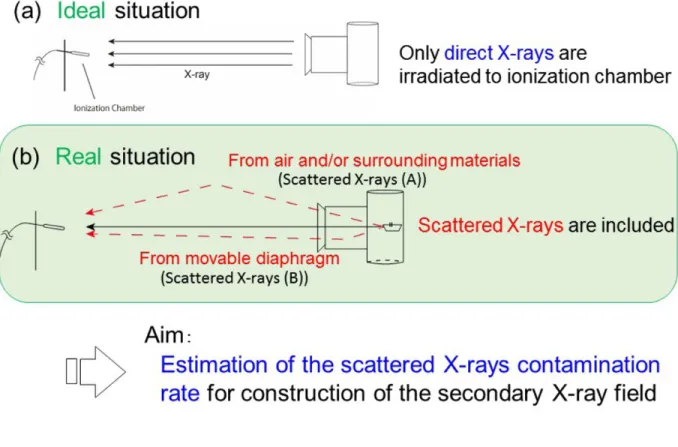
Figure 2 (a) shows the ideal situation of X-ray exposure in which only direct X-rays are
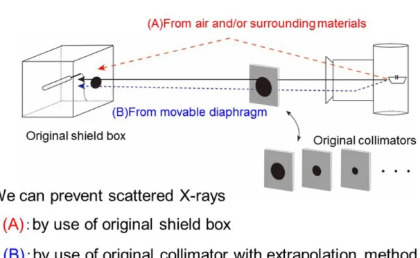
irradiated to the ionization chamber. On the other hand, scattered X-rays are included in the real situation as shown in Fig. 2 (b); (A) and (B) indicate scattered X-rays caused by air and movable diaphragm, respectively. The diagnostic X-ray equipment used consists of an X-ray tube and a movable diaphragm. It is well known that the movable diaphragm generates scattered X-rays [6-8]. Figure 3 shows the concept of our study. Using a shield box, scattered X-rays from the air are reduced, and using an extrapolation method by means of collimators, the scattered X-rays from the movable diaphragm is estimated.
The aim of this study is to estimate the contamination rate of the scattered X-rays to the direct X-rays for construction of the precise secondary X-ray field by means of medically used diagnostic X-ray equipment.
Fig. 1: Background of our study. It is necessary to calibrate the ionization chamber at the
secondary X-ray field by means of diagnostic X-ray equipment.
Fig. 2: Concept of air-kerma measurements. In an ideal situation, scattered X-rays in the
air and the movable diaphragm become contamination.
Fig. 3: The proposed method. We developed a shield box to prevent scattered X-rays,
and a collimator method is applied to extrapolation.
Methods and materials
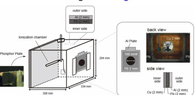
Figure 4 shows a schematic drawing of the shield box which was newly developed for
the present study. The shield box consists of 2 mm thick lead plates and an ionization chamber which can be set in the inner position. The front of this box has an entrance window (through-hole) with a diameter of 100 mm, and at the rear, a surface phosphor plate can be set to confirm both the irradiation area and the position of ionization chamber using an X-ray beam.
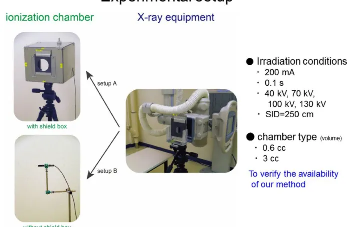
Figure 5 shows images of our experiment. Our experimental apparatus consists of
diagnostic X-ray equipment (Toshiba Medical Systems Corporation, MRAD-A 50S/70), collimator, shield box, ionization chambers and dosimeter (EMF Japan, EMF521). The ionization chamber is located in a shield box which we called "setup A". To compare with the setup A, the experiment was carried out using "setup B" in which shield box is excluded. In these situations, the collimator for applying the extrapolation method [9-10] is placed on the side containing the movable diaphragm. The collimators are composed of 2 mm thick lead and 2 mm thick aluminum plates which are 210 mm high and 165 mm wide. We bored a through-hole at its center. Diameters of the through-hole are 25-90 mm. As shown in the figure, an acrylic guide is set on a tripod stand to adjust these collimators easily. In the proposed method using the shield box as shown in Fig. 3, the extrapolation method is applied for the estimation of scattered X-rays from the movable diaphragm. In contrast, if the shield box is not used, the extrapolation method is applied to the estimation of scattered X-rays for both the movable diaphragm and air.
Distances between the X-ray source to collimator and ionization chamber are 35 cm and 250 cm, respectively. The tube current-time product is 100 mAs, and tube voltages are 40-130 kV. We used ionization chambers having different detection volumes of 0.6 cc (PTW, N 30001) and 3 cc (PTW, DC-300).
Fig. 4: A schematic drawing of the original shield box. The ionization chamber is held
by a clamp which is fixed to the upper side. In order to check the exposure region, a phosphor plate can be inserted at the rear.
Fig. 5: Experimental setup. Two different conditions are carried out as represented in this
figure. The proposed method (setup A) uses a shield box (ionization chamber is placed inside it), and to compare this another experimental setup (setup B) is performed.
Results
The contamination rate of the scattered X-rays was deduced by an extrapolation method. In Fig. 6, we exemplify the typical result at 70 kV. In this method, a true exposure dose without scattered X-rays can be determined by the extrapolation of experimental data which are plotted as a function of diameter of collimators. For fitting to the experimental data, a linear function was applied. In the center graph of Fig. 6, open and closed circles show experimental results for setup B and setup A, respectively. The difference between these data (approximately 1%) indicates contamination of scattered X-rays from air (indicated by (A)); the effect using the shield box is clearly observed. Then we adopted the extrapolated value to setup A. The difference between the extrapolated value and each experimental data shows the contribution of scattered X-rays from the movable diaphragm (indicated by (B)). In this graph, each data point has statistical uncertainty, which is defined as the standard deviation [11] of the values for five measurements. Additionally, the left plot shows extrapolated data concerning error for both the statistical uncertainty and calibration factor of the ionization chamber used. The error of the calibration factor is approximately 5%, which is much larger than the contamination rate of scattered X-rays. If we will calibrate a different ionization chamber using our secondary X-ray field, the ionization chamber can be calibrated with an at least 5% uncertainty. Although the calibration factor has a larger uncertainty when compared with contamination of scattered X-rays, we considered that the experiment using our shield box should be performed and extrapolation should be taken into account.
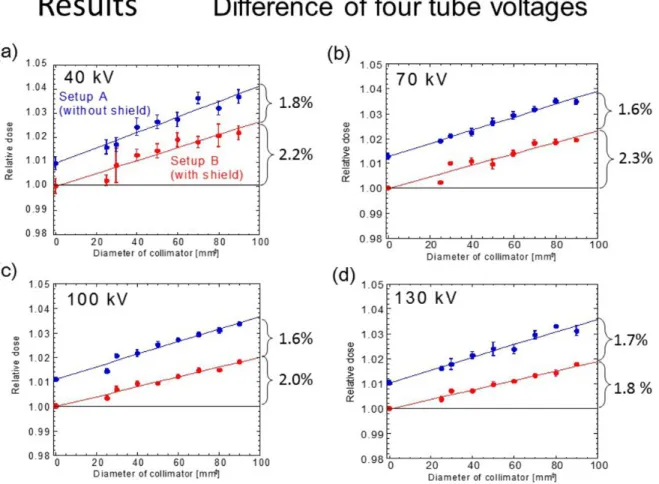
Figure 7 shows a comparison of all experimental results between the four tube voltages.
The Y axis shows relative dose which is normalized by the extrapolated value. The four results show a similar trend as represented in Fig.6, and that extrapolated method works well. From these results we found that the contamination of scattered X-rays from air (A) are 1.6-1.8% for 40-130 kV and those from the movable diaphragm (B) are 1.8-2.3%. Although these estimated values are not general, the values become one of the rough indications when experiments will be performed using the diagnostic X-ray equipment installed in the clinic.
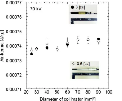
Figure 8 shows a comparison of experiments at 70 kV between two different ionization
chambers. The result indicates that our method is independent to the detection volumes of the ionization chambers.
Fig. 6: Extrapolation method. To fit the linear function to the experimental data, an
extrapolated value is obtained. The extrapolated data includes no contamination from scattered X-rays. Their errors include only statistical uncertainty (center graph), and error of air-kerma includes both statistical and systematic uncertainties (left graph).
Fig. 7: Experimental results and extrapolated values for four tube voltages. © - Tokushima/JP
Fig. 8: The comparison of experimental results between two different ionization
chambers; 0.6 cc chamber and 3 cc chamber. The results are consistent.
Conclusion
In conclusion, we evaluated the contamination rates of the scattered X-rays for construction of a secondary X-ray field by means of general diagnostic X-ray equipment (see Fig. 9). In the experimental room there is additional scattered X-rays caused by surrounded materials compared with other standard ray fields. To prevent scattered X-rays, we proposed the use of an original shield box and of applying a collimators method. Our equipment is conveniently portable, therefore we considered that our equipment is useful to calibrate ionization chambers using X-ray equipment set at clinical examination rooms. We applied the method to a general experimental room in Japan, and found that the contamination rates from the air and the movable diaphragm were less than 1.8% and 2.3%, respectively. The precision and accuracy of the extrapolation method are approximately 0.7% in the measured value [C], and 5% in the air-kerma [J/kg]. The large error of the calibration factor contributes to the air-kerma value obtained. Our method is more precise than the error found in a calibration factor. If a precisely calibrated ionization chamber is available, we can calibrate other ionization chambers with a small degree of error.
Fig. 9: Summary of the present study. © - Tokushima/JP
Personal information
Name: Itsumi Maehata
Affiliation: School of Health Sciencies, Tokushima University, Japan
Email: [email protected]
References
[1] Amy Berrington de Gonzalez, Sarah Darby. Risk of cancer from diagnostic X-ray:
estimates for the UK and 14 other countries. The Lancet. 2004;363:345-351.
[2] Uffmann M, Prokop CS. Digital radiography: The balance between image quality and
required radiation dose. European Journal of Radiology. 2009;72:202-208.
[3] Takegami K, Hayashi H, Okino H, Kimoto N, Maehata I, Kanazawa Y, Okazaki T,
Kobayashi I. Practical calibration curve of small-type optically stimulated luminescence (OSL) dosimeter for evaluation of entrance skin dose in the diagnostic X-ray region. Radiological Physics and Technology. 2015; accepted.
[4] Grosswendt B. Backscatter factors for x-rays generated at voltages between 10 and
100 keV. Physics in Medical and Biology. 1954;29(5):579-591.
[5] Klevenhagen SC. Experimentally determined backscatter factors for x-rays
generated at voltages between 16 and 140 kV. Physics in Medical and Biology. 1989;34(12):1871-1882.
[6] Hayashi H, Taniuchi S, Kamiya N, et al. Development of a Pin-hole Camera Using a
Phosphor Plate, and Visualization of the Scattered X-ray Distribution and Optical Image. Japanese Journal of Radiological Technology. 2012;68(3):307-311.
[7] Takegami K, Hayashi H, Konishi Y, et al. Development of multistage collimator
for narrow beam production using filter guides of diagnostic X-ray equipment and improvement of apparatuses for practical training. Medical Imaging and Information Sciences. 2013;30(4):101-107.
[8] Hayashi H, Takegami K, Okino H, et al. Procedure to measure angular dependences
[10] Trout ED, Kelley JP, and Lucas AC. Determination of Half-Value Layer.
The American Journal of Roentgenology, Radium Therapy, and Nuclear Medicine. 1960;84(4):729-740.
[11] GF Knoll. Radiation Detection and Measurement. New York: John Willy and Sons,