Kudo et al., 1
Periostin promotes invasion and anchorage-independent growth
in the metastatic process of head and neck cancer
Yasusei Kudo1, Ikuko Ogawa 2, Shojiro Kitajima1, Masae Kitagawa1, Hidehiko Kawai3, Patrick M. Gaffney4, Mutsumi Miyauchi1, Takashi Takata1, 2
1
Department of Oral Maxillofacial Pathobiology, Division of Frontier Medical Science, Graduate School of Biomedical Sciences, Hiroshima University, 2Center of Oral Clinical Examination, Hiroshima University Hospital,
3
Department of Regulatory Radiobiology, Research Institute for Radiation Biology and Medicine, Hiroshima University, Hiroshima 734-8553, Japan; 4Division of Hematology, Oncology and Transplantation, Institute of Human Genetics, Department of Medicine, University of Minnesota Medical School, Minneapolis, MN 55455.
Note: I. Ogawa and S. Kitajima contributed equally to this work.
Requests for reprints: Yasusei Kudo and Takashi Takata, Department of Oral Maxillofacial Pathobiology, Division of Frontier Medical Science, Graduate School of Biomedical Sciences, Hiroshima University, 1-2-3 Kasumi, Minami-ku, Hiroshima 734-8553, Japan. Phone: +81 82-257-5634; Fax +81 82-257-5619; E-mail: [email protected] and [email protected].
Running Title: Periostin promotes invasion and anchorage-independent growth in head and neck cancer
Key Words: Periostin, invasion, metastasis, head and neck cancer, anchorage-independent growth
Kudo et al., 2 Abstract
Head and neck squamous cell carcinoma (HNSCC) is one of the most common types of human cancer. Typically HNSCC cells show persistent invasion that frequently leads to local recurrence and distant lymphatic metastasis. However, molecular mechanisms associated with invasion and metastasis of HNSCC remain poorly understood. Here we identified Periostin as an invasion promoting factor in HNSCC by comparing the gene expression profiles between parent HNSCC cells and a highly invasive clone. Indeed, Periostin
overexpression promoted the invasion and anchorage independent growth both in vitro and in
vivo in HNSCC cells. Moreover, Periostin overexpressing cells spontaneously metastasized
to cervical lymph nodes and to the lung through their aggressive invasiveness in an orthotopic mouse model of HNSCC. Interestingly, Periostin was highly expressed in HNSCCs in
comparison with normal tissues, and the level of Periostin expression was well correlated with the invasiveness of HNSCC cases. In summary, these findings suggest that Periostin plays an important role for invasion and anchorage independent growth in the metastatic process of HNSCC.
Kudo et al., 3 Introduction
Head and neck squamous cell carcinoma (HNSCC) is one of the most common types of human cancer, with an annual incidence of more than 500,000 cases worldwide (1).
HNSCC is associated with severe disease- and treatment-related morbidity and has a 5-year survival rate of approximately 50%; this rate has not improved in more than 2 decades (2). Review of the literature indicates that the most important factor for the high mortality rate is the advanced stage of the disease at the time of diagnosis and treatment. In the prognosis of HNSCC, the extent of lymph node metastasis is a major determinant. Like most epithelial cancers, HNSCC develops through the accumulation of multiple genetic and epigenetic alterations in a multistep process. Recent molecular studies have advanced our
understanding of the disease and provided a rationale to develop novel strategies for early detection, classification, prevention and treatment. Attempts to identify the genes involved in the metastasis is pivotal for the early prediction of HNSCC behavior. However, the identity and time of onset of the alterations that endow cancer cells with these metastatic functions are largely unknown. The process of metastasis consists of sequential and selective steps including proliferation, induction of angiogenesis, detachment, motility, invasion into
circulation, aggregation and survival in the circulation, cell arrest in distant capillary beds and extravasation into organ parenchyma (3). The development of a metastasis depends on interplay between host factors and intrinsic characteristics of cancer cells, and the metastatic lesion represents the end point of many destructive events that only a few cells can survive (4). Moreover, neoplasms contain a variety of subpopulations of cells with differing
metastatic potential, and the possible existence of highly metastatic clones may exist within a primary tumor (4). Indeed, we previously isolated highly invasive clones from parent HNSCC cell line by using an in vitro invasion assay method (5).
Kudo et al., 4 Here we compared the transcriptional profile of parent cells and a highly invasive clone by microarray analysis in order to identify genes that differ in their expression. We identified Periostin (osteoblast specific factor 2 (fasciclin I-like)) as the gene demonstrating the highest fold change expression in the invasive clone. Periostin is a secreted protein, which was originally identified from osteoblasts (6, 7). In the present study, we demonstrated that Periostin was involved in the invasion and anchorage independent growth in HNSCC.
Materials and Methods
Gene array analysis. The Amersham CodeLink system using the UniSet Human I
Expression Bioarray, containing 10458 genes probes, was used to compare the transcription profiles between parent cells and high invasive clone. This array contains a broad range of genes derived from publicly available, well-annotated mRNA sequences. The CodeLink array is unique in being capable of detecting minimal differences in gene expression, as low as 1.3-fold with 95% confidence, because of the novel three-dimensional aqueous gel matrix, which the empirically tested 30-mer oligonucleotides are deposited on (8). This substantially reduces background, enhances sensitivity, and allows for the detection and quantification of subtle regulatory relationships among genes in parent HNSCC cells and highly invasive clone. Total RNA was isolated from cultures of confluent cells using the RNeasy Mini Kit (Qiagen) according to the manufacture’s instructions. Preparations were quantified and their purity was determined by standard spectrophometric methods. Data were expressed as the average differences between the perfect match and mismatch probes for the Periostin gene (Supplemental data 1).
Total RNA from 41 primary HNSCC and 13 normal tissues was labeled and hybridized to Affymetrix U133A Gene Chips as previously reported (9). To explore the genes that are
Kudo et al., 5 coordinately expressed with Periostin in HNSCC tumors, we performed a similarity search using a Pearson correlation metric as implemented in tGeneData Analyst Pro 1.0 software (Supplemental data 2).
Cell culture. MSCC-1 and MSCC-Inv1 cells were previously established in our laboratory (5, 10). These cells were maintained in Keratinocyte-SFM (Invitrogen) under a condition of 5 % CO2 in air at 37 ˚C. Other HNSCC cell lines HSC2, HSC3 and Ca9-22 were provided by Japanese Collection of Research Bioresources Cell Bank. They were maintained in RPMI-1640 (Nissui Pharmaceutical Co., Tokyo, Japan) supplemented with 10 % heat-inactivated FBS (Invitrogen) and 100 U/ml penicillin-streptomycin (Gibco) under conditions of 5 % CO2 in air at 37 ˚C. For growth assay, cells were plated onto 24 well plates (Falcon), and trypsinized cells counted by Cell Counter (Coulter Z1).
RT-PCR. Total RNA was isolated from cultures of confluent cells using the RNeasy Mini Kit (Qiagen). Preparations were quantified and their purity was determined by standard
spectrophometric methods. cDNA was synthesized from 1 4µg total RNA according to the ReverTra Dash (Toyobo Biochemicals, Tokyo, Japan). Primer sequences included the following; human Periostin, 5’-gatggagtgcctgtggaaat-3’ (forward) and 5’-aacttcctcacgggtgtgtc-3’ (reverse) (product size, 239bp); human GAPDH, 5’-tccaccaccctgttgctgta-5’-aacttcctcacgggtgtgtc-3’ (forward) and 5’-accacagtccatgccatcac-3’ (reverse) (product size, 450bp). Aliquots of total cDNA were amplified with 1.25 U of rTaq-DNA polymerase (Qiagen), and amplifications were performed in a PC701 thermal cycler (Astec, Fukuoka, Japan) for 30-35 cycles after an initial 30 sec denaturation at 94 ˚C, annealed for 30 sec at 60 ˚C, and extended for 1 min at 72 ˚C in all primers. The amplification reaction products were resolved on 1.5% agarose/TAE gels
Kudo et al., 6 (Nacalai tesque, Inc., Kyoto, Japan), electrophoresed at 100 mV, and visualized by ethidium-bromide staining.
Western blot analysis. Western blotting was carried out as we described previously (11). A polyclonal anti-Periostin antibody was generated by immunizing the rabbits with specific peptides (EGEPEFRLIKEGETC) for Periostin and purified through an affinity column. An anti-His polyclonal antibody (Cell Signaling Technology Inc.) and ß-actin monoclonal antibody (Sigma) were also used. Thirty µg of protein was subjected to 10 % polyacrylamide gel electrophoresis followed by electroblotting onto a nitrocellulose filter. For detection of the immunocomplex, the ECL western blotting detection system (Amersham) was used
Generation of Periostin-overexpressing HNSCC cells. A Periostin expression plasmid, pcDNA3.1 encoding a hexa-histidine tagged Periostin cDNA, was kindly provided by Dr. X-F. Wang (Duke University). The periostin/pcDNA3.1 plasmid or the vector alone was
introduced into HSC2 and HSC3 cells, and the stable clones were obtained by G418 selection (500 µg/ml, Gibco) in the culture medium. Cell transfections were performed using FuGENE 6 (Roche) according to the manufacture’s instruction. Four Periostin-expressing clones and one control clones of each cells were chosen for the subsequent experiments.
In vitro invasion assay. In vitro invasion assay was performed as described previously (5).
Briefly, invasion was measured by use of a 24 well cell culture insert with 8 mm pores (3097, Falcon, Becton Dickinson, Franklin Lakes, NJ). The filter was coated with 20 µg of EHS extract (Iwaki Garasu, Tokyo, Japan), which was reconstituted basement membrane substance. The lower compartment contained 0.5 ml of serum-free medium. After
Kudo et al., 7
trypsinization, 1.5 x 10⁵ cells were resuspended in 100 µl of serum-free medium and placed in the upper compartment of the cell culture insert for 6-24 hours. Cells were placed in the upper compartment of the cell culture insert for 6-24 hours. For invasiveness after Periostin siRNA treatment, we used 50 µg of EHS extract in this experiments, because MSCC-Inv1 cells have a high invasive activity. After incubation, we collected the penetrating cells onto the lower side of the filter to isolate highly invasive clones by the method of Kalebic et al. with minor
modification (12). To examine the invasiveness, cells were fixed with formalin and stained with hematoxylin and eosin. The invasiveness of the cells was determined by counting of the penetrating cells onto the lower side of the filter through the pores under a microscope at x100 magnification. We assayed 3 times and randomly selected 3 fields were counted for each assay.
Generation of recombinant Periostin. Full-length human Periostin cDNA was subcloned into pIZ/V5-His vector (Invitrogen). pIZ/V5-His vector containing Periostin was transfected into High-Five insect cells by using Cellfectin reagent (Invitrogen). Stable clones were obtained by Zeocin selection in the culture medium. A Ni-nitrilotriacetic acid column was used to purify recombinant Periostin according to the manufacturer's instructions (Invitrogen).
Silencing by Small Interfering RNA. Stealth siRNA (Oligo ID, HSS116398, Invitrogen) is a 25-bp duplex oligoribonucleotide with a sense strand corresponding to nucleotides 62-86 of the reported human Periostin mRNA sequence. The sense sequence of HSS116398 is
5'-CCCUAUAAACGCCAACAAUCAUUAU-3'. MSCC-Inv1 cells were transfected by 150 pmol of siRNA in 1 ml of OPTI-MEM per the instruction of the manufacturer. Following siRNAs
Kudo et al., 8 A scrambled sequence that does not show significant homology to rat, mouse or human gene sequences was used as a control.
Cell adhesion assay. Flat-bottomed 96-well ELISA plates (Costar) were incubated overnight at 4°C with 10 1g/ml of Periostin protein in the presence of anti-1vß3 or anti-1vß5 integrin antibody (10 1g/ml) and blocked for 1 hour at room temperature with PBS containing 2% bovine serum albumin. Cells were suspended in medium at a density of 3 x 10⁵ cells/ml, and 0.1 ml of the cell suspension was added to each well of the coated plates. After incubation for 1 hour at 37°C, unattached cells were removed by rinsing with PBS. Attached cells were then trypsinized and counted by Cell Counter.
Soft-Agar Colony Formation Assay. Assays of colony formation in soft agar were performed using standard methods. Briefly, 4 ml underlayers consisting of 0.5 % agar medium with 20 % FBS were prepared in 60 mm dishes. Periostin overexpressing cells and control cells were trypsinized, centrifuged, and resuspended in 0.2 % agar medium with 20 % FBS. 1 x 10⁴ cells were then plated onto the previously prepared underlayers. The cells were kept wet by adding a small amount of RPMI-1640 medium with 10 % FBS. The cells were incubated at 37 °C in a humidified 5% CO2 atmosphere for 3 weeks. Then, colonies
were photographed and counted.
Xenograft assays in nude mice. To examine whether Periostin expression in HNSCC cells affects the anchorage independent growth in vivo, Periostin-overexpressing HSC2 cells (1 2 107 in 500 1l of Hanks’ balanced salt solution) were injected s.c. into multiple sites in athymic (nude) mice. The control groups were injected with the same number of vector-transfected
Kudo et al., 9 HSC2 cells. The protocol of the experiment was approved by the Committee of Research Facilities for Laboratory Animal Science, Hiroshima University. The animals were monitored for tumor formation every week and sacrificed 1 month later. Tumor length (L) and width (W) were measured at the end of the experiment, and tumor volume was calculated by the
formula of (L x W2)/2.
Orthotopic implantation. Periostin-overexpressing and control HSC2 cells (5 2 105 in 50 1l of Hanks’ balanced salt solution) were injected into the tongue of male athymic (nude) mice. The animals were monitored for tumor formation every 3 day and sacrificed 2 weeks later. Tongue tumors, cervical lymph nodes and lungs were removed and fixed in 4 % formalin. Specimens were embedded in paraffin, cut into 4 1m-thick sections and stained with hematoxylin and eosin. They were histologically evaluated for diagnosis, regional lymph node metastasis and pulmonary metastasis.
Tissue samples. Tissue samples of HNSCC were retrieved from the Surgical Pathology Registry of Hiroshima University Hospital from 1998 to 2004, after approval by the Ethical Committee of our institutions. 10 % buffered-formalin fixed and paraffin embedded tissues were used for immunohistochemical examination. The histological grade and stage of tumor were classified according to the criteria of the Japan Society for Head and Neck Cancer. Fresh samples were taken from the neoplastic tissues and non-neoplastic tissues for RT-PCR analysis.
Immunohistochemical staining. Immunohistochemical detection of Periostin in HNSCC cases was performed on 4.5 1m sections mounted on silicon-coated glass slides, using a
Kudo et al., 10 streptavidin-biotin peroxidase technique as described previously (11). The expression of
Periostin was graded as ++ (over 30 % of tumor cells showed strong or diffuse
immunopositivity), + (10-30 % of tumor cells showed moderate or patchy immunopositivity) and - (less than 10 % of the tumor cells showed weak or focal immunopositivity or no staining).
Three pathologists (Y.K., I.O., and T.T.) made all the assessments. Possible correlation
between variables of the analyzed tumor samples was tested for association by the Fisher's exact test. A P value < 0.05 was required for significance.
Kudo et al., 11 Results
Identification of Periostin as an overexpressed gene in a highly invasive HNSCC cell line. We previously established a HNSCC cell line, MSCC-1, from lymph node metastasis (10). Moreover, we isolated a highly invasive clone MSCC-Inv1 from MSCC-1 cells by using an in vitro invasion assay device (5). In the present study, we compared the transcriptional profile of parent cells (MSCC-1) and a highly invasive clone (MSCC-Inv1) by microarray analysis in order to identify genes that differ in their expression (Fig.1A). Several genes were selectively overexpressed in the highly invasive clone (Fig. 1A and Supporting Data 1). Among these genes, the most overexpressed gene was Periostin. The finding of higher expression of Periostin in the highly invasive clone than the parent cells was confirmed by RT-PCR and immunoblotting (Fig. 1B and C). For immunobloting, we generated a
polyclonal Periostin antibody. Moreover, MSCC-Inv1 cells secreted significant amounts of Periostin to media in comparison with MSCC-1 cells (Fig. 1D). In the present study, Periostin was chosen to investigate its ability to promote the invasion of HNSCC.
Overexpression of Periostin promotes invasion of HNSCC cells in vitro. To demonstrate the involvement of Periostin in the invasion of HNSCC, we generated the Periostin overexpressing cells. At first, we examined the Periostin mRNA in 5 HNSCC cell lines. Among above 5 cell lines, only MSCC-1 and MSCC-Inv1 cells highly expressed Periostin (Fig. 2A). Then, we transfected Periostin into HSC2 and HSC3 cells with slight expression of Periostin mRNA and obtained stable clones expressing Periostin (Fig. 2B). In the following analyses, we used clone #2 of HSC2 and clone #3 of HSC3 cells, which showed Periostin overexpression. Although Periostin overexpression did not promote the cell proliferation (Fig. 2C), it dramatically enhanced the invasiveness of both HSC2 and HSC3
Kudo et al., 12
cells (Fig. 2D2C). Invasiveness was well correlated with ectopic expression levels of Periostin (Fig. 2C). Moreover, treatment with recombinant Periostin protein also enhanced the invasiveness of HSC2 cells in a concentration dependent manner (Fig. 2E). To confirm the correlation between Periostin and invasion, we reduced the expression of Periostin by small interference RNA (siRNA) treatment in MSCC-Inv1 cells (Fig. 2F2D). Periostin siRNA treatment remarkably inhibited the invasion (Fig. 2G2D). Overall, these results indicate that Periostin plays an important role in invasion of HNSCC.
Periostin enhanced anchorage independent growth of HNSCC cells both in vitro and in vivo. Similar to Periostin transfected HSC2 cells, treatment with recombinant Periostin protein also enhanced the invasiveness of HSC2 cells in a concentration dependent manner (Fig. 3A). Periostin contains the four internal repeats of fasciclin I (FAS1) domain that represents an ancient cell adhesion domain common to plants and animals (13). In mammals, there are four proteins containing FAS1 domains, specifically, two secretary proteins, Periostin and ßig-h3, and two membrane proteins, FEEL-1 and -2. FAS1 of ßig-h3 bears motifs interacting with integrins 13ß1 and 1vß5 (14, 15) and mediates endothelial cell adhesion and migration via integrin 1vß3 (16). Similarly to ßig-h3, recombinant Periostin supports adhesion of ovarian epithelial cells that can be inhibited by monoclonal antibodies against 1vß3 or 1vß5 integrin, but not by anti-ß1 integrin antibody (17). We also found that treatment of specific anti-1vß3 and anti-1vß5 integrin antibodies inhibited the adhesion of HSC2 cells to the culture wells precoated with Periostin, indicating that interference with the function of integrins has an effect on the ability of Periostin to mediate cell adhesion of HNSCC cells (Fig. 3A3B).
Kudo et al., 13 It has been suggested that alterations in cellcell adhesion molecules, integrins or
integrin-associated signaling molecules in cancer cells may be involved in anchorage-independent growth (18). Therefore, we hypothesize that Periostin may affect the
anchorage-independent growth of HNSCC through the interaction with integrins. To test this hypothesis, we examined the anchorage independent growth of Periostin overexpressing cells in vitro by using soft-agar colony formation assay. In the following analysis, we used a clone #2 of HSC2 cells which showed higher invasiveness. The Periostin overexpressing cells formed considerably larger colonies in the soft agar in comparison with control cells (Fig.
3B3C). In addition, the number of colonies with Periostin overexpressing cells was higher than those with control cells (Fig. 3C). To determine the anchorage independent growth in
vivo, Periostin overexpressing HNSCC cells were injected subcutaneously into nude mice.
After 28 days, the growth characteristics of resulting tumors were analyzed. Interestingly, transplantation of the Periostin overexpressing cells showed comparatively large tumor volume than that of the control cells (Fig. 3Da-c). The volume of tumors derived from Periostin overexpressing cells was about sixteen fold higher than that of tumors from control cells (Fig. 3E3Dd). Tumor volume was 187±57.1 and 11.4±10.4 in the Periostin
overexpressed and control cells, respectively.
Periostin overexpressing cells frequently metastasize to lymph nodes and lung through their aggressive invasiveness. As demonstrated above experiments, Periostin enhanced invasion and anchorage independent growth of HNSCC cells. To further evaluate if Periostin overexpression affects metastasis in vivo, we orthotopically implanted the
Periostin overexpressing cells into the tongues of nude mice (Fig. 4A). An orthotopic implant technique has been previously used to examine the lymphatic metastatic activity of human
Kudo et al., 14 HNSCC derived from different patients (19, 20). After 2 weeks of implantation, mice were
sacrificed. Tumors were observed in the tongues of mice implanted with both Periostin overexpressing cells and control cells (Fig. 4Ba and Ca). Strikingly, in mice implanted with Periostin overexpressing cells the tumors were larger than in mice implanted with control cells. Histologically, Periostin overexpressing tumors demonstrated a poorly differentiated
phenotype characterized by a diffuse and trabecular growth pattern without keratinization. In contrast, control tumors demonstrated characteristics consistent with a well differentiated phenotype with tumor islands with keratin pearl formation (Fig. 4Bb and Cb). Moreover, Periostin overexpressing tumors showed remarkable invasiveness including destruction of mandibular bone and lymphocytic infiltration (Fig. 4Ca and Cc). Interestingly, the Periostin overexpressing cells spontaneously metastasized to cervical lymph nodes (Fig. 4Cd) and lung (Fig. 4Ce). Overall, 6 of 11 mice implanted with Periostin overexpressing cells showed metastasis to regional lymph nodes and/or lung, but no metastasis was observed in mice implanted with control cells (0 of 10) (Fig. 4D). These findings suggest that Periostin overexpression may be involved in metastasis through aggressive invasiveness.
Overexpression of Periostin is frequently found in HNSCC cases and associated with the invasiveness of HNSCC. To determine if the upregulation of Periostin is a common feature of HNSCC in human subjects, we performed RT-PCR analysis on 3 normal tissues and 9 HNSCC samples. As shown in Fig. 5Aa, the expression levels of Periostin mRNA in cancer tissues was higher than in normal tissues. The average of Periostin expression levels (Periostin/Gapdh ratio) was 3-fold higher in tumors than normal tissues (Fig. 5B5Ab).
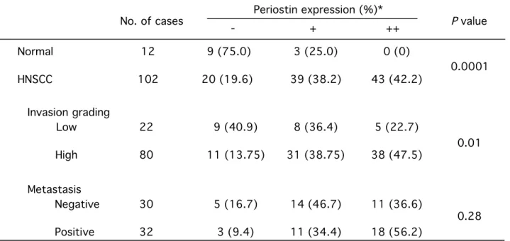
Next, we examined the expression of Periostin in 12 normal oral mucosae and 62 HNSCC cases by immunohistochemistry. HNSCC cells showed highly expression of
Kudo et al., 15 Periostin in comparison with normal oral mucosae (Fig. 5C 5Ba and b and Table 1). In 62
HNSCC cases, 26 (41.9 %) cases showed high expression of Periostin. Then, we
investigated the relationship between Periostin expression and the invasiveness in 62 102
HNSCC cases. For evaluation of invasiveness of HNSCC, we used the grading of mode of invasion (Grade 1 has a well defined borderline, Grade 2 has a less-marked borderline, Grade 3 has groups of cells and no distinct borderline, and Grade 4 has diffuse growth) as firstly described by Jacobsson et al. (21). We defined two groups, low (Grade 1 and 2) and high (Grade 3 and 4). Interestingly, higher expression of Periostin was significantly
associated with the grading of mode of invasion (P<0.05) (Fig.5D 5Bc and d and Table 1). In particular, cancer cells at invasive front expressed Periostin at higher levels (Fig. 5E5Be). We also examined the association between Periostin expression and metastasis in 40 62
HNSCC cases with available clinical information. 60 56.2 % of HNSCC cases with metastasis showed high expression of Periostin (Table 1).
To further evaluate the expression of Periostin in patients with HNSCC we compared the expression in a previously published microarray dataset of 41 HNSCC patients and 13 normal controls (9). Similar to our data, Periostin was expressed at higher levels in HNSCC tissues, in comparison with normal oral mucosal tissues (Fig. 5F5C). Moreover, HNSCC cases with angiolymphatic invasion showed higher expression of Periostin (Fig. 5F5C). To further explore the genes that are coordinately expressed with Periostin in HNSCC tumors, we performed a similarity search using a Pearson correlation metric as implemented in tGeneData Analyst Pro 1.0 software. The expression of selected genes demonstrating the highest coordinate expression with periostin in normal oral mucosal tissue and HNSCC tissues was then visualized by hierarchical clustering (Fig. 5G5D). FAP, SULF1, COL5A2,
Kudo et al., 16 COL3A1, COL10A1, COL4A1, FN1 and INHBA were well correlated with Periostin expression
(Fig. 5G 5D and Supplemental data 2).
Discussion
It is believed that neoplasms contain a variety of subpopulations of cells with differing
metastatic potential, and the presence of highly metastatic clones may exist within a primary tumor (4). Recent data demonstrate that reinjection of metastatic cell populations can lead to enrichment in the metastatic phenotype by work on experimental metastasis with cancer cell lines (22-25). Further, metastasis related genes were identified by comparing the gene expression profiles between parent and metastatic cell populations using microarray analysis (22-25). We previously established HNSCC cell line from metastatic cervical lymph node of HNSCC and then isolated highly invasive clones from this cell line by in vitro invasion assay (5, 10). By using these cell lines, we demonstrated that methylation of E-cadherin and degradation of ß-catenin were involved in the invasion of HNSCC through the loss of cell-cell adhesion (5). Thus, we previously could obtain the highly invasive phenotype by isolation of cell populations using an in vitro invasion assay. Therefore, we thought that comparing the gene expression profile of the parent and highly invasive clone could be a good approach to identify genes that influence the invasion of HNSCC. Here, we identified several genes, which encode secretary or cell surface proteins implicated in invasion, cell adhesion,
angiogenesis and growth factor as candidate genes for the invasion of HNSCC by comparing the gene expression profiles between parent HNSCC cells and a highly invasive clone using microarray analysis. Among these genes, Periostin was found to be the most highly
expressed gene in invasive HNSCC cells. As expected, overexpression of Periostin
Kudo et al., 17 was abolished by Periostin siRNA treatment. These observations strongly indicate that
Periostin plays an important role in the invasion of HNSCC. In addition, we found that Periostin overexpression did not promote proliferation, but growth in soft agar and in vivo growth as tumor xenografts, demonstrating that Periostin enhances anchorage independent growth of HNSCC cells.
Periostin contains an N-terminal secretory signal peptide, followed by a cysteine-rich domain, four internal homologous repeats, and a C-terminal hydrophilic domain. The four internal repeats region of Periostin shares a homology with the axon guidance protein FAS1 containing sequences that allows binding of integrins and glycosaminoglycans in vivo (26). FAS1 domains of ßig-h3, which shares a significant structural homology with Periostin, bear motifs interacting with integrins 13ß1 and 1vß5 (14, 15) and mediate endothelial cell
adhesion and migration via integrin 1vß3 (16). Similarly to ßig-h3, we found that interference with the function of integrins by specific anti-1vß3 and anti-1vß5 integrin antibodies had an effect on the ability of Periostin to mediate cell adhesion in HNSCC cells. Taken together, these data strongly suggest that FAS1 domain of Periostin binds to integrins. It is well known that integrin mediates cell-extracellular matrix (ECM) interaction and that integrin-mediated adhesion regulates a variety of intracellular events (27). Therefore, we
hypothesize that Periostin-integrin interaction may inhibit the ECM-integrin interaction and trigger the intracellular signaling and activation of certain genes that are involved in invasion and anchorage independent growth of HNSCC. This hypothesis is supported by recent report that Periostin activated the Akt/PKB pathway via the 1vß3 integrin to promote cellular survival in colon cancer (28). We suggest that overexpression of Periostin may confer on HNSCC cells the ability to survive in the absence of anchorage by inhibiting anoikis-related apoptotic pathways, thus allowing Periostin overexpressing HSC2 cells to form colonies in
Kudo et al., 18 soft agar and tumors in nude mice. However, in order to clarify the underlying mechanism of
invasion and anchorage independent growth by Periostin, further studies are required.
As mentioned above, Periostin promoted invasion and anchorage independent growth in HNSCC cells. These striking phenotypes seem to be an important for cancer metastasis. Interestingly, Periostin overexpression dramatically induced metastasis to lymph nodes and to the lung in an orthotopic implant model of HNSCC, demonstrating spontaneous metastasis from tongue. This orthotopic implant model of HNSCC seems clinically relevant because its tumor progression containing metastasis mimic the clinical scenario. Moreover, Periostin overexpressing tumors showed forming a larger tumor mass and remarkable invasiveness including destruction of mandibular bone and lymphatic infiltration. Therefore, we suggest that aggressive invasiveness and anchorage independent growth by Periostin overexpression may consequently lead to metastasis. To confirm the role of Periostin in metastasis as described above, we would like to examine the effect of Periostin siRNA in vivo in the future.
Indeed, immunohistochemical analysis revealed that Periostin expression was well
associated with the pattern of invasion and lymph node metastasis in HNSCC cases.
Moreover, 56.2 % of HNSCC cases with metastasis showed higher expression of Periostin, but we could not find statistical significance between Periostin expression and metastasis. In near future, we will examine the correlation between Periostin expression and metastasis in a large number of HNSCC cases. Interestingly, Bao et al. also demonstrated that a colon cancer cell line with low metastatic potential engineered to overexpress Periostin displayed a striking phenotype of greatly accelerated tumor metastatic growth as xenografts in the animal model system of metastasis (28). Importantly, higher expression of Periostin was frequently observed in HNSCC tissues in comparison with normal tissues. By evaluation of Periostin expression in HNSCC patients, a strong correlation of Periostin expression with FAP, SULF1,
Kudo et al., 19 COL5A2, COL3A1, COL10A1, COL4A1, FN and INHBA was observed. Although we have to
confirm this correlation, this finding gives an imagination that Periostin expression appears to be correlated with a stromal reaction likely related to invasion and metastasis. In addition, previous studies have shown that Periostin expression is up-regulated in various types of tumor including HNSCC (29), colon (28, 30), breast (31), lung (32) and ovarian cancer (17). Taken together, highly expression of Periostin may be a common event of tumor development in various types of cancer and can be a useful marker to predict its malignancy. In fact, serum levels of Periostin were elevated in patients with breast cancer, non-small cell lung cancer and thymoma (33-35). Therefore, we will examine the detection of Periostin in saliva and blood from the patients with HNSCC in the future. In conclusion, our studies have revealed a critical role of Periostin for invasion and anchorage independent growth in the metastatic process. These findings provide new and important information on the progression of HNSCC. The finding that Periostin influences metastatic potential
demonstrated in this study raises the possibility that it could be used as a molecular target in anti-metastasis therapy of HNSCC patients.
Acknowledgements
We thank Dr. X-F. Wang for providing Periostin expression vector. We also thank Dr. H. Kuniyasu and Dr. S. Siriwardena for helpful discussions and critical reading the paper and Ms. Hatano for assistance of manuscript preparation. Supported by in part by grants-in-aid from the Ministry of Education, Science and Culture of Japan to YK and TT, and grants from Takeda Co. and Tsuchiya foundation to YK.
Kudo et al., 20 References
1. Mao L, Hong WK, Papadimitrakopoulou VA. Focus on head and neck cancer. Cancer Cell 2004; 5: 311-6.
2. Forastiere A, Koch W, Trotti A, Sidransky D. Head and neck cancer. N Engl J Med 2001; 345: 1890–900.
3. Fidler IJ. Critical factors in the biology of human cancer metastasis: Twenty-eighth GHA Clowes Memorial Award Lecture. Cancer Res 1990; 50: 6130-8.
4. Fidler IJ. Tumor heterogenity and the biology of cancer invasion and metastasis. Cancer Res 1978; 38: 2651-60.
5. Kudo Y, Kitajima S, Ogawa I, Hiraoka M, Salgolzaei S, Keikhaee MR, et al. Invasion and metastasis of oral cancer cells require methylation of E-cadherin and/or degradation of membranous ß-catenin. Clin Cancer Res 2004; 10: 5455-63.
6. Horiuchi K, Amizuka N, Takeshita S, Takamatsu H, Katsuura M, Ozawa H, et al.
Identification and characterization of a novel protein, Periostin, with restricted expression to periosteum and periodontal ligament and increased expression by transforming growth factor beta. J Bone Miner Res 1999; 14: 1239–49.
7. Takeshita S, Kikuno R, Tezuka K, Amann E. Osteoblast-specific factor 2: cloning of a putative bone adhesion protein with homology with the insect protein fasciclin I. Biochem J 1993; 294: 271–8.
8. Ramakrishnan R, Dorris D, Lublinsky A, Nguyen A, Domanus M, Prokhorova A, et al. An assessment of Motorola CodeLink microarray performance for gene expression profiling applications. Nucleic Acids Res 2002; 30: e30.
Kudo et al., 21 9. Ginos MA, Page GP, Michalowicz BS, Patel KJ, Volker SE, Pambuccian SE, et al.
Identification of a Gene Expression Signature Associated with Recurrent Disease in Squamous Cell Carcinoma of the Head and Neck. Cancer Res 2004; 64: 55-63. 10. Kudo Y, Kitajima S, Sato S, Ogawa I, Miyauchi M, Takata T. Establishment of an oral
squamous cell carcinoma cell line with high invasive and p27 degradation activity from lymph node metastasis. Oral Oncol 2003; 39: 515-20.
11. Kitajima S, Kudo Y, Ogawa I, Bashir T, Kitagawa M, Miyauchi M, et al. Role of Cks1 overexpression in oral squamous cell carcinomas: cooperation with Skp2 in promoting p27 degradation. Am J Pathol 2004; 165: 2147-55.
12. Kalebic T, Williams JE, Talmadge JE, Kao-Shan CS, Kravitz B, Locklear K, et al. A novel method for selection of invasive tumor cells: derivation and characaterization of highly metastatic K1735 melanoma cells based on in vitro and in vivo invasive capacity. Clin Exp Metastasis 1998; 6: 301-18.
13. Huber O, Sumper M. Algal-CAMs: isoforms of a cell adhesion molecule in embryos of the alga Volvox with homology to Drosophila fasciclin I. EMBO J 1994; 13: 4212-22.
14. Kim JE, Kim SJ, Lee BH, Park RW, Kim KS, Kim IS. Identification of motifs for cell adhesion within the repeated domains of transforming growth factor-ß-induced gene, ßig-h3. J Biol Chem 2000; 275: 30907-15.
15. Kim JE, Jeong HW, Nam JO, Lee BH, Choi JY, Park RW, et al. Identification of motifs in the fasciclin domains of the transforming growth factor-ß-induced matrix protein ßig-h3 that interact with the 1vß5 integrin. J Biol Chem 2002; 277: 46159–65.
16. Nam JO, Kim JE, Jeong HW, Lee SJ, Lee BH, Choi JY, et al. Identification of the 1vß3 integrin-interacting motif of ßig-h3 and its anti-angiogenic effect. J Biol Chem 2003; 278: 25902-17.
Kudo et al., 22 17. Gillan L, Matei D, Fishman DA, Gerbin CS, Karlan BY, Chang DD. Periostin secreted by
epithelial ovarian carcinoma is a ligand for v3 and v5 integrins and promotes cell motility. Cancer Res 2002; 62: 5358–64.
18. Frisch SM, Ruoslahti E. Integrins and anoikis. Curr Opin Cell Biol 1997; 9: 701–6.
19. Osaki T, Tatemoto Y, Yoneda K, Yamamoto T. Tumorigenicity of cell lines established from oral squamous cell carcinoma and its metastatic lymph nodes. Eur J Cancer B Oral Oncol 1994; 30B: 296-301.
20. Kawashiri S, Kumagai S, Kojima K, Harada H, Yamamoto E. Development of a new invasion and metastasis model of human oral squamous cell carcinomas. Eur J Cancer B Oral Oncol 1995; 31B: 216-21.
21. Jacobsson PA, Eneroth GM, Killander D, Moberger G, Martensson B. Histologic
classification and grading of malignancy in carcinoma of the larynx. Acta Radiol 1973; 12: 1-7. 22. Clark EA, Golub TR, Lander ES, Hynes RO. Genomic analysis of metastasis reveals an
essential role for RhoC. Nature 2000; 406: 9532-5.
23. Kang Y, Siegel PM, Shu W, Drobnjak M, Kakonen SM, Cordon-Cardo C, et al. A multigenic program mediating breast cancer metastasis to bone. Cancer Cell 2003; 3: 537-49.
24. Yang J, Mani SA, Donaher JL, Ramaswamy S, Itzykson RA, Come C, et al. Twist, a master regulator of morphogenesis, plays an essential role in tumor metastasis. Cell 2004; 117: 927-39.
25. Minn AJ, Gupta GP, Siegel PM, Bos PD, Shu W, Giri DD, et al. Genes that mediate breast cancer metastasis to lung. Nature 2005; 436: 518-24.
26. Elkins T, Hortsch M, Bieber AJ, Snow PM, Goodman CS. Drosophila fasciclin I is a novel homophilic adhesion molecule that along with fasciclin III can mediate cell sorting. J Cell Biol 1990; 110: 1825-32.
Kudo et al., 23 27. Meredith J, Schwartz M. Integrins, adhesion and apoptosis. Trends Cell Biol 1997; 7: 146–
50.
28. Bao S, Ouyang G, Bai X, Huang Z, Ma C, Liu M, et al. Periostin potently promotes metastatic growth of colon cancer by augmenting cell survival via the Akt/PKB pathway. Cancer Cell 2004; 5: 329-39.
29. Gonzalez HE, Gujrati M, Frederick M, Henderson Y, Arumugam J, Spring PW, et al.
Identification of 9 genes differentially expressed in head and neck squamous cell carcinoma. Arch Otolaryngol Head Neck Surg 2003; 129: 754-9.
30. Tai IT, Dai M, Chen LB. Periostin induction in tumor cell line explants and inhibition of in vitro cell growth by anti-periostin antibodies. Carcinogenesis 2005; 26: 908-15.
31. Shao R, Bao S, Bai X, Blanchette C, Anderson RM, Dang T, et al. Acquired expression of periostin by human breast cancers promotes tumor angiogenesis through up-regulation of vascular endothelial growth factor receptor 2 expression. Mol Cell Biol 2004; 24: 3992-4003. 32. Sasaki H, Lo KM, Chen LB, Auclair D, Nakashima Y, Moriyama S, et al. Expression of
Periostin, homologous with an insect cell adhesion molecule, as a prognostic marker in non-small cell lung cancers. Jpn J Cancer Res 2001; 92: 869-73.
33. Sasaki H, Yu CY, Dai M, Tam C, Loda M, Auclair D, et al. Elevated serum periostin levels in patients with bone metastases from breast but not lung cancer. Breast Cancer Res Treat 2003; 77: 245-52.
34. Sasaki H, Dai M, Auclair D, Fukai I, Kiriyama M, Yamakawa Y, et al. Serum level of the periostin, a homologue of an insect cell adhesion molecule, as a prognostic marker in nonsmall cell lung carcinomas. Cancer 2001; 92: 843-8.
Kudo et al., 24 35. Sasaki H, Auclair D, Kaji M, Fukai I, Kiriyama M, Yamakawa Y, et al. Serum level of the
periostin, a homologue of an insect cell adhesion molecule, in thymoma patients. Cancer Lett 2001; 172: 37-42.
Kudo et al., 25 Figure legends
Figure 1. Periostin is identified as an invasion related gene in HNSCC. (A) Schematic representation of the isolation process of highly invasive clone. Highly invasive clone (MSCC-Inv1) was isolated from parent cells (MSCC-1) by in vitro invasion assay. The transcriptional profile of MSCC-1 cells and MSCC-Inv1 cells were compared by microarray analysis. Highly expressed genes in the highly invasive clone are listed. Among these genes, the mostly overexpressed gene in the highly invasive clone is Periostin (redbold). (B) Confirmation of higher expression of Periostin mRNA in the highly invasive clone by RT-PCR. Amplification was performed for 30 cycles. (C) Confirmation of higher expression of
Periostin protein in the highly invasive clone by Western blot analysis. ß-actin expression was used as a loading control. (D) The production of Periostin by MSCC-1 and MSCC-Inv1 cells. The production of Periostin was examined by Western blot analysis with conditioned culture media.
Figure 2. Periostin promotes the invasion of HNSCC cells. (A) Expression of Periostin mRNA in HNSCC cell lines. Expression of Periostin mRNA in HSC2, HSC3, Ca9-22, and
MSCC-1 and MSCC-Inv1 cells by RT-PCR was examined. Amplification was performed for 35 cycles. (B) Generation of Periostin overexpressing cells. HSC2 and HSC3 cells were engineered to overexpress Periostin by transfection with pcDNA3.1-His tagged Periostin. We obtained each 4 stable clones expressing Periostin. Ectopic expression of Periostin was examined by immunobloting with anti-His antibody. The whole lysates from all samples were blotted with ß-actin for loading control. (C) Cell proliferation (upper panel) and invasion (lower panel) of Periostin overexpressing HNSCC cells. Cells were plated on 24 well plates and trypsinized cells were counted by Cell Counter at 0, 2 and 4 day. (D) Invasion of
Kudo et al., 26
Periostin overexpressing HNSCC cells. The invasiveness of the cells was determined by in
vitro invasion assay. (E) Treatment with Recombinant Periostin enhanced invasiveness in HNSCC. Recombinant Periostin protein (0, 1 and 10 mg/ml) was treated in HSC2 cells. The invasiveness of the cells was determined by in vitro invasion assay. (FD) Knockdown of Periostin in MSCC-Inv1 cells. Periostin siRNA were transfected. Upper panel shows that Tthe effectiveness at knockdown the expression of Periostin was validated at the mRNA by RT-PCR and at the protein level by immunobloting with anti-Periostin antibody. (G) Lower panel shows that Kknockdown of Periostin inhibits the invasiveness of HNSCC cells. The invasiveness of the cells was determined by in vitro invasion assay.
Figure 3. Periostin is involved in the anchorage independent growth of HNSCC in vitro and in
vivo. (A) Treatment with Recombinant Periostin enhanced invasiveness in HNSCC. Recombinant Periostin protein (0, 1 and 10 mg/ml) was treated in HSC2 cells. The invasiveness of the cells was determined by in vitro invasion assay. (B) Interaction between Periostin and Integrin in HNSCC cells. HSC2 cells (3 x 10⁴ cells/well) were cultured in a 96-well plate precoated with 10 1g/ml of recombinant Periostin in the presence of anti-1vß3 or anti-1vß5 integrin antibody (10 1g/ml) for 1 hour. Non-coated wells were used as a control. Adhesive cells were counted after washing. (BC) Colony formation of Periostin overexpressing HNSCC cells in soft agar. After 3 weeks, colonies were photographed.
Upper panel shows Rrepresentative photographs of colonies are shown. (C) Lower panel shows Nnumber of colonies of Periostin overexpressing HNSCC cells. The colonies under a phase contrast microscope were counted at x10 magnification. We assayed twice and randomly selected 5 fields were counted for each assay. (D) Representative results showing the tumor growth of xenografts in immunocompromised mice. Periostin overexpressing and
Kudo et al., 27 control HSC2 cells (1 2 107 cells) were individually injected subcutaneously into 2 sites in
each 5 nude mice. After 1 month, the growth characteristics of the resulting tumors were analyzed. (a) Representative photographs of tumors. (b) Higher magnification of photographs of tumors. (c) Gross appearance of the tumor masses. (Ed) The volume of tumors. Tumor length (L) and width (W) were measured and tumor volume was calculated by the formula of (L x W2)/2.
Figure 4. Periostin promotes metastasis of HNSCC mediated by aggressive invasiveness in
vivo. (A) Schema of orthotopic implantation of HNSCC cells with or without Periostin
overexpression. Periostin overexpressing and control cells (5 2 105 cells) were orthotopically implanted into tongue of nude mice. After 2 weeks, tongue tumors, cervical lymph nodes and lungs were dissected. (B) Representative hematoxyline and eosin-stained images of histopathological sections from mice injected with control cells. (a) Histology of tumor mass in the tongue. (b) High power of (a). Tumor mass is enclosed with dotted line. Original magnifications: x5 (a); x25 (b). (C) Representative hematoxyline and eosin-stained images of histopathological sections from mice injected with Periostin overexpressing cells. (a) Histology of tumor mass in the tongue. (b) High power of (a). (c) Histology of invading tumor in lymphatics. (d) Histology of lymph node metastasis. (e) Histology of lung
metastasis. Arrow indicates invaded tumor (c) and metastasized tumor (d and e). Original magnifications: x5 (a); x25 (b-e). (D) Summary of the metastasis of Periostin overexpressing and control cells injected mice.
Figure 5. Higher expression of Periostin is frequently observed in HNSCC. (A) (a)
Kudo et al., 28 Graph shows the Periostin/GAPDH ratios which was measured by densitometry. (Bb) The
average of Periostin/GAPDH ratios. The average of Periostin/GAPDH ratios in HNSCC cases (T) and in normal tissues (N) is 1.86±0.40 and 0.51±0.22, respectively. (CB) Immunohistochemical analysis of Periostin. Expression of Periostin was examined by immunohistochemistry in HNSCC (Cancer) (a) and normal oral mucosae (Normal) (b). (D) To know the Ccorrelation between Periostin expression and Invasiveness invasiveness of HNSCC, we. To evaluated the invasiveness of HNSCC by using invasion grading, , we defined two groups as as low (Grade 1 and 2) and high (Grade 3 and 4). Representative cases of low (c) and high (d) groups are shown. (E) Periostin expression in invasive front of HNSCC. Representative case of higher expression of Periostin in invasive front is shown (e). (FC) Clustering of HNSCC samples to demonstrate relative expression of Periostin. Total RNA from 41 primary HNSCC and 13 normal tissues was labeled and hybridized to Affymetrix U133A Gene Chips as previously reported (9). The average of signal intensity of Periostin in 41 HNSCC and 13 normal tissues in microarray analysis. Left graph shows signal intensity of Periostin in normal and HNSCC tissues. Right graph shows signal intensity of Periostin in HNSCC cases with or without angiolymphatic invasion. The average of signal intensity of Periostin is shown at the bottom of the graph. (GD) Clustering of 41 HNSCC and 13 normal tissues to demonstrate relative expression of Periostin. We analyzed by hierarchical
clustering using a filtered set of 6800 variably expressed genes. The gene cluster that includes Periostin is highlighted in blue in the horizontal dendrogram.
Supplemental data
Microsoft Excel file 1. Raw expression data of microarray analysis between parent cells and
Kudo et al., 29 Microsoft Excel file 2. Raw data of correlation analysis for Periostin.
Table 1. Periostin expression in normal oral mucosae and HNSCC and correlation with invasion and metastasis.
*The expression of Periostin was graded as ++ (over 30 % of tumor cells showed strong or diffuse
immunopositivity), + (10-30 % of tumor cells showed moderate or patchy immunopositivity) and - (less than 10 % of the tumor cells showed weak or focal immunopositivity or no staining).
No. of cases Periostin expression (%)* - + ++ Normal HNSCC 12 102 9 (75.0) 20 (19.6) 3 (25.0) 39 (38.2) 0 (0) 43 (42.2) Invasion grading Low High 22 80 9 (40.9) 11 (13.75) 8 (36.4) 31 (38.75) 5 (22.7) 38 (47.5) Metastasis Negative Positive 30 32 5 (16.7) 3 (9.4) 14 (46.7) 11 (34.4) 11 (36.6) 18 (56.2) P value 0.01 0.28 0.0001
GAPDH Periostin M S C C -1 M S C C -I n v 1 M S C C -1 M S C C -I n v 1 ß-actin Periostin M S C C -1 M S C C -I n v 1 Periostin Parent oral cancer cell
(MSCC-1)
Isolation
Highly invasive clone (MSCC-Inv1)
Microarray analysis MSCC-1 vs MSCC-Inv1
Probe_Name Ratio Description
NM_006475.1 65.175345 OSTEOBLAST SPECIFIC FACTOR 2 (FASCICLIN I-LIKE) (OSF-2/Periostin) NM_003641.1 18.556444 INTERFERON INDUCED TRANSMEMBRANE PROTEIN 1 (9-27) (IFITM1)
NM_032642.1 18.173555 WINGLESS-TYPE MMTV INTEGRATION SITE FAMILY, MEMBER 5B (WNT5B), TRANSCRIPT VARIANT 1 NM_001387.1 16.965932 DIHYDROPYRIMIDINASE-LIKE 3 (DPYSL3)
AA810805 13.177289 603639245F1 HOMO SAPIENS CDNA, 5' END NM_005397.1 12.508605 PODOCALYXIN-LIKE (PODXL)
X16323 11.012348 MRNA FOR HEPATOCYTE GROWTH FACTOR (HGF) NM_007036.2 9.4544153 ENDOTHELIAL CELL-SPECIFIC MOLECULE 1 (ESM1)
NM_002575.1 9.446 SERINE (OR CYSTEINE) PROTEINASE INHIBITOR, CLADE B (OVALBUMIN), MEMBER 2 (SERPINB2) NM_000055.1 8.7671114 BUTYRYLCHOLINESTERASE (BCHE)
NM_001920.1 8.5095798 DECORIN (DCN)
NM_006528.1 7.0713753 TISSUE FACTOR PATHWAY INHIBITOR 2 (TFPI2) AK023450 6.958723 CAPILLARY MORPHOGENESIS PROTEIN 2 (CMG2) NM_007363.2 6.6625375 NON-POU-DOMAIN-CONTAINING, OCTAMER-BINDING (NONO) NM_032495.1 6.2201358 HYPOTHETICAL PROTEIN SMAP31 (SMAP31)
NM_001257.1 6.0081838 CADHERIN 13, H-CADHERIN (HEART) (CDH13) BC009269 5.8917143 MRNA FOR PUTATIVE OXIDOREDUCTASE
NM_002192.1 5.5939091 INHIBIN, BETA A (ACTIVIN A, ACTIVIN AB ALPHA POLYPEPTIDE) (INHBA) NM_000627.1 5.4214464 LATENT TRANSFORMING GROWTH FACTOR BETA BINDING PROTEIN 1 (LTBP1)