INTRODUCTION
Baseball is one of the most popular sports in the world. Although baseball is relatively safe, numerous reports suggested that there has been a rapid rise in injury rates among youth baseball players (1 - 4). It is important to demonstrate the nature of such injuries. In Tokushima, we started a check - up for child and adolescent baseball players in the field from 1981. From the findings of the check -up, we investigated the frequency of pain in each part of the body. The most frequent area of pain was the elbow joint, which was found in about 30% of players (5). Matsuura et al. (6) followed 449 youth baseball players without prior elbow pain for one season and reported that 30% of youth players had elbow pain each year, and nearly 72.3% of players with elbow pain had abnormal findings on physical examination such as limitation in the range of motion, tenderness, and pain on valgus stress test. Furthermore, 81.4% of the subjects with abnormal findings on physical examination ex-hibited radiographic abnormalities : medial epicondylar fragmen-tation was observed in 97.2% and osteochondritis dissecans (OCD) of the capitellum was seen in 2.8%. Hang et al. (4) reported results that were similar to our findings. They explained that 52% of the 343 Taiwanese Little League baseball players (mean age, 11.6 years ; range, 9.5 - 12.0 years) had episodes of soreness of the throwing elbow. Radiographic examination of all players revealed
separation or fragmentation of the medial epicondyle in 76% and OCD of the capitellum in only 1 catcher. Although medial epicon-dylar fragmentation was prevalent in either report, the prognosis of medial epicondylar fragmentation is relatively good (7, 8). By contrast, the prognosis of OCD of the capitellum is often poor. However, it was difficult to detect OCD of the capitellum based on subjective complaints and physical examination, because most players with OCD of the capitellum were asymptomatic or mini-mally symptomatic. Ultrasound imaging, which does not involve exposure to radiation, is inexpensive, and is a portable type of di-agnostic imaging, is useful for detecting OCD of the capitellum. Harada et al. (9) found 33 subjects with medial epicondylar frag-mentation (21.6%) and 2 with OCD of the capitellum (1.3%) among 153 players aged 9 to 12 years using ultrasound imaging in the field. Matsuura et al. (10) reported that the prevalence of OCD of the capitellum on ultrasonography in 1040 young baseball players aged 10 to 12 years was 2.1%.
The purpose of this study was to report the outcome of an elbow check - up system for child and adolescent baseball players in 2013. In particular, we investigated the situation of elbow injuries in youth baseball players with elbow pain and ultrasonographic find-ings of the capitellum in all players.
MATERIALS AND METHODS
The Institutional Review Board of Tokushima University Hos-pital approved the study protocol, and all parents and coaches pro-vided informed consent.
A total of 1605 players (mean age, 10.1 years ; range, 6 - 12 years)
ORIGINAL
Outcome of an elbow check-up system for child and
adolescent baseball players
Toshiyuki Iwame1, Tetsuya Matsuura2, Naoto Suzue2, Shinji Kashiwaguchi3, Takenobu Iwase4, Shoji Fukuta2, Daisuke Hamada2, Tomohiro Goto2, Takahiko Tsutsui2, Keizo Wada2, Hiroshi Egawa1, Akihiro Nagamachi5, and Koichi Sairyo2
1Department of Orthopaedic Surgery, Tokushima Prefectural Central Hospital, Tokushima, Japan,2Department of Orthopedics, Tokushima University, Tokushima, Japan,3Department of Orthopaedic Surgery, Tokyo Shinjuku Medical Center, Tokyo, Japan,4Department of Orthopae-dic Surgery, Tokushima National Hospital, Tokushima, Japan,5Department of Orthopaedic Surgery, Mitoyo General Hospital, Kagawa, Japan
Abstract : Purpose : Our aim was to examine the outcome of an elbow check -up system for youth baseball players. In particular, we investigated the nature of elbow injuries in youth baseball players with elbow pain and ultra-sonographic findings of the capitellum. Materials and Methods : A total of 1605 players participating in the re-gional summer championship in July 2013 underwent a questionnaire survey, physical examination, ultrasound imaging, and radiographic examination. Results : A total of 499 (31.1%%) players reported episodes of elbow pain, of whom 320 (64.1%%) had abnormal findings on physical examination, and 115 (35.9%%) agreed to undergo radiog-raphy. Among them, 98 (85.2%%) exhibited radiographic abnormalities. On the initial ultrasonography screening, 60 (3.7%%) players had an abnormal finding and 55 (91.7%%) agreed to undergo radiography. Among them, 26 (47.3%%) were found to have osteochondritis dissecans (OCD) of the capitellum on radiographs. Conclusions : About 30% of youth baseball players had episodes of elbow pain, and 64.1%% of players with elbow pain had abnormal findings on physical examination. Furthermore, 85.2%% of subjects who underwent radiographic examination exhibited radiographic abnormalities. About 4%% of young baseball players had an abnormal finding on initial ultra-sonography screening, and nearly 50%% of them had OCD of the capitellum on radiographs. J. Med. Invest. 63 : 171-174, August, 2016
Keywords : Childhood and adolescent, Baseball, Elbow check-up
Received for publication January 4, 2016 ; accepted February 4, 2016. Address correspondence and reprint requests to Tetsuya Matsuura, MD, PhD, Department of Orthopedics, Tokushima University 3 - 18 - 15 Kuramoto, Tokushima 770 - 8503, Japan and Fax : +81 - 88 - 633 - 0178.
The Journal of Medical Investigation Vol. 63 2016
belonging to junior baseball teams that participated in the regional summer championship in July 2013 were the subjects of this in-vestigation.
1. Elbow Injuries in Players with Elbow Pain
Questionnaires were distributed to the team coaches, and infor-mation was filled out by subjects with the assistance of their coaches and/or parents. Questionnaire items included age, player position, and any past episodes of pain during or after throwing practice. The author (T.M.) reviewed the questionnaire with indi-vidual subjects to increase their understanding of the questions and check the accuracy of the information.
Physical examination of the elbow was recommended to players who complained of elbow pain on the questionnaire and was per-formed in the field. Range of motion, tenderness, and the valgus stress test were included in the physical examination. Limitation of range of motion was regarded as positive if the difference be-tween the throwing and non - throwing arms was more than 5!.
Radiographic examination was recommended to players who had positive findings on the physical examination. Radiography of the elbow was performed in two directions : anteroposterior with the elbow flexed at 45!and laterally. Films were reviewed at the office by one of the authors (T.M.).
2. Ultrasonographic Findings of the Capitellum
Both elbows were sonographically examined in all 1605 players in the field. Ultrasonography of the lateral aspect of the elbow was performed by two of the authors (T.I. and N.S.) using the following equipment : a FAZONE M sonography diagnostic imaging system (ZONARE Medical Systems Inc) with a 5 - to 10 - MHz linear array transducer (FAZONE CB ; Fujifilm Corp), the MyLabFive portable ultrasound system (Esaote Europe BV) with a 6 - to 18 - MHz linear array transducer, and the M- Turbo ultrasound system (SonoSite Inc) with a 6 - to 13 - MHz linear array transducer. Anterior view images were taken with the subject seated and the elbow fully extended. Posterior view images were taken with the elbow fully flexed to obtain a sufficient view of the anterior aspect of the cap-itellum. Sonographic findings were graded as follows according to Matsuura et al. (11) : grade 0, normal ; grade 1a, irregular sur-face of the subchondral bone ; grade 1b, cystic lesion of the sub-chondral bone surface ; grade 2, irregularity of the subsub-chondral bone ; and grade 3, discontinuity of the subchondral bone. We de-fined grade 1a, 2, and 3 as abnormal findings. Grade 1b was not considered abnormal because Matsuura et al. (10) reported that cystic appearance of the subchondral bone surface was thought to be a variation of normal development during ossification.
Radiographic examination was recommended to players who had positive findings on ultrasound. Radiography of the elbow was performed in four directions : anteroposterior with the elbow ex-tended and flexed at 45!,lateral, and oblique. One of the authors (T.M.) diagnosed the radiographic findings and graded the stage using the classification of Matsuura et al. (11).
RESULTS
1. Elbow Injuries in Players with Elbow Pain
Figure 1 summarizes the overall results of the examination. Of the 1605 players, 499/1605 (31.1%) reported episodes of pain in the throwing elbow during throwing practice. The remaining 1106 players had no prior elbow pain.
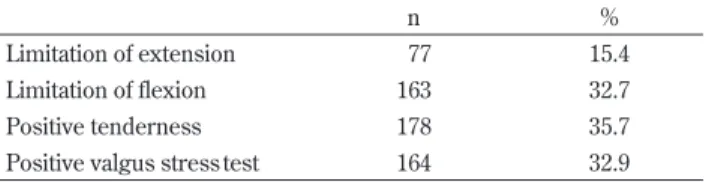
Of the 499 subjects who reported elbow pain, 320/499 (64.1%) had abnormal findings on the physical examination. Among them, a limitation of extension was found in 77/499 (15.4%) subjects and a limitation of flexion was found in 163/499 (32.7%) subjects. Ten-derness of the elbow was found in 178/499 subjects (35.7%) and the
valgus stress test was positive in 164/499 subjects (32.9%) (Table 1). Of the 320 subjects who had abnormal findings on physical ex-amination, 115/320 (35.9%) agreed to undergo radiography. Among them, 98/115 (85.2%) exhibited radiographic abnormalities : medial epicondylar injury was observed in 84, and OCD of the capitellum was seen in 13, and physeal injury of the olecranon was seen in 1 subject.
2. Ultrasonographic Findings of the Capitellum
Figure 2 summarizes the overall results of the examination. On initial ultrasonography screening, 60/1605 (3.7%) of players had an abnormal finding and 55/60 (91.7%) agreed to undergo radiog-raphy. Among them, 32/55 (58.2%) were found to have an abnormal finding of the capitellum on radiographs, and OCD of the capitel-lum was evident in 26/55 subjects (47.3%). Of the 26 subjects, 22/ 26 (84.6%) had no elbow pain and 12/26 (46.2%) had no positive sign on the physical examination.
Of the 26 players diagnosed with OCD of the capitellum, based on the radiographic stage using the classification of Matsuura et al. (11), 19 had stage I (radiolucent area), 6 had stage II (non -displaced fragments), and 1 had stage III (loose bodies and sclerotic changes).
Figure 1. Outcomes of Elbow Examination
Table 1. Outcomes of Physical Examination of Players with Elbow Pain
n %
Limitation of extension 77 15.4
Limitation of flexion 163 32.7
Positive tenderness 178 35.7
Positive valgus stress test 164 32.9
DISCUSSION
We investigated the outcome of an elbow check - up system by administering a questionnaire, physical examination, radiographic examination, and ultrasound imaging in this study.
A total of 31.1% of 1605 youth baseball players had episodes of elbow pain. Among them, 64.1% had abnormal findings on physical examination. Furthermore, 85.2% of subjects who underwent ra-diographic examination showed abnormal signs on radiographs. Our results were almost equivalent to the previous report by Matsuura et al. (6) Hang et al. (4) reported that 52% of young base-ball players (mean age, 11.6 years ; range, 9.5 - 12.0 years) had epi-sodes of soreness of the throwing elbow. The reason why our results (31.1%) were lower than that reported by Hang et al. (4) was that our study included all elementary school students (mean age, 10.1 years ; range, 6 - 12 years) in contrast to their study.
The results of ultrasonography screening showed that 3.7% of all subjects had an abnormal finding and 1.6% of youth baseball players were found to have OCD of the capitellum on radiographs. Matsuura et al. (10) reported that the prevalence of OCD of the capitellum on ultrasonography in 1040 young baseball players (mean age, 10.7 years ; range, 10 - 12 years) was 2.1%. Our results (1.6%) were lower than that reported by Matsuura et al. (10), and the cause seemed to be the inclusion of all elementary school students in our study. Harada et al. (9) found 2 cases of OCD (1.3%) among 153 players aged 9 to 12 years using ultrasound imaging. The results were consistent with our findings.
Of the 26 subjects with a diagnosis of OCD of the capitellum, 22/ 26 (84.6%) had no elbow pain and 12/26 (46.2%) had no abnormal sign on physical examination. Therefore, OCD of the capitellum in these subjects would not have been detected if ultrasonography of the lateral aspect of the elbow had not been performed. Matsuura et al. (11) reported that the average age at which stage I OCD was diagnosed was 11.5 years, suggesting that the typical age of onset
was 10 to 12 years in baseball players, and conservative treatment resulted in healing in 90.5% of subjects with a stage I lesion. In this study, 19 of 26 subjects (73.1%) with OCD had a stage I lesion based on radiographic examination. These results suggest that screening for OCD of the capitellum using ultrasonography is useful for early detection and provides an opportunity for successful conservative treatment.
However, 1 subject had positive signs on physical examination and was diagnosed with OCD of the capitellum (stage II) on radio-graphs, despite an abnormal finding was not detected on ultra-sound. Ultrasound imaging is profoundly affected by the tech-niques and experience of the examiners.
This study has several limitations. One major limitation is that only 35.9% of players who had abnormal findings on the physical examination agreed to undergo radiography. Among the players who did not undergo radiographic examination, the rate of elbow injuries was thought to be high. Another limitation is that although we confirmed the physical and ultrasonographic abnormalities on plain radiography, radiography was not performed for those subjects with no abnormalities on physical examination and ultra-sound imaging. Another disadvantage was that the inter - observer relationship was not investigated by the two sonographers in the ultrasound screening.
CONCLUSIONS
About 30% of youth baseball players had episodes of elbow pain, and 64.1% of players with elbow pain had abnormal findings on physical examination. Furthermore, 85.2% of subjects who under-went radiographic examination exhibited radiographic abnor-malities.
About 4% of young baseball players had an abnormal finding on initial ultrasonography screening, and nearly 50% of them had OCD of the capitellum on radiographs. About 85% of the subjects with OCD of the capitellum had no elbow pain.
REFERENCES
1. Torg JS, Moyer RA : Non - union of a stress fracture through the olecranon epiphyseal plate observed in an adolescent base-ball player. A case report. J Bone Joint Surg 59 : 264 - 265, 1977 2. Guggenheim JJ, Stanley RF, Woods GW, Tullos HS : Little League survey : the Houston study. Am J Sports Med 4 : 189 -200, 1976
3. Larson RL, Singer KM, Bergstrom R, Thomas S : Little League survey : the Eugene study. Am J Sports Med 4 : 201 - 209, 1976 4. Hang DW, Chao CM, Hang YS : A clinical and roentgenographic study of Little League elbow. Am J Sports Med 32 : 79 -84, 2004
5. Matsuura T : Epidemiology of sports injury in child and ado-lescent athletes [In Japanese]. In : Muto Y, Kashiwaguchi S, Uchio Y eds. Handbook of screening for musculoskeletal sys-tem. Nankodo Inc, Tokyo, pp.25 - 29, 2007
6. Matsuura T, Suzue N, Kashiwaguchi S, Arisawa K, Yasui N : Elbow injuries in youth baseball players without prior elbow pain. A 1 - year prospective study. Orthop J of Sports Med 2013 7. Matsuura T, Ikata T, Kashiwaguchi S, Iwase T : A follow up study of humeral medial epicondyle lesion among young baseball players [In Japanese]. Jpn J Orthop Sports Med 17 : 263 -269, 1997
8. Harada M, Takahara M, Hirayama T, Sasaki J, Mura N, Ogino T : Outcome of nonoperative treatment for humeral medial epicondylar fragmentation before epiphyseal closure in young baseball players. Am J Sports Med 40 : 1583 - 1590, 2012 Figure 2. Outcomes of Elbow Ultrasonography Screening
9. Harada M, Takahara M, Sasaki J, Mura N, Ito T, Ogino T : Using sonography for the early detection of elbow injuries among young baseball players. AJR Am J Roentgenol 187 : 1436 - 1441, 2006
10. Matsuura T, Suzue N, Iwame T, Nishio S, Sairyo K : Preva-lence of osteochondritis dissecans of the capitellum in young
baseball players. Results based on ultrasonographic findings. Orthop J of Sports Med 2014
11. Matsuura T, Kashiwaguchi S, Iwase T, Takeda Y, Yasui N : Conservative treatment for osteochondrosis of the humeral capitellum. Am J Sports Med 36 : 868 - 872, 2008