Relationship of oral conditions to the incidence of infective endocarditis in periodontitis patients with valvular heart disease: a cross-sectional study
Masami Ninomiya1, Mari Hashimoto1, Kouji Yamanouchi2, Yoshiaki Fukumura3, Toshihiko Nagata1, Koji Naruishi1*
1Department of Periodontology and Endodontology, Tokushima University Graduate School of Biomedical Sciences, 3-18-15 Kuramoto, Tokushima 770-8504, Japan
2Yamanouchi Dentistry and Oral Surgery
3Department of Cardiovascular Surgery, Tokushima Red Cross Hospital
Running title: Relationship of IE and oral conditions
Funding: This study was supported by a Grant-in-Aid for Scientific Research (B) (No. 15H05054), Scientific Research (C) (No. 26463135) and Scientific Research (C) (No. 16K11832) from the Japan Society for the Promotion of Science.
Disclosure: No Disclosures to Report.
*Corresponding author: Dr. Koji Naruishi, D.D.S., Ph.D.
Department of Periodontology and Endodontology, Tokushima University Graduate School of Biomedical Sciences, 3-18-15 Kuramoto, Tokushima 770-8504, Japan
Phone: +81-88-631-3111; Fax: +81-88-633-7009 e-mail: naruishi@tokushima-u.ac.jp
This is a post-peer-review, pre-copyedit version of an article published in Clinical Oral Investigations. The final authenticated version is available online at: https://doi.org/10.1007/s00784-019-02973-2.
ABSTRACT
Objectives Infective endocarditis (IE) is a life-threatening infectious disease, but the pathogenesis of the disease remains uncertain. The objective of this study was to examine whether oral infectious conditions are associated with the occurrence of IE in valvular heart disease (VHD) patients.
Materials and Methods A total of 119 periodontitis (P) patients with or without VHD were enrolled, and cross-sectional analyses were performed. Patients were classified as follows: 1) mild-to-moderate P without VHD, 2) mild-to-moderate P with VHD, 3) severe P without VHD, or 4) severe P with VHD. A total of 78 VHD patients were classified as 1) VHD without IE or 2) VHD with IE. Conditional logistic regression analysis was performed to compute the odds ratio (OR) and 95% confidence interval (CI).
Results No significant differences were observed between patients with or without VHD in oral conditions. A significant increase in the percentage of alveolar bone loss in VHD patients with IE was observed compared with that of patients without IE. The ratio of both Porphyromonas gingivalis (Pg) IgG titer>1.68 and Pg fimA type II genotype in patients with IE was significantly higher than in patients without IE. There was a significant correlation between the occurrence of IE and clinical oral findings (number of remaining teeth: OR, 0.17; rate of alveolar bone loss>40%: OR, 11.8).
Conclusions VHD patients with IE might have severe periodontitis compared with patients without IE, although further investigation will be needed because this is based on only 7 VHD patients with IE.
Clinical relevance The patients with IE had fewer remaining teeth, more advanced bone resorption compared with those of patients without IE. These findings suggest a possible association between the occurrence of IE and periodontal infection.
Keywords: Infective endocarditis (IE); Valvular heart disease (VHD); Periodontitis; Oral examination
Introduction
Periodontitis is a polymicrobial infectious disease that results in loss of teeth by various causes such as inflammation-mediated bone resorption [1]. More than 500 individual species of microbes have been identified in the human mouth [2], and periodontists have understood the clinical significance of microbial examination for periodontitis since the end of the 1980s [3]. Infection with periodontal bacteria causes humoral immunological responses and elevates the levels of serum immunoglobulin (Ig) G antibody against bacteria [4]. Persistent low-grade infection of Gram-negative bacteria, such as Porphyromonas gingivalis (Pg), from periodontitis is associated with increased atherosclerosis, diabetes mellitus, and other systemic diseases disseminated through the blood stream [5,6]. Therefore, clinical oral examination focused on microbial infection is essential for general health.
Valvular heart disease (VHD) reduces the heart’s functional capacity, and patients with VHD have increased risks of perioperative adverse events [7,8]. In particular, Infective endocarditis (IE) is an infection of the endocardial lining of the heart with pre-existing lesions or on intra-cardiac foreign materials [9]. IE treatment requires prolonged antibiotic therapy, and half of patients with IE undergo valvular surgery [10]. Unfortunately, the mortality of IE remains high: approximately 20% for in-hospital mortality [11]. Although Gram-positive cocci, such as Staphylococcus aureus, is a leading cause of IE [12], Isoshima et al. detected a bacterial DNA from the Gram-negative periodontal pathogen Pg in cardiac tissue of patients with IE using an in
vitro DNA amplification assay [13]. Therefore, the risk of IE caused by bacteremia from
guidelines have reduced indications for antibiotic therapy in the prevention of IE and highlight the clinical significance of oral hygiene [11]. Systemic micro-inflammation by periodontitis may impair vascular function, because previous report showed that impaired endothelial function recovered by intensive periodontal therapy [14]. However, the clinical relationship between the occurrence of VHD and oral conditions, including periodontal conditions, remains unclear.
Poor oral health may have a profound effect on general health, and it is important to examine periodontal infections. Rigorous evaluation of oral conditions, including periodontal infection, may help clarify the risks of IE, especially in patients with VHD. Therefore, we examined whether oral conditions are associated with IE in VHD patients using cross-sectional analyses.
METHODS
Subjects and evaluation
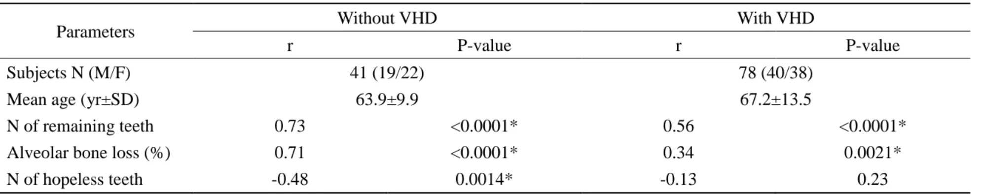
A total of 119 periodontitis patients with or without VHD who visited Tokushima Red Cross Hospital or Tokushima University Hospital between 2012 and 2017 were enrolled (without VHD: 19 males, 22 females, average age, 63.9±9.9 yr; with VHD: 40 males, 38 females; average age, 67.2±13.5 yr) (Fig.1). All participants provided written informed consent. The incidence of VHD and IE was diagnosed by specialists of cardiovascular surgery, and all VHD patients were referred to the dental office to screen for and reduce oral bacterial infections. Oral conditions were evaluated by 3 trained dentists, and the all examinations were performed using the same evaluation standards in order to lessen the inter-examiner error. The number of remaining teeth were counted by inspection. Hopeless teeth were diagnosed by periodontitis specialists according to a previous report [15]. Periodontitis patients were divided into mild-to-moderate and severe groups according to descriptions from the Center for Disease Control and Prevention and the American Academy of Periodontology [16]. For moderate periodontitis, the following definition was proposed: ≥2 interproximal sites with AL ≥4 mm (not on same tooth), or ≥2 interproximal sites with PD ≥5 mm (not on same tooth). For severe periodontitis, the following definition was also proposed: ≥2 interproximal sites with AL ≥6 mm (not on same tooth) and ≥1 interproximal site with PD ≥5 mm. Assessment of alveolar bone loss was performed by calculating bone loss as a proportion of the tooth root length on X-ray films using a Schei ruler [17]. Interproximal alveolar bone loss of all remaining teeth was measured at the deepest point on the mesial or distal surface of each tooth radiographically as the % of bone loss
from the cement enamel junction to the tooth apex. The titers of IgG antibodies against periodontal bacteria, such as Pg, Aggregatibacter actinomycetemcomitans (Aa),
Prevotella intermedia (Pi), and Eichenerra corrodens (Ec), in the patient's blood were
measured using the DEMECAL® blood examination system (Leisure, Inc., Tokyo, Japan). We collected the blood plasma of each patient, and the sample were mailed immediately to the company at room temperature. The Pg gene in the saliva of patients was evaluated using the BML periodontal bacteria detection system (BML, Inc., Tokyo, Japan). We collected the whole unstimulated salivary flows before oral cleaning, and the saliva sample were mailed immediately to the company at room temperature. All examinations were performed when the patients were in a stable condition after receiving acute medical care. Exclusion criteria were as follows: 1) patients who did not agree to the oral examination, 2) death within 2 weeks after hospitalization, and 3) isolated cases with various medical reasons.
This study was conducted according to the guidelines described in the Declaration of Helsinki and was approved by the ethics committee of Tokushima Red Cross Hospital (No. 459).
Statistical analysis
Statistical differences in the oral conditions of 119 patients with and without VHD were examined using cross-sectional analyses. Subjects were classified as follows: 1) mild-to-moderate periodontitis without VHD (N=17), 2) mild-to-moderate periodontitis with VHD (N=29), 3) severe periodontitis without VHD (N=24), or 4) severe periodontitis with VHD (N=49). A total of 78 VHD patients were classified as 1) VHD without IE (N=71) or 2) VHD with IE (N=7). Differences in parameters between groups
were analyzed by a Mann-Whitney U test, chi-square test, and Pearson's correlation coefficient test because the data were not normally distributed. In the 78 VHD patients, the odds ratio (OR) of the clinical parameters for the incidence of IE derived from bivariate analysis and the 95% confidence interval (CI) were obtained by conditional logistic regression analysis. Statistical analyses were performed using JMP® 8 ver. 8.0.2 (SAS Institute Japan, Tokyo), and a P-value less than 0.05 was considered significant.
RESULTS
Correlation between plasma IgG titer against Pg and oral conditions
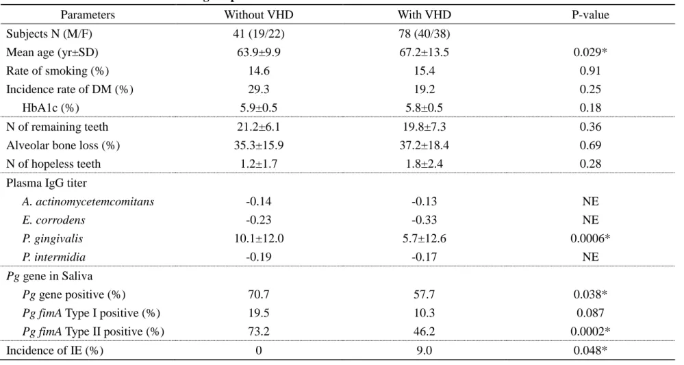
Statistical correlations between plasma IgG titer against Pg and oral conditions in periodontitis patients with and without VHD were analyzed (without VHD: number of remaining teeth, r=0.73, P<0.0001; percentage of alveolar bone loss, r=0.71, P<0.0001; number of hopeless teeth, r=-0.48, P=0.0014; with VHD: number of remaining teeth, r=0.56, P<0.0001; percentage of alveolar bone loss, r=0.34, P=0.0021, Pearson's correlation coefficient test) (Table 1). There was no statistical relationship between plasma IgG titer against Pg and the number of hopeless teeth, but a trend was observed in patients with VHD.
Statistical differences in the clinical and microbial findings of subjects with and without VHD in each periodontitis group
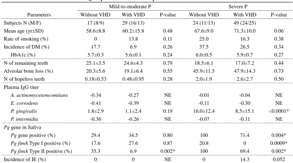
At first, we examined the statistical differences between periodontitis patients with and without VHD in clinical and microbial findings. As shown in Table 2, the mean age of patients with VHD was significantly higher than in patients without VHD (P=0.029, Mann-Whitney U test). There were no significant differences between patients with and without VHD in several oral conditions (number of remaining teeth: P=0.36; percentage of alveolar bone loss: P=0.69; number of hopeless teeth: P=0.28, Mann-Whitney U test). No significant differences were observed between patients with and without VHD in the rate of smoking (P=0.91, chi-square test) and the incidence of DM (P=0.25, chi-square test). The plasma IgG titer against Pg in patients with VHD was significantly lower than in patients without VHD (P=0.0006, Mann-Whitney U test). In addition, the Pg gene
was detected in the saliva of patients with VHD significantly less than in patients without VHD (P=0.038, chi-square test). Furthermore, the Pg fimA type II genotype in patients with VHD was also lower than in patients without VHD (P=0.0002, chi-square test). There was no significant difference in the fimA type I genotype between patients with and without VHD (P=0.087, chi-square test). An increase of plasma IgG titers against Aa, Ec, and Pi in patients with and without VHD was not observed.
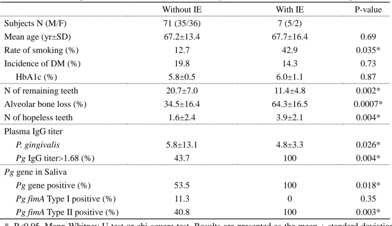
Next, we examined the statistical differences in the clinical and microbial findings of subjects with and without VHD in each periodontitis group. As shown in Table 3, in the mild-to-moderate periodontitis group, no statistical differences were observed between patients with and without VHD in oral findings (percentage of alveolar bone loss, P=0.55, number of hopeless teeth, Mann-Whitney U test). No significant difference was observed between patients with and without VHD in microbial findings (plasma IgG titer against Pg, P=0.19, Mann-Whitney U test; detection rate of the Pg gene, P=0.80, chi-square test). On the other hand, the fimA type II genotype was significantly lower in saliva samples of periodontitis patients with VHD than in patients without VHD (P=0.002, chi-square test), although there was no significant difference in the fimA type I genotype (P=0.87, chi-square test). An increase of plasma IgG titer against Aa, Ec, and Pi in patients with and without VHD was not observed. IE was not observed, even in the patients with VHD.
In the severe periodontitis group, there were no statistical differences between patients with and without VHD in oral findings (percentage of alveolar bone loss (P=0.73, number of hopeless teeth, P=0.50, Mann-Whitney U test). Plasma IgG titer against Pg in patients with VHD was significantly lower than in patients without VHD (P<0.0001, Mann-Whitney U test). In addition, there was a statistical difference in the
detection rate of the Pg gene in saliva between patients with and without VHD (P=0.004, chi-square test). Furthermore, both the fimA type I and type II genotypes in saliva samples of severe periodontitis patients with VHD were lower than in patients without VHD (fimA type I: P=0.0009, fimA type II: P=0.002, chi-square test). An increase of plasma IgG titer against Aa, Ec, and Pi in patients with and without VHD was not observed. Furthermore, there was a higher incidence of IE in severe periodontitis patients with VHD compared with that of patients without VHD, but it was not significantly different (P=0.052, chi-square test).
Relationships between the clinical and microbial findings and the incidence of IE in periodontitis patients with VHD
For periodontitis patients with VHD (Table 4), there was a significant decrease in the number of remaining teeth in patients with IE compared with that of patients without IE (P=0.002, Mann-Whitney U test). Furthermore, there were significant differences between patients with and without IE in the percentage of alveolar bone loss (P=0.0007, Mann-Whitney U test) and the number of hopeless teeth (P=0.004, Mann-Whitney U test). The rate of smoking in VHD patients with IE was significantly higher than in VHD patients without IE (P=0.035, chi-square test). There were no significant differences between patients with and without IE in DM-related parameters (incidence of DM: P=0.73, chi-square test; HbA1c: P=0.87, Mann-Whitney U test). The plasma IgG titer against Pg in VHD patients with IE was significantly lower than in VHD patients without IE (P=0.026, Mann-Whitney U test), but the ratio of Pg IgG titer>1.68 in VHD patients with IE was significantly higher than in VHD patients without IE (P=0.004, chi-square test). The detection rate of the Pg gene in saliva of
VHD patients with IE was significantly higher than in patients without IE (P=0.018, chi-square test). Furthermore, the fimA type II genotype, but not the fimA type I genotype, was significantly increased in saliva samples of VHD patients with IE compared to that in patients without IE (P=0.003, chi-square test).
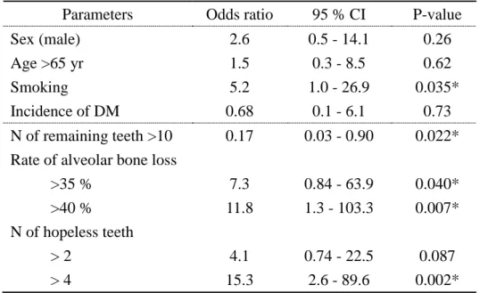
Conditional logistic regression analysis of clinical findings relating to the occurrence of IE
To explore the factors for IE, we calculated the ORs for bivariate relationships (Table 5). There were significant relationships between several oral conditions and the occurrence of IE (number of teeth: OR, 0.17, 95% CI, 0.03–0.90, P=0.022; alveolar bone loss >40%: OR, 11.8, 95% CI, 1.3–103.3, P=0.007: number of hopeless teeth >4: OR, 15.3, 95% CI, 2.6–89.6, P=0.002). Smoking was also an important parameter to evaluate risk of occurrence of IE in VHD patients (OR, 5.2, 95% CI, 1.0–26.9, P=0.035). No significant relationship between the incidence of DM and IE was observed (OR, 0.68, 95% CI, 0.1–6.1, P=0.73).
DISCUSSION
Periodontitis is an infectious disease caused by periodontal bacteria such as Pg [18]. Plasma IgG titers against Pg in periodontitis patients are significantly higher than those of healthy subjects [19]. As shown in Table 1, we demonstrated that the plasma IgG titers against Pg significantly correlated to several oral conditions. Furthermore, there was a positive correlation between percentage of alveolar bone loss and Pg IgG titer. This result indicates that the many remaining teeth of subjects in this study were not healthy periodontally because of continuous Pg infection. Next, we found that the plasma IgG titer against Pg in the severe periodontitis group was significantly higher than in the mild-to-moderate periodontitis group, and the Pg gene in the saliva of patients with severe periodontitis corresponded to the increase in plasma IgG titer (see supplemental Table). Therefore, increased Pg infection is a good parameter to measure the progression of periodontal inflammation. However, the relationship between oral infectious status and the occurrence of VHD remains unclear, and persistent bacteremia by Pg invasion into the blood stream may be a significant virulent factor for the development of many systemic diseases such as atherosclerosis or cardiovascular diseases [20, 21].
We examined the clinical and microbial differences of patients with and without VHD (Table 2). There were no significant differences between patients with and without VHD in oral conditions such as percentage of alveolar bone loss, number of both remaining teeth and hopeless teeth. There were also no significant differences in both the rate of smoking and incidence of DM, which are strong risk factors for periodontitis. No differences of periodontal conditions between patients with and without VHD were
observed, which suggests that VHD is not a risk factor for periodontitis. The blood culture system is a standard technique to diagnose microbial infections, but blood culture has several clinical disadvantages, such as a 24–48 hour minimum time to detect and identify causative bacteria and that prior antibiotic administration markedly reduces the sensitivity of living bacteria. By contrast, polymerase chain reaction (PCR) of microbial DNA is a rapid technique with a processing time of only a few hours, and intact dead or living bacteria can be detected [22]. Additionally, the PCR assay requires only small amounts of sample, which may be the greatest clinical benefit. Previously, Nakagawa et al. showed that both disease associated and non-disease associated genotypes exist in Pg, and that fimA genotyping is useful for the prediction of periodontitis development [23]. The Pg fimA gene is classified into 6 genotypes; fimA type II is associated with periodontitis, and fimA type I is associated with periodontal health [23]. We found that the fimA type II genotype was higher than the fimA type I genotype in patients with and without VHD by PCR. Importantly, the detection rate of both the Pg gene and fimA type II genotype in the saliva of patients with VHD decreased significantly compared with those of patients without VHD, which corresponded to a decrease in Pg IgG titer in patients with VHD (Table 2). These findings are not surprising because antimicrobial therapy has frequently been used to prevent the occurrence of blood stream infections or to adjust inappropriate antibiotics [11].
Next, we examined whether there were statistical differences in the clinical and microbial findings between patients with and without VHD in both the mild-to-moderate and severe periodontitis groups (Table 3). In both periodontitis groups, no significant differences were observed between patients with and without VHD in
several parameters including the rate of smoking, incidence of DM, number of teeth, and percentage of alveolar bone loss, although un-regulative recruitment-selection bias might be considered in these parameters. For the severe periodontitis group, the plasma IgG titer against Pg in patients with VHD decreased significantly compared with that of patients without VHD. The detection rate of the Pg gene and fimA type II genotype also decreased significantly in patients with VHD compared with those in patients without VHD. Although frequent use of antibiotics in VHD patients may decrease microbial values, examination of Pg infection was still useful to evaluate microbial periodontal infection because Pg infection in the severe periodontitis group increased significantly compared with that in the mild-to-moderate periodontitis group even in VHD patients (see Supplemental Table).
IE occurs in patients with previously detected heart disease or a history of open heart surgery [11], but the clinical data regarding unrecognized cardiac lesions with IE are limited. Early detection of IE is clinically needed because IE is a life-threatening heart disease and therapy should begin soon after diagnosis. Although only 7 VHD patients with IE were examined, we found a higher occurrence of IE in patients with VHD, especially with severe periodontitis. Therefore, we focused on the relationship of oral findings to the occurrence of IE in VHD patients. Importantly, we found a decrease in the number of remaining teeth and an increase of alveolar bone loss in VHD patients with IE than in patients without IE (Table 4). Furthermore, because the rate of smoking in VHD patients with IE was significantly higher than in patients without IE, smoking may be a significant factor for the occurrence of IE in VHD patients. On the other hand, there were no significant differences between patients with and without IE in DM-related parameters, although the incidence of DM is often associated with
progression of periodontal inflammation [6]. Severe periodontitis in VHD patients with IE might be independent of poor glycemic control in patients. Furthermore, conditional logistic regression analyses revealed that several oral parameters were significantly associated with the occurrence of IE (Table 5). Interestingly, we found that there was an inhibitory relationship between the number of teeth and the occurrence of IE (OR: 0.17) and a positive relationship between severe alveolar bone loss and the occurrence of IE (OR: 11.8). Although further investigation is needed to understand the clinical significance of dental treatment for the prevention of IE, treatment of periodontitis is important to prevent loss of teeth. Periodontal conditions in VHD patients with IE is more severe than in patients without IE. We found that the plasma IgG titer against Pg in VHD patients with IE was significantly lower than in VHD patients without IE, but the ratio of Pg IgG titer>1.68 in VHD patients with IE was significantly higher than in VHD patients without IE. The plasma IgG titers against Pg lower than a cut-off value of 1.68 were defined as negative for periodontitis infection levels, and this blood test is used to screen for chronic periodontitis [19]. In addition, the detection rate of both the
Pg gene and fimA type II genotype increased significantly in VHD patients with IE.
Previously, Nakano et al. reported that the Pg gene was detected in cardiovascular specimens of patients including those with IE, and the fimA type II genotype was most frequently detected [24]. This report supports our findings that there is a positive relationship between Pg infection and the occurrence of IE in VHD patients. Furthermore, Nakano et al. also reported that the detection rate of the Aa gene in patients with IE was higher than in patients without IE using a PCR assay [25], but we did not find an increase of plasma IgG titer against Aa using ELISA. This discrepancy may be explained by differences in the sensitivity of the method between the two
studies.
Prophylactic administration of antimicrobials prior to periodontal treatment is recommended in several countries to prevent the occurrence of IE, but recent American Heart Association recommendations strongly suggest that dental procedures are not an important cause of IE [26]. On the other hand, Tonneti et al. reported that periodontal infection control by intensive periodontal treatment improved endothelial function [14]. Although oral health may be associated with improved endothelial function, which results in the prevention of IE, further studies using multivariate analysis with a large sample size are needed to clarify the precise relationship between oral status and the incidence of IE.
Our study had several limitations. First, the conclusions are based on 7 patients with IE because of the rare occurrence of the disease, which is one of the major limitations of the present study. Second, although we found several factors related to periodontal infection, we did not examine causality because this was a cross-sectional study. A multicenter, prospective study with a large number of patients would be needed to confirm these results, although periodontal infection may affect to the development of IE throughout blood stream. Third, although our models were adjusted for several factors, such as age and gender, no adjustment was made for other factors that may impact the occurrence of VHD or IE such as congenital abnormalities, rheumatoid diseases, hypertension, and the use of artificial heart valves. Finally, although confounding factors should be considered in this study, it is difficult to disentangle the factors that affect VHD/IE and periodontitis because they may be linked. These risk adjustments would increase the validity of future studies.
Conclusions
We found that patients with IE had fewer remaining teeth, larger numbers of teeth requiring extraction, and more advanced bone resorption compared with those of patients without IE. Furthermore, an increase in Pg infection was associated with the occurrence of IE in VHD patients. These results suggest a possible association between the occurrence of IE and periodontal infection. Oral microbial examinations in collaboration with dentists may be clinically useful to evaluate the possible risks of IE in VHD patients, although further investigation will be needed because the results were based on only 7 VHD patients with IE.
Acknowledgements
The authors thank the medical staff of Tokushima Red Cross Hospital and Tokushima University Hospital for their assistance.
Compliance with Ethical Standards
Conflict of interest: The authors declare that they have no conflict of interest.
Funding: This study was supported by a Grant-in-Aid for Scientific Research (B) (No. 15H05054), Scientific Research (C) (No. 26463135) and Scientific Research (C) (No. 16K11832) from the Japan Society for the Promotion of Science.
Ethical approval: This study was conducted according to the guidelines described in Declaration of Helsinki, and was approved by the ethics committee of Tokushima Red Cross Hospital (No. 459).
Informed consent: Informed consent was obtained from all individual participants included in the study.
References
1. Armitage GC (2004) Periodontal diagnoses and classification of periodontal diseases. Periodontol 2000 34:9-21.
2. Han YW, Wang X (2013) Mobile microbiome: oral bacteria in extra-oral infections and inflammation. J Dent Res 92:485-91.
3. Zambon JJ (1996) Periodontal diseases: microbial factors. Ann Periodontol 1:879-925.
4. Lamster IB, Smith QT, Celenti RS, Singer RE, Grbic JT (1994) Development of a risk profile for periodontal disease: microbial and host response factors. J Periodontol 65:511-20.
5. Chistiakov DA, Orekhov AN, Bobryshev YV (2016) Links between atherosclerotic and periodontal disease. Exp Mol Pathol 100:220-35.
6. Grossi SG (2001) Treatment of periodontal disease and control of diabetes: an assessment of the evidence and need for future research. Ann Periodontol 6:138-45. 7. Thonghong T, De Backer O, Søndergaard L (2018) Comprehensive update on the
new indications for transcatheter aortic valve replacement in the latest 2017 European guidelines for the management of valvular heart disease. Open Heart 5:e000753.
8. Coffey S, Cairns BJ, Iung B (2016) The modern epidemiology of heart valve disease. Heart 102:75-85.
9. Abegaz TM, Bhagavathula AS, Gebreyohannes EA, Mekonnen AB, Abebe TB (2017) Short- and long-term outcomes in infective endocarditis patients: a systematic review and meta-analysis. BMC Cardiovasc Disord 17(1):291.
10. Song JK (2015) Infective endocarditis involving an apparently structurally normal valve: new epidemiological trend? Korean J Intern Med 30:434-42.
11. Millot S, Lesclous P, Colombier ML, Radoi L, Messeca C, Ballanger M, et al (2017) Position paper for the evaluation and management of oral status in patients with valvular disease. Arch Cardiovasc Dis 110:482-94.
12. Han SM, Sorabella RA, Vasan S, Grbic M, Lambert D, Prasad R, Wang C, Kurlansky P, Borger MA, Gordon R, George I (2017) Influence of Staphylococcus
aureus on outcomes after valvular surgery for infective endocarditis. J Cardiothorac
Surg 12:57.
13. Isoshima D, Yamashiro K, Matsunaga K, Shinobe M, Nakanishi N, Nakanishi I, Omori K, Yamamoto T, Takashiba S (2017) Assessment of pathogenesis of infective endocarditis by plasma IgG antibody titer test against periodontal bacteria. Clin Case Rep 5:1580-6.
14. Tonetti MS, D'Aiuto F, Nibali L, Donald A, Storry C, Parkar M, Suvan J, Hingorani AD, Vallance P, Deanfield J (2007) Treatment of periodontitis and endothelial function. N Engl J Med 356:911-20.
15. Avila G, Galindo-Moreno P, Soehren S, Misch CE, Morelli T, Wang HL (2009) A novel decision-making process for tooth retention or extraction. J Periodontol 80:476-91.
16. Eke PI, Page RC, Wei L, Thornton-Evans G, Genco RJ (2012) Update of the case definitions for population-based surveillance of periodontitis. J Periodontol 83:1449-54.
17. Bassiouny MA, Grant AA (1975) The accuracy of the Schei ruler: a laboratory investigation. J Periodontol 46:748-52.
18. Darveau RP, Tanner A, Page RC (1997) The microbial challenge in periodontitis. Periodontol 2000 14:12-32.
19. Kudo C, Naruishi K, Maeda H, Abiko Y, Hino T, Iwata M, et al (2012) Assessment of the plasma/serum IgG test to screen for periodontitis. J Dent Res 91:1190-5. 20. Teeuw WJ, Slot DE, Susanto H, Gerdes VE, Abbas F, D'Aiuto F, Kastelein JJ, Loos
BG (2014) Treatment of periodontitis improves the atherosclerotic profile: a systematic review and meta-analysis. J Clin Periodontol 41:70-9.
21. Aarabi G, Zeller T, Seedorf H, Reissmann DR, Heydecke G, Schaefer AS, Seedorf U (2017) Genetic susceptibility contributing to periodontal and cardiovascular disease. J Dent Res 96:610-7.
22. Maeda H, Fujimoto C, Haruki Y, Maeda T, Kokeguchi S, Petelin M, Arai H, Tanimoto I, Nishimura F, Takashiba S (2003) Quantitative real-time PCR using TaqMan and SYBR Green for Actinobacillus actinomycetemcomitans,
Porphyromonas gingivalis, Prevotella intermedia, tetQ gene and total bacteria.
FEMS Immunol Med Microbiol 39:81-6.
23. Nakagawa I, Amano A, Ohara-Nemoto Y, Endoh N, Morisaki I, Kimura S, Kawabata S, Hamada S (2002) Identification of a new variant of fimA gene of
Porphyromonas gingivalis and its distribution in adults and disabled populations
with periodontitis. J Periodontal Res 37:425-32.
24. Nakano K, Inaba H, Nomura R, Nemoto H, Takeuchi H, Yoshioka H, Toda K, Taniguchi K, Amano A (2008) Distribution of Porphyromonas gingivalis fimA genotypes in cardiovascular specimens from Japanese patients.Oral Microbiol Immunol 23:170-2.
25. Nakano K, Inaba H, Nomura R, Nemoto H, Tamura K, Miyamoto E, Yoshioka H, Taniguchi K, Amano A, Ooshima T (2007) Detection and serotype distribution of
Actinobacillus actinomycetemcomitans in cardiovascular specimens from Japanese
patients. Oral Microbiol Immunol 22:136-9.
26. Baddour LM, Wilson WR, Bayer AS, Fowler VG Jr, Tleyjeh IM, Rybak MJ, et al (2015) Infective endocarditis in adults: diagnosis, antimicrobial therapy, and management of complications: A scientific statement for healthcare professionals from the American Heart Association. Circulation 132:1435-86.
Figure legend
Figure 1. Flow chart of subjects. All of valvular heart disease (VHD) patients were diagnosed and recruited at Department of Cardiovascular Surgery, Tokushima Red Cross Hospital (N=78). All of periodontitis patients were diagnosed and recruited at Department of Periodontics, Tokushima University Hospital (N=41). The microbial examinations of all subjects (N=119) were performed using outsourced laboratory system. Subjects were divided to 4 groups as follows: mild-to-moderate periodontitis (Moderate P) with or without VHD, severe periodontitis (Severe P) with or without VHD.
Ninomiya et al.
Fig.1
78 VHD patients (including 7 IE patients) (40 males, 38 females)
Dentistry in hospital Dept. of Cardiovascular Surgery
Diagnosis (VHD or IE)/ Informed Consent
Tokushima Red Cross Hospital
41 periodontitis patients (19 males, 22 females)
• Oral examination • taking X-ray
Referral
119 patients (59 males, 60 females)
Outsourced Laboratory Tests Mail
Collect sample (blood/ saliva)
Dept. of Periodontics, Tokushima University Hospital Referral
78 VHD patients (including 7 IE patients) (40 males, 38 females)
Diagnosis (periodontitis)/ Informed Consent
• Blood test for IgG titer • Saliva test for bacterial DNA
Moderate P without VHD: N=17 Moderate P with VHD: N=29 Severe P without VHD: N=24 Severe P with VHD: N=49
Table 1. Correlation between Pg IgG titer and oral conditions in patients with or without VHD
Parameters Without VHD With VHD
r P-value r P-value
Subjects N (M/F) 41 (19/22) 78 (40/38)
Mean age (yr±SD) 63.9±9.9 67.2±13.5
N of remaining teeth 0.73 <0.0001* 0.56 <0.0001*
Alveolar bone loss (%) 0.71 <0.0001* 0.34 0.0021*
N of hopeless teeth -0.48 0.0014* -0.13 0.23
*, P<0.05, Pearson's correlation coefficient test. r, correlation coefficient. VHD, valvular heart disease; N, number; SD, standard deviation.
Table 2. Clinical and microbial findings in patients with or without VHD
Parameters Without VHD With VHD P-value
Subjects N (M/F) 41 (19/22) 78 (40/38)
Mean age (yr±SD) 63.9±9.9 67.2±13.5 0.029*
Rate of smoking (%) 14.6 15.4 0.91
Incidence rate of DM (%) 29.3 19.2 0.25
HbA1c (%) 5.9±0.5 5.8±0.5 0.18
N of remaining teeth 21.2±6.1 19.8±7.3 0.36
Alveolar bone loss (%) 35.3±15.9 37.2±18.4 0.69
N of hopeless teeth 1.2±1.7 1.8±2.4 0.28
Plasma IgG titer
A. actinomycetemcomitans -0.14 -0.13 NE E. corrodens -0.23 -0.33 NE P. gingivalis 10.1±12.0 5.7±12.6 0.0006* P. intermidia -0.19 -0.17 NE Pg gene in Saliva Pg gene positive (%) 70.7 57.7 0.038*
Pg fimA Type I positive (%) 19.5 10.3 0.087
Pg fimA Type II positive (%) 73.2 46.2 0.0002*
Incidence of IE (%) 0 9.0 0.048*
*, P<0.05, Mann-Whitney U test or chi-square test. Results are presented as the mean ± standard deviation (SD). VHD, valvular heart disease; P, periodontitis; IE, infective endocarditis; DM, diabetes mellitus; fimA, fimbrial gene; Pg, P. gingivalis; N, number; SD, standard deviation; NE, not examined.
Table 3. Clinical and microbial findings in patients with different periodontitis states with or without VHD
Mild-to-moderate P Severe P
Parameters Without VHD With VHD P-value Without VHD With VHD P-value
Subjects N (M/F) 17 (8/9) 29 (16/13) 24 (11/13) 49 (24/25)
Mean age (yr±SD) 58.6±8.8 60.2±15.8 0.48 67.6±9.0 71.3±10.0 0.06
Rate of smoking (%) 0 13.8 0.11 25.0 16.3 0.38
Incidence of DM (%) 17.7 6.9 0.26 37.5 26.5 0.34
HbA1c (%) 5.7±0.3 5.6±0.1 0.24 6.0±0.5 5.9±0.7 0.27
N of remaining teeth 25.1±3.5 24.6±4.3 0.79 18.5±6.1 17.0±7.2 0.44
Alveolar bone loss (%) 20.3±5.6 19.1±6.4 0.55 45.9±11.5 47.9±14.3 0.73
N of hopeless teeth 0.18±0.53 0.48±0.95 0.28 2.0±1.9 2.6±2.7 0.50
Plasma IgG titer
A. actinomycetemcomitans -0.34 -0.27 NE -0.01 -0.04 NE E. corrodens -0.41 -0.39 NE -0.11 -0.30 NE P. gingivalis 1.8±2.9 1.1±2.4 0.19 16.0±12.4 8.5±15.1 <0.0001* P. intermidia -0.36 -0.26 NE -0.07 -0.11 NE Pg gene in Saliva Pg gene positive (%) 29.4 34.5 0.80 100 71.4 0.004*
Pg fimA Type I positive (%) 17.6 27.6 0.87 20.8 0 0.0009*
Pg fimA Type II positive (%) 35.3 6.9 0.002* 100 69.4 0.002*
Incidence of IE (%) 0 0 NE 0 14.3 0.052
*, P<0.05, Mann-Whitney U test or chi-square test. Results are presented as the mean ± standard deviation (SD). VHD, valvular heart disease; P, periodontitis; IE, infective endocarditis; DM, diabetes mellitus; fimA, fimbrial gene; Pg, P. gingivalis; N, number; SD, standard deviation; NE, not examined.
Table 4. Relationship of the clinical and microbial findings to the occurrence of IE in VHD patients
Without IE With IE P-value
Subjects N (M/F) 71 (35/36) 7 (5/2)
Mean age (yr±SD) 67.2±13.4 67.7±16.4 0.69
Rate of smoking (%) 12.7 42.9 0.035*
Incidence of DM (%) 19.8 14.3 0.73
HbA1c (%) 5.8±0.5 6.0±1.1 0.87
N of remaining teeth 20.7±7.0 11.4±4.8 0.002*
Alveolar bone loss (%) 34.5±16.4 64.3±16.5 0.0007*
N of hopeless teeth 1.6±2.4 3.9±2.1 0.004*
Plasma IgG titer
P. gingivalis 5.8±13.1 4.8±3.3 0.026*
Pg IgG titer>1.68 (%) 43.7 100 0.004*
Pg gene in Saliva
Pg gene positive (%) 53.5 100 0.018*
Pg fimA Type I positive (%) 11.3 0 0.35
Pg fimA Type II positive (%) 40.8 100 0.003*
*, P<0.05, Mann-Whitney U test or chi-square test. Results are presented as the mean ± standard deviation (SD). VHD, valvular heart disease; IE, infective endocarditis; DM, diabetes mellitus; fimA, fimbrial gene; Pg,
P. gingivalis; N, number; SD, standard deviation. The cut-off value of IgG titer against Pg for screening
Table 5. Odds ratio of clinical findings for the occurrence of IE Parameters Odds ratio 95 % CI P-value
Sex (male) 2.6 0.5 - 14.1 0.26
Age >65 yr 1.5 0.3 - 8.5 0.62
Smoking 5.2 1.0 - 26.9 0.035*
Incidence of DM 0.68 0.1 - 6.1 0.73
N of remaining teeth >10 0.17 0.03 - 0.90 0.022* Rate of alveolar bone loss
>35 % 7.3 0.84 - 63.9 0.040*
>40 % 11.8 1.3 - 103.3 0.007*
N of hopeless teeth
> 2 4.1 0.74 - 22.5 0.087
> 4 15.3 2.6 - 89.6 0.002*
A total of 78 VHD patients were analyzed by conditional logistic regression analysis. *P<0.05, chi-square test. CI, confidence interval.
Supplemental Table. Clinical and microbial findings in patients with or without VHD: statistical differences between moderate and severe periodontitis
Without VHD With VHD
Parameters Mild-to-
moderate P Severe P P-value
Mild-to-
moderate P Severe P P-value
Subjects N (M/F) 17 (8/9) 24 (11/13) 29 (16/13) 49 (24/25) Age (yr±SD) 58.6±8.8 60.2±15.8 0.48 67.6±9.0 71.3±10.0 0.06 Rate of smoking (%) 0 25.0 0.026* 13.8 16.3 0.76 Incidence rate of DM (%) 17.7 37.5 0.17 6.9 26.5 0.033* HbA1c (%) 5.7±0.3 6.0±0.5 0.38 5.6±0.1 5.9±0.7 0.12 N of remaining teeth 25.1±3.5 18.5±6.1 0.004* 24.6±4.3 17.0±7.2 <0.0001*
Alveolar bone loss (%) 20.3±5.6 45.9±11.5 0.0001* 19.1±6.4 47.9±14.3 0.0001*
N of hopeless teeth 0.18±0.53 2.0±1.9 0.027* 0.48±0.95 2.6±2.7 <0.0001*
Plasma IgG titer
A. actinomycetemcomitans -0.34 -0.01 NE -0.27 -0.04 NE E. corrodens -0.41 -0.11 NE -0.39 -0.30 NE P. gingivalis 1.8±2.9 16.0±12.4 <0.0001* 1.1±2.4 8.5±15.1 0.032* P. intermidia -0.36 -0.07 NE -0.26 -0.11 NE Pg gene in Saliva N of Pg gene positive 5 24 <0.0001* 10 35 0.0001*
N of Pg fimA type I positive 3 5 0.78 8 0 0.0001*
N of Pg fimA type II positive 6 24 0.0001* 2 34 <0.0001*
*, P<0.05, Mann-Whitney U test or chi-square test. VHD, valvular heart disease; P, periodontitis; IE, infective endocarditis; DM, diabetes mellitus; FimA, fimbrial gene; Pg, P. gingivalis; N, number; SD, standard deviation; NE, not examined.
We examined statistical differences in oral status between moderate and severe periodontitis in patients with or without VHD. There were 41 periodontitis patients without VHD with the following characteristics: The percentage of alveolar bone loss and number of hopeless teeth in the severe periodontitis group were significantly higher than in the moderate periodontitis group (alveolar bone loss: P=0.0001; number of hopeless teeth: P=0.027, Mann-Whitney U test). The rate of smoking was higher in the severe periodontitis group than in the moderate periodontitis group (P=0.026, chi-square test). The plasma IgG titer against Pg in the severe periodontitis group was significantly higher than in the moderate periodontitis group (P<0.0001, Mann-Whitney U test). Similar to the increase in IgG titer against Pg, the Pg gene was detected in the saliva of 100% of patients in the severe periodontitis group. Furthermore, we detected the fimA type II genotype in all 24 saliva samples of patients with severe periodontitis, but the fimA type II genotype was detected in only a few samples of patients with moderate periodontitis (35.3%). There was no significant difference in the fimA type I genotype between moderate and severe periodontitis groups (P=0.78, chi-square test). An increase in the plasma IgG titer against Aa, Ec, and Pi was not observed.
There were 78 periodontitis patients with VHD with the following characteristics: The percentage of alveolar bone loss and number of hopeless teeth in the severe periodontitis group were significantly higher than in the moderate periodontitis group (alveolar bone loss: P=0.0001; number of hopeless teeth: P<0.0001, Mann-Whitney U test). There was a significant difference in the incidence of DM between
the moderate and severe periodontitis groups (P=0.033, chi-square test), but no significant difference was observed in HbA1c levels. There was no significant difference in the rate of smoking between the moderate and severe periodontitis groups (P=0.76, chi-square test). The plasma IgG titer against Pg in the severe periodontitis group was significantly higher than in the moderate periodontitis group (P=0.032, Mann-Whitney U test). Similar to the increase in IgG titer against Pg, the Pg gene was detected in the saliva of 69.4% of patients with severe periodontitis. In 35 Pg gene-positive saliva samples, the fimA type II genotype was predominant (97.1%), while the fimA type II genotype was detected only in a few samples from patients with moderate periodontitis (6.9%). There was a significant difference in the
fimA type I genotype between moderate and severe periodontitis groups (P=0.0001, chi-square test). An increase in the plasma IgG titer