Department of Stomatognathic Function and Occlusal Reconstruction, Graduate School of Health Biosciences, Tokushima University, Tokushima 770–8504, Japan
Preliminary study of neural excitation with positron
emission tomography after tooth mechanical stimulation
Katsuhiro OMOTO, Takuma IWASA, Shaista Afroz, Mayu UEDA,
Kazuo OKURA, Yoshizo MATSUKA
Abstract : Dentists often cannot objectively find abnormalities in patients who complain of discomfort or abnormal sensation in their dental occlusion. We hypothesized that abnormal neural transmission from the tooth is related to this occlusal discomfort sensation. Chronic tooth contact habits may induce neural excitation from the tooth to the central nervous system, and may aggravate the sensation of discomfort in the central nervous system. However, the details of neural transmission from the tooth to the central nervous system are still unclear. In this study, we stimulated a rat premolar mechanically and observed activated bran sites using positron emission tomography (PET) and 18F-2-fluoro-2-deoxy-D-glucose (FDG). We anesthetized 5-7-week-old
male rats using isoflurane inhalation anesthesia and stimulated the upper right premolar mechanically with an electric von Frey system (Model 1601C, IITC Instruments) by measuring mechanical pressure. Before the tooth mechanical stimulation, we injected FDG through the rat’s caudal vein and then used a stimulation intensity of 100, 200 or 300 g. We recorded FDG accumulation with PET. The PET brain images were separated into four parts (right higher, left higher, right lower and left lower) for analysis and the peak value of striatal uptake (SUV) in each part was analyzed. The PET images showed that the accumulated FDG in the right lower part of the brain was higher with 300 g tooth stimulation than with 100 or 200 g. The data showed that the tooth stimulation site in the lower part of the brain was activated with tooth stimulation by comparing it with the other parts. We also measured SUV in the right and left sensory areas, motor area, hippocampus, trigeminal ganglia (TG) and spinal cord. The TG and sensory area showed more FDG accumulation compared with mouth opening.
Key words : Tooth stimulation, neural excitation, positron emission tomography
原 著
Introduction
Occasionally, dentists see some patients with
complaints like “I cannot chew well,” or “My
dental occlusion is uncomfortable”. The dentists
often cannot find objective abnormalities in these patients who complain discomfort or abnormal sensation in their dental occlusion (Clark and Simmons 2003, Hara et al. 2012).
Tamaki et al. called this problem “occlusal
discomfort syndrome” and reported the possible
etiology of occlusal discomfort syndrome (Tamaki et al. 2013). Following these authors, occlusal discomfort syndrome occurs in both a broad and a narrow sense. The definition of the broad sense is, “A comprehensive syndrome of pathology characterized by discomfort related to occlusion. Obvious occlusal disharmony (idiopathic) may or may not be associated,” and that of the narrow sense is, “An idiopathic syndrome of pathology characterized by discomfort related to occlusion, but having
nothing to do with occlusion.” (Tamaki et al. 2013). The narrow sense might be the same pathology associated with occlusal dysesthesia defined by Clark and Simmons (Clark and Simmons 2003). It is difficult for dentists to manage a narrow sense occlusal discomfort patient, and dentists occasionally perform occlusal adjustment by grinding the teeth without any patient dental occlusal problems. The occlusal adjustment might be made worse in these conditions.
It has been reported that psychological factors worsen the symptoms of occlusal discomfort in patients who do not have clear abnormal occlusion (Reeves and Merrill 2007). Also, a systematic review found that the risk factors of occlusal discomfort syndrome may be 1) psychiatric disorders, 2) phantom phenomenon and neuroplasticity, and 3) alteration in proprioceptive input transmission (Hara et al. 2012). If the patient does not feel that the occlusal contacts are stable after the prosthesis is set, the prosthesis may continually bother the patient, who will then feel as if they have to check it frequently. The checking procedure may induce tooth clenching, and the resulting periodontal nerve abnormal sensory transmission
will go straight to the central nervous system, inducing the patient’s feeling of discomfort.
We hypothesized that abnormal neural transmission from the tooth is related to this sensation of occlusal discomfort (Fig. 1). Chronic tooth contact habits may induce neural excitation from the tooth to the central nervous system, and may aggravate the discomfort sensation. However, details of the neural transmission from the tooth to the central nervous system are still unclear.
We found that chronic trigeminal neural stimulation increased neurotransmitter release from trigeminal ganglia (TG) (Kitamura et al. 2009) and decreased sensory threshold (Kumada et al. 2012, Matsuka et al. 2012). Also, botulinum toxin blocking the neurotransmitter release recovered the sensory threshold (Kitamura et al. 2009, Kumada et al. 2012, Omoto et al. 2015). Moreover, our study found that local anesthesia injection into the periodontal area of occlusal discomfort patients reduced the patients’ occlusal discomfort sensation (Matsuka et al. 2007).
Previous studies in human functional MRI (fMRI) have found that tooth stimulation induces brain activation (SI, SII, insula, inferior frontal
Fig. 1 Hypothesis on the disorder of the nerve communication in the occlusal discomfort sensation. Chronic tooth contact habit may induce sensitivity change of the mechanical nerve receptor and trigeminal neural transmission. The information goes through synaptic transmission in the spinal tract
gyrus, inferior parietal lobe or postcentral gyrus) (Miymoto et al. 2006, Trulsson et al. 2010, Habre-Hallage et al. 2014). The aim of this study was to observe the detail of functional change in the brain after tooth stimulation, and to achieve this we stimulated rat premolar mechanically and observed activated brain sites using positron emission tomography (PET).
Materials and methods
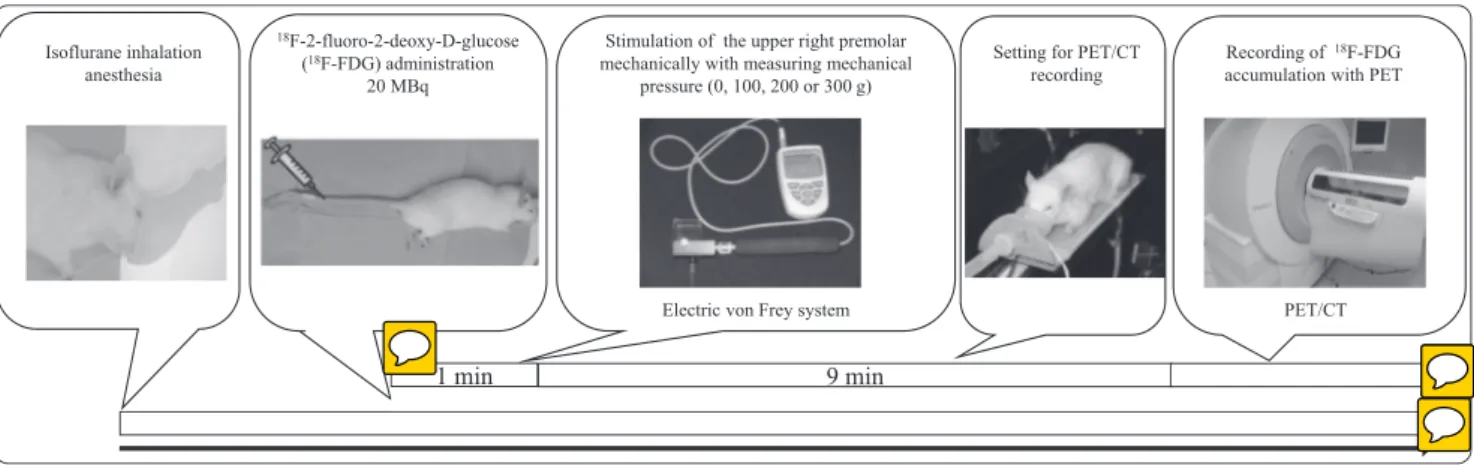
All experimental procedures were carried out in accordance with the NIH guidelines on animal care, and the animal protocol was approved by
Tokushima University (#13125). All
experimental procedures were performed under anesthesia with isoflurane (1.5-2%) (Fig. 2). We
injected 18F-2-fluoro-2-deoxy-D-glucose (FDG)
into the rat (100 - 200 g) tail vein. The FDG was measured the activity and adjusted the activity to 20 MBq / 0.1-0.2 mL on each experimental day. After the FDG injection, the rat right upper premolar was pressed by an electronic von Frey anesthesiometer (Model 1601C, IITC Life Science, Woodland Hills, CA, USA) while measuring the pressing force (100, 200, 300 g) for 1 min. Then brain images of the rat were taken with a Siemens Inveon small-animal PET scanner (Siemens Healthcare, Knoxville, TN, USA). Because we speculated that stretched
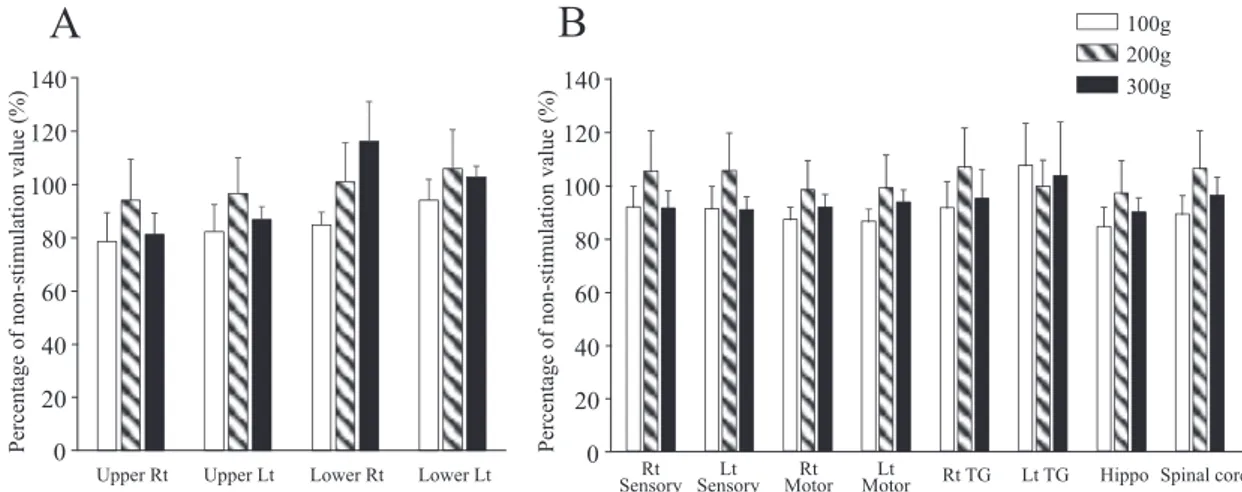
mouth opening induces brain function, we recorded PET with only mouth opening and without tooth stimulation. Stretched mouth opening was achieved by stretching the upper and lower incisors with threads. Since the deepness of systemic anesthesia is related to brain function (Mizuma et al. 2010), we tried to keep the deepness of the isoflurane anesthesia the same in all rats. The deepness of the systemic anesthesia was checked by pinching the rats’ tail. One stimulation strength was applied on one day and the other stimulation strength was applied on the other day, so tooth stimulation occurred on multiple days for each rat. The stimulation strength was selected randomly. During data analysis, the PET brain images were separated into four parts (upper right, upper left, lower right and lower left) (Fig. 3). The peak value of striatal uptake (SUV) in each part was analyzed. We measured SUV in the right and left sensory area (parietal lobe), motor area (frontal lobe), hippocampus, TG and spinal cord. The tooth-stimulated PET data were divided from the mouth-stretching PET data without tooth stimulation.
Statistical analysis was done using R software (The R Foundation for Statistical Computing, Austria). Analysis of variance and Student’s t-test were used in this study.
Results
The PET/CT images showed that FDG was accumulated in the brain (Fig. 4A). The FDG uptake was 20 s and the FDG level increased gradually during the next 5 min (Fig. 4B). Since mouth opening itself induced FDG accumulation, we compared the tooth stimulation data with the mouth opening data (Fig 5). We found that the accumulated FDG was higher in the lower part of the brain than in the upper part (Fig. 5A). Also, 200 or 300 g tooth stimulation showed higher accumulation than 100 g, and 300 g stimulation showed the highest FDG level in the lower right part of the brain. However, 300 g
tooth stimulation was lower than 200 g in the upper part and lower left part (Fig. 5A). The lower part of the brain showed statistically higher FDG accumulation than the upper part of the brain.
We analyzed in detail the data points that showed high FDG accumulation (Fig. 5B). Hippocampus and right TG were included in the lower part of the brain. The data showed that the TG and sensory area had more FDG accumulation compared with the mouth-opening data. Sensory area and right TG showed that 200 g tooth stimulation had higher accumulation than 300 g stimulation, and right TG showed more FDG accumulation than left TG.
Discussion
So far, there are no animal studies that investigate how tooth stimulation information transmits to the central nervous system. In this study, we investigated which parts of the brain showed FDG accumulation after rat tooth stimulation. We found: 1) FDG accumulation in rat brain with PET/CT images; 2) quick FDG uptake during the first 20 s and a gradually increasing FDG level during the next 5 min; 3) that the accumulated FDG was higher in the
Upper right Upper left
Sensory area Sensory area
Motor area Motor area
Lower right Lower left
Hippocampus Hippocampus Trigeminal ganglia Trigeminal ganglia
Fig. 3 PET brain images were separated into four parts for data analysis.
PET/CT photography image
Uptake change in the brain after injection of 18F-FDG 0 0.2 0.4 0.6 0.8 1 0 1 2 3 4 5 S U V ( g/ m l) Time (min)
A
B
lower part of the brain compared with the upper part, 4) 200 and 300 g tooth stimulation showed higher accumulation than 100 g, and 5) the TG and sensory area showed more FDG accumulation compared with mouth opening. There were some limitations to this study: the number of the animals was not sufficient, and is difficult to indicate the brain areas clearly using PET/CT. PET/MRI is better for defining the location.
Injected FDG from the tail vein circulates through the whole body through the vein, and passes the blood-brain barrier (Reivich et al. 1983, Brooks et al. 1986). FDG can detect brain function, and it has been reported that tactile stimulation of the hand and fingers causes asymmetrical increases in local cerebral glucose metabolism, confirming topographic maps of the postcentral gyrus (Reivich et al. 1983).
We found that tooth stimulation induced FDG accumulation, and that the accumulated FDG was higher in the lower parts of the brain, particularly the lower right part, than in the upper part of the brain. This result showed that tooth stimulation affects brain function, and that there was an accumulation of FDG in the lower right of the brain. The detailed analysis showed
that the sensory area and TG showed some FDG accumulation, and the data showed that the sensory function was activated after tooth stimulation. Because left TG showed some accumulation, there may be crosstalk between right and left sensory information.
Because the TG and sensory areas showed more FDG accumulation compared with mouth opening, and 200 or 300 g tooth stimulation showed higher accumulation than 100 g, FDG accumulation is related to tooth stimulation strength. However, at many points, FDG accumulation with 300 g stimulation was lower than 200 g stimulation, which may show that 200 g tooth stimulation is enough to activate the periodontal nerve.
In the future, it would be better to increase the number of samples, to use awake animals (Mizuma et al. 2010), and to consider changing the amount of FDG.
Conclusion
In this study, we found FDG accumulation in the rat brain using PET/CT images, and the accumulated FDG was higher in the lower part of the brain than the upper part. The FDG
0 20 40 60 80 100 120 140
Upper Rt Upper Lt Lower Rt Lower Lt
P er ce nt ag e of n on -s ti m ul at io n va lu e (% ) 0 20 40 60 80 100 120 140 Rt
Sensory SensoryLt MotorRt MotorLt Rt TG Lt TG Hippo Spinal cord
100g 200g 300g
A
B
P er ce nt ag e of n on -s ti m ul at io n va lu e (% )Fig. 5 FDG accumulation in the several parts in the brain with tooth stimulation. Ratio of FDG accumulation data with tooth stimulation to that without tooth stimulation during stretched mouth opening. A: Rat brain FDG data after tooth stimulation divided into four parts. There was statistical
difference between upper left and lower left data with ANOVA (p < 0.022). B: FDG accumulation in several points in the brain. The number of rats was four in each experiment.
accumulation was dependent on tooth stimulation strength, and TG and sensory areas showed more FDG accumulation.
Acknowledgment
Supported by Grant No. 26293412 from the Ministry of Education, Science and Culture in Japan.
Conflicts of Interest
The authors declare no conflict of interest.
References
Brooks, D.J., Beaney, R.P. et al. (1986) Glucose transport across the blood-brain barrier in normal human subjects and patients with cerebral tumours studied using [11C]3-O-methyl-D-glucose and positron emission tomography. J. Cereb. Blood. Flow Metab. 6, 230–239.
Clark, G., Simmons, M. (2003) Occlusal dysesthesia and temporomandibular disorders: is there a link? Alpha Omegan 96, 33–39.
Habre-Hallage, P., Dricot, L. et al. (2014) Cortical activation resulting from the stimulation of periodontal mechanoreceptors measured by functional magnetic resonance imaging (fMRI). Clin. Oral Investig. 18, 1949–1961.
Hara, E.S., Matsuka, Y. et al. (2012) Occlusal dysesthesia: a qualitative systematic review of the epidemiology, aetiology and management. J. Oral Rehabil. 39, 630–638.
Kitamura, Y., Matsuka, Y. et al. (2009) Botulinum toxin type a (150 kDa) decreases exaggerated neurotransmitter release from trigeminal ganglion neurons and relieves neuropathy behaviors induced by infraorbital nerve
constriction. Neuroscience 159, 1422–1429. Kumada, A., Matsuka, Y. et al. (2012) Intradermal
injection of Botulinum toxin type A alleviates infraorbital nerve constriction-induced thermal hyperalgesia in an operant assay. J. Oral Rehabil. 39, 63–72.
Matsuka, Y., Ogawa, T. et al. (2007) Involvement of periodontal mechanoreceptor in occlusal dysesthesia patient. J. Prosth. Res. (Japanese) 51, 364.
Matsuka, Y., Yokoyama, T. et al. (2012) Application of purified botulinum type a neurotoxin to treat experimental trigeminal neuropathy in rats and patients with urinary incontinence and prostatic hyperplasia. J. Toxicol. 2012, Article ID 648384, http://dx.doi.org/10.1155/2012/648384 Miyamoto, J.J., Honda, M. et al. (2006) The
representation of the human oral area in the somatosensory cortex: a functional MRI study. Cereb. Cortex 16, 669–675.
Mizuma, H., Shukuri, M. et al. (2010) Establishment of in vivo brain imaging method in conscious mice. J. Nucl. Med. 51, 1068-1075.
Omoto, K., Maruhama, K. et al. (2015) Cross-Excitation in Peripheral Sensory Ganglia Associated with Pain Transmission. Toxins (Basel) 7, 2906–2917.
Reeves, J.L., Merrill, R.L. (2007) Diagnostic and treatment challenges in occlusal dysesthesia. Calif. Dent. Assoc. 35, 198–207.
Reivich, M., Gur, R. et al. (1983) Positron emission tomographic studies of sensory stimuli, cognitive processes and anxiety. Hum. Neurobiol. 2, 25–33.
Tamaki, K., Ishigaki, S,. et al. (2013) Occlusal discomfort syndrome. Ann. Jap. Prosthod. Soc. 5, 369–386.
Trulsson, M., Francis, S.T. et al. (2010) Brain activations in response to vibrotactile tooth stimulation: a psychophysical and fMRI study. J. Neurophysiol. 104, 2257–2265.