INTRODUCTION
The three major imaging modalities used for the diagnosis of musculoskeletal disorders are plain radiography, computed tomogra-phy (CT), and magnetic resonance imaging (MRI). Ultrasonogra-phy has recently become a notable fourth imaging modality as a result of various technical advances such as the improvement of linear high- frequency probe. Generally, plain radiographs and CT are useful for bone injuries such as fractures, while MRI is useful for bone and soft tissue disorders. Each of these examinations has its merits and demerits and they are often used to complement each other. However, ultrasonography can show both bone and soft tis-sue from various angles as needed, providing greater detail in many cases compared with other imaging modalities.
Athletes often experience overuse injuries. Pain typically occurs in the joints under high load in a particular sport, such as the elbow joint in baseball players and the ankle and foot in soccer players. To diagnose these injuries, ultrasonography is often more useful than plain radiography, CT, or MRI.
In this report, we introduce some characteristic ultrasonographic findings in sports injuries affecting the lower extremity.
OSGOOD-SCHLATTER DISEASE
Osgood - Schlatter disease was first described by Robert Os-good and Carl Schlatter in 1903 (1, 2). The main cause of this in-jury is repetitive traumatic traction at the tibial tuberosity by the quadriceps muscle. The traction force causes destruction of the apophysis. Onset of Osgood - Schlatter disease typically occurs in
adolescents aged 10 to 14 years, and affects mainly boys involved in sports such as soccer, basketball, and volleyball (3). Character-istic symptoms include pain on motion and tenderness and swel-ling of the tibial tuberosity.
Diagnosis is generally made based on clinical symptoms, but imaging methods can show certain characteristic findings. Plain radiography or CT images show an abnormality in the secondary ossification center, ranging from irregularity of the apophysis with separation from the tibial tuberosity to fragmentation. On the other hand, ultrasonography can show greater detail for the area sur-rounding the attachment of the patellar tendon. It can detect every pathological feature in the clinical course including cartilage swel-ling, fragmentation of the tuberosity ossification center, patellar ten-don lesions, and reactive bursitis (3 - 5). Using these ultrasonographic findings, Czyrny classified Osgood - Schlatter disease in three types (2) (Fig. 1) : type 1, delamination of the internal ossification center ; type 2, delamination tear or fracture of the epiphyseal part of the tibial tuberosity ; and type 3, delamination tear of the ossification center resulting in irregular deformation of the tuberosity. The presence of bursitis is also considered a characteristic finding. In many cases of Osgood - Schlatter disease, neovascularization at the insertion of the tibial tuberosity is noted. The amount of the blood flow tends to correlate to the degree of pain (6) (Fig. 2). These types and findings sometimes affect the strategy of treatment of this injury.
PATELLAR TENDINOPATHY (JUMPER’S KNEE)
Jumper’s knee is an insertional tendinopathy of the extensor apparatus of the knee, which may affect athletes participating in various sports at every level. This syndrome is characterized by pain at the insertion of the quadriceps tendon at the upper pole of the patella (20%), the insertion of the patellar tendon at the lower pole of the patella (70%), or the tibial tuberosity (10%) (7). Diag-nosis is typically based on medical history and clinical findings.REVIEW
State-of-the-art ultrasonographic findings in lower extremity
sports injuries
Naoto Suzue(1), Tetsuya Matsuura(1), Toshiyuki Iwame(2), Kosaku Higashino(1), Toshinori Sakai(1), Daisuke Hamada(1),
Tomohiro Goto(1), Yoichiro Takata(1), Toshihiko Nishisho(1), Yuichiro Goda(1), Takahiko Tsutsui(1), Ichiro Tonogai(1),
Ryo Miyagi(1), Mitsunobu Abe(1), Masatoshi Morimoto(1), Kazuaki Mineta(1), Tetsuya Kimura(1), Akihiro Nitta(1),
Tadahiro Higuchi(1), Shingo Hama(1), Subash C. Jha(1), Rui Takahashi(1), Shoji Fukuta(1), and Koichi Sairyo(1)
(1)Department of Orthopedics, the University of Tokushima, Tokushima, Japan,(2)Department of Orthopaedic Surgery, Tokushima Prefectural Central Hospital, Tokushima, Japan
Abstract : Athletes sometimes experience overuse injuries. To diagnose these injuries, ultrasonography is often more useful than plain radiography, computed tomography (CT), or magnetic resonance imaging (MRI). Ultra-sonography can show both bone and soft tissue from various angles as needed, providing great detail in many cases. In conditions such as osteochondrosis or enthesopathies such as Osgood Schlatter disease, Sinding -Larsen -Johansson disease, bipartite patella, osteochondritis dissecans of the knee, painful accessory navicular, and jumper’s knee, ultrasonography can reveal certain types of bony irregularities or neovascularization of the surrounding tissue. In patients of enthesopathy, ultrasonography can show the degenerative changes at the in-sertion of the tendon. Given its usefulness in treatment, ultrasonography is expected to become essential in the management of overuse injuries affecting the lower limb in athletes. J. Med. Invest. 62 : 109-113, August, 2015
Keywords :ultrasonography, sports injury, over use, osteochondrosis, enthesopathy
Received for publication December 10, 2014 ; accepted February 8, 2015. Address correspondence and reprint requests to Naoto Suzue, MD, PhD, Department of Orthopedics, the University of Tokushima 3 - 18 - 15 Kuramoto, Tokushima 770 - 8503, Japan and Fax : +81 - 88 - 633 - 0178.
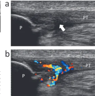
Imaging techniques such as grayscale and color Doppler sonogra-phy are valuable tools for confirming the diagnosis and providing guidance for treatment. Grayscale ultrasonography shows thick-ening and a hypoechoic area at the proximal part of the patellar tendon (5, 8) (Fig. 3a). Alterations in the tendon structure at its insertion on the bone may also be seen, which are correlated to mucoid degeneration (3). Furthermore, Weinberg et al. (9) and Terslev et al. (10) showed that the presence of neovessels can be demonstrated by color Doppler ultrasonography in some patients with jumper’s knee (Fig. 3b).
Treatment of jumper’s knee is mainly conservative in the first and second stages of severity, relying on the administration of non - steroidal anti - inflammatory drugs for 1 - 2 months, dry ice compresses three times a day, and bandages. This condition heals in about 6 - 12 months ; however, if the symptoms do not resolve, surgical therapy might be necessary (3).
Recently, several new conservative therapies, such as platelet-rich plasma (PRP), extracorporeal shock wave therapy (ESWT), sclerosing, and fenestration, have been introduced. In these cases, ultrasonography can be a useful tool. Autologous growth factors are delivered locally in the form of PRP by injection to the tendon repair site, and PRP has gained popularity as a potentially useful regenerative therapy for jumper’s knee. When PRP injection is performed, ultrasonography is helpful for accurate identification of the injection site, which shows pathological changes such as a hypoechoic or hypervascular area (7, 11 - 14). ESWT has also be-come a widely used treatment (7, 15, 16). While the mechanisms underlying its effects on tendinopathies remain unclear, analgesic and stimulating effects of ESWT on tissue regeneration have been suggested. Recent advances in ESWT machines enable identifica-tion of the treatment target using ultrasonographic guidance. With respect to sclerosing treatments, immunohistochemical analyses of tendon biopsies from hypervascular regions have shown nerves located close to blood vessels, which is possibly why ultrasound -guided sclerosing treatments have been effective (17 - 20). In addi-tion, treatment by ultrasound - guided patellar tendon fenestration or arthroscopic shaving has also been reported (17, 18, 21).
SINDING-LARSEN-JOHANSSON DISEASE
Sinding - Larsen - Johansson disease is a juvenile osteochondro-sis and its predilection age tends to slightly younger than that of Osgood - Schlatter disease. The former affects the distal pole of the patella at the proximal insertion of the patellar tendon. Conse-quently, the symptoms of Sinding - Larsen - Johansson disease are similar to those of jumper’s knee. Sinding-Larsen-Johansson dis-ease is clinically characterized by pain localized at the distal pole of the patella, increasing during flexion combined with loading of the knee joint. Other clinical features include swelling of the in-frapatellar soft tissues and functional limitations (3, 22). The disease Figure 1. Osgood - Schlatter disease, longitudinal ultrasonographic
im-ages. (a) : type 1. A clean delamination tear of the ossification center (12 year - old male, soccer). Arrow heads, the delaminated layer of the ossification center. (b) : type 2. A significant anterior displacement of the attachment of the patellar tendon (13 year - old male, soccer). Double arrow, distance of anterior displacement of the fractured cartilage. (c) : type 3. An irregular deformation of the tuberosity (13 year - old male, baseball). Arrow, an ectopic ossification formed within patellar tendon. PT , patellar tendon, E , epiphysis, asterisk, bursitis.
Figure 2. Neovascularization at the insertion of the tuberosity (12 year -old male, soccer). Color Doppler ultrasonography shows neovasculari-zation surrounding the insertion of patellar tendon. PT , patellar tendon, E , epiphysis.
Figure 3. Jumper’s knee, longitudinal ultrasonographic images (25 year -old male, soccer). (a) : Grayscale ultrasonography shows thickening and a hypoechoic area (arrow) at the proximal part of the patellar tendon. (b) : Color Doppler ultrasonography shows the vascular flow. PT , patel-lar tendon, P , patella.
is caused by repetitive microtraumas and excessive prolonged stress occurring on a specific skeletal region that is both mechani-cally and biologimechani-cally weak, when the stress exceeds the intrinsic resistance.
In Sinding - Larsen - Johansson disease, ultrasonography shows cartilage and patellar tendon swelling at its proximal insertion and fragmentation at the patellar distal pole (22, 23) (Fig. 4). Some types of fragmentation may also be seen. Although Sinding Larsen -Johansson disease is a relatively common injury in adolescent athletes, few reports have examined it in detail.
BIPARTITE PATELLA
Bipartite patella is also a juvenile osteochondrosis, with a predi-lection age even younger than that of Sinding - Larsen - Johansson disease. In 1975, Green described the cases of three boys with painful bipartite patella in whom pain was relieved by excision of the accessory ossification centers (24). Since this report was pub-lished, bipartite patella has been observed as a cause of anterior knee pain in adolescents and young athletes. Bipartite patella is classified into three groups. In the first group, the patella is divided by a transverse split line into an upper large bone fragment and a lower small bone fragment. In the second group, the patella is divided by a longitudinal split line into an outer one - fourth bone fragment and an inner three - fourths bone segment. In the third group, the patella is divided into a relatively large inner lower bone fragment and small upper outer bone fragment (25). The most common symptom is pain at the separated fragments during or after strenuous activity. Localized tenderness over the separated fragments is the most common physical finding.
Ultrasonographic examination of the bipartite patella shows an irregularity in the bony contours, with occurrence of a cleft between the main part of the patellar body and the large superolateral ac-cessory ossicle. The signal detected in the interface between bones is less echogenic than bone, reflecting its fibrocartilaginous nature (26). Furthermore, neovascularization can be noted on color Dop-pler ultrasonography surrounding the ossicle (Fig. 5), at the inser-tion of the vastus lateralis muscle or the area between the fragment and the patella. While bipartite patella is also a common injury, few reports have described it in detail.
OSTEOCHONDRITIS DISSECANS OF THE KNEE
Osteochondritis dissecans (OCD) has been defined as a local-ized process wherein a focus of subchondral bone and adjacent ar-ticular cartilage separates from the surrounding bone (27 - 31). Given that the knee is the most common location for OCD developmentand that this lesion is typically found in individuals who participate in sports, the hypothesis of repetitive microtrauma as the causative pathology has gained favor. However, the cause of OCD remains controversial, as does the most appropriate treatment for the vary-ing degrees of OCD lesions (29). The yearly incidence of OCD is reportedly between 0.02% and 0.03% (27), with a male to female ratio of 2 : 1 (32). This is one of the severest injuries affecting ath-letes because if fragmentation has progressed to the terminal stage, structural regeneration is not possible.
OCD is detected using radiography, CT, and MRI. MRI is con-sidered the most useful modality because the immature condyle of the femur can sometimes shows ossification variants (33). Ultra-sonography can show the fragmentation and discontinuity of the subchondral bone of the femoral condyle (34) (Fig. 6) ; however, it is difficult to distinguish between various degrees of OCD and its ossification variants using ultrasonography. Therefore, further study and research are needed to clarify these differences.
On the other hand, ultrasonography can be used in the treatment of OCD, as reported by Berná- Serna et al., who showed good re-sults using ultrasonography - guided percutaneous drilling (35).
SYMPTOMATIC ACCESSORY NAVICULAR
Generally, the accessory navicular is one of the most common accessory ossicles of the foot. The prevalence of the injury is thought to be 4 - 21% in the asymptomatic population. This condi-tion has a higher incidence in women, and 50 - 90% of cases have bilateral ossicles (36). This entity is usually asymptomatic, and Figure 4. SindingLarsen Johansson disease, longitudinal ultrasono
-graphic image (10 year - old male, soccer). Fragmentation at the patellar distal pole (arrows) . PT , patellar tendon, P , patella.
Figure 5. Bipartite patella, longitudinal ultrasonographic image (10 year - old male, basketball). Color Doppler ultrasonography shows neovas-cularization surrounding the ossicle. VL, vastus lateralis muscle, P , pa-tella, asterisk, accessory ossicle.
Figure 6. Osteochondritis dissecans of the lateral femoral condyle, longitudinal ultrasonographic image (12 year - old male, soccer). (a) : Normal femoral condyle. (b) : ultrasonography shows the fragmentation of the subchondral bone (arrow) . FC , femoral condyle, C , cartilage.
pain may occur following a sprain in many cases. The accessory navicular is classified into three types (37). Type 1 is defined as a small accessory bone within the distal posterior tibialis tendon with no cartilage connection to the navicular tuberosity, appearing to be a sesamoid bone. Type 2 refers to a triangular secondary ossi-fication center of the navicular tuberosity, which is connected to the navicular by fibrocartilage. Many cases of symptomatic accessory navicular belong to type 2. Type 3 is considered to result from os-seous fusion of the ossification center, resulting in a prominent navicular tuberosity.
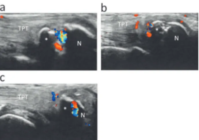
Occasionally, the posterior tibialis tendinitis is the cause of me-dial foot pain (38). Ultrasonography is a highly effective method for the evaluation of tendinous abnormalities, since evidence of a diffusely edematous tendon with surrounding fluid or frank rupture can easily be detected with high- resolution equipment (39). Thus, ultrasonography can show not only the shape of the accessory navicular and navicular tuberosity but also the presence of swelling of the tibialis posterior tendon or inflammation of the surrounding tissues (38). Several types of inflammation are observed as abnor-mal blood flow in ultrasonography. For example, abnorabnor-mal blood flow may be noted between the accessory navicular and the navicu-lar tuberosity as well as at the enthesis of the tibialis posterior ten-don. Ultrasonography allows the discrimination of various condi-tions in the symptomatic accessory navicular (Fig. 7).
CONCLUSION
Ultrasonography can reveal characteristic findings in overuse sports injuries that occur not only in adolescents but also in adults. To date, radiography, CT, and MRI have been the primary imag-ing modalities for these injuries ; however, recent advances have allowed ultrasonography to be considered in their ranks. Despite the rapid advances in ultrasonography, some injuries need further study. Certainly, ultrasonography has some limitation. For exam-ple, it depends on the operator’s skill whether clear images can be described. Moreover ultrasonography cannot show the inside of bony cortex or the parts surrounded with bones. However in consideration of these limitations, given the usefulness of ultra-sonography, especially in treatment, it is expected to become an essential for overuse injuries affecting the lower limb in athletes.
CONFLICT OF INTEREST
We have no conflict of interests to disclose.
REFERENCES
1. Osgood RB : Lesions of the tibial tubercle occurring during adolescence. Clin Orthop Relat Res 1993 : 6, 1903
2. Czyrny Z : Osgood - Schlatter disease in ultrasound diagnostics-a pictoridiagnostics-al essdiagnostics-ay. Med Ultrdiagnostics-ason 12(4) : 323 - 335, 2010 3. Draghi F, Danesino GM, Coscia D, Precerutti M, Pagani C :
Overload syndromes of the knee in adolescents : Sonographic findings. J Ultrasound 11(4) : 151 - 157, 2008
4. Chiang Y- P, Wang T- G, Hsieh S - F : Application of Ultrasound in Sports Injury. J Med Ultrasound 21(1) : 1 - 8, 2013 5. Blankenbaker DG, De Smet AA : The role of ultrasound in the
evaluation of sports injuries of the lower extremities. Clin Sports Med 25(4) : 867 - 897, 2006
6. Sailly M, Whiteley R, Johnson A : Doppler ultrasound and tibial tuberosity maturation status predicts pain in adolescent male athletes with Osgood - Schlatter’s disease : a case series with comparison group and clinical interpretation. Br J Sports Med 47(2) : 93 - 97, 2013
7. Vetrano M, Castorina A, Vulpiani MC, Baldini R, Pavan A, Ferretti A : Platelet- rich plasma versus focused shock waves in the treatment of jumper’s knee in athletes. Am J Sports Med 41(4) : 795 - 803, 2013
8. Gisslèn K, Gyulai C, Söderman K, Alfredson H : High preva-lence of jumper’s knee and sonographic changes in Swedish elite junior volleyball players compared to matched controls. Br J Sports Med 39(5) : 298 - 301, 2005
9. Weinberg EP, Adams MJ, Hollenberg GM : Color Doppler sonography of patellar tendinosis. Am J Roentgenol 171(3) : 743 - 744, 1998
10. Terslev L, Qvistgaard E, Torp - Pedersen S, Laetgaard J, Danneskiold - Samsoe B, Bliddal H : Ultrasound and Power Doppler findings in jumper’s knee -preliminary observations. Eur J Ultrasound 13(3) : 183 - 189, 2001
11. Tok F, Öz!cakar L, De Muynck M, Kara M, Vanderstraeten G :
Musculoskeletal ultrasound for sports injuries. Eur J Phys Rehabil Med 48(4) : 651 - 663, 2012
12. Charousset C, Zaoui A, Bellaiche L, Bouyer B : Are multiple platelet- rich plasma injections useful for treatment of chronic patellar tendinopathy in athletes? a prospective study. Am J Sports Med 42(4) : 906 - 11, 2014
13. Ferrero G, Fabbro E, Orlandi D, Martini C, Lacelli F, Serafini G : Ultrasound - guided injection of platelet- rich plasma in chronic Achilles and patellar tendinopathy. J Ultrasound 15 (4) : 260 - 266, 2012
14. Smith J, Sellon JL : Comparing PRP injections with ESWT for athletes with chronic patellar tendinopathy. Clin J Sport Med 24(1) : 88 - 89, 2014
15. van Leeuwen MT, Zwerver J, van den Akker - Scheek I : Ex-tracorporeal shockwave therapy for patellar tendinopathy : a review of the literature. Br J Sports Med 43(3) : 163 - 168, 2009 16. Vulpiani MC, Vetrano M, Savoia V, Di Pangrazio E, Trischitta D, Ferretti A : Jumper’s knee treatment with extracorporeal shock wave therapy : a long - term follow - up observational study. J Sports Med Phys Fitness 47(3) : 323 - 328, 2007 17. Willberg L, Sunding K, Forssblad M, Fahlström M, Alfredson
H : Sclerosing polidocanol injections or arthroscopic shaving to treat patellar tendinopathy/jumper’s knee? A randomised controlled study. Br J Sports Med 45(5) : 411 - 415, 2011 18. Sunding K, Willberg L, Werner S, Alfredson H, Forssblad M,
Fahlström M : Sclerosing injections and ultrasound - guided Figure 7. Symptomatic accessory navicular, longitudinal ultrasono
-graphic images. (a) : Neovascularization between the accessory navicu-lar (asterisk) and the navicunavicu-lar tuberosity (N) (12 year - old male, soccer). (b) : Neovascularization at the enthesis of the tibialis posterior tendon (TPT) (11 year - old male, soccer). (c) : Neovascularization at the both site of the accessory navicular (11 year - old female, soccer).
arthroscopic shaving for patellar tendinopathy : good clinical re-sults and decreased tendon thickness after surgery -a medium-term follow - up study. Knee Surg Sports Traumatol Arthrosc 6, 2014
19. Hoksrud A, Bahr R : Ultrasound - guided sclerosing treatment in patients with patellar tendinopathy (jumper’s knee). 44 -month follow - up. Am J Sports Med 39(11) : 2377 - 2380, 2011 20. Hoksrud A, Torgalsen T, Harstad H, Haugen S, Andersen TE, Risberg MA : Ultrasound - guided sclerosis of neovessels in pa-tellar tendinopathy : a prospective study of 101 patients. Am J Sports Med 40(3) : 542 - 547, 2012
21. Kanaan Y, Jacobson JA, Jamadar D, Housner J, Caoili EM : Sonographically guided patellar tendon fenestration : prog-nostic value of preprocedure sonographic findings. J Ultra-sound Med 32(5) : 771 - 777, 2013
22. De Flaviis L, Nessi R, Scaglione P, Balconi G, Albisetti W, Derchi LE : Ultrasonic diagnosis of Osgood - Schlatter and Sinding - Larsen - Johansson diseases of the knee. Skeletal Radiol 18(3) : 193 - 197, 1989
23. Valentino M, Quiligotti C, Ruggirello M : Sinding Larsen -Johansson syndrome : A case report. J Ultrasound 15(2) : 127 - 129, 2012
24. Green WT : Painful bipartite patellae. A report of three cases. Clin Orthop Relat Res 1975 (110) : 197 - 200, 1975
25. Oohashi Y : Developmental anomaly of ossification type pa-tella partita. Knee Surg Sports Traumatol Arthrosc 15, 2014 26. Blankstein A, Cohen I, Salai M, Diamant L, Chechick A, Ganel
A : Ultrasonography : an imaging modality enabling the diag-nosis of bipartite patella. Knee Surg Sports Traumatol Arthrosc 9(4) : 221 - 224, 2001
27. Crawford DC, Safran MR : Osteochondritis dissecans of the knee. J Am Acad Orthop Surg 14(2) : 90 - 100, 2006
28. Moretti B, Notarnicola A, Moretti L, Giordano P, Patella V : A volleyball player with bilateral knee osteochondritis dissecans treated with extracorporeal shock wave therapy. Chir Organi Mov 93(1) : 37 - 41, 2009
29. Grimm NL, Tisano B, Carey JL : Three osteochondritis dis-secans lesions in one knee : a case report. Clin Orthop Relat Res 471(4) : 1186 - 1190, 2013
30. Swain JH, Grimm NL, Shea KG : Juvenile osteochondritis dis-secans of the knee. J Orthop Sports Phys Ther 40(8) : 534, 2010
31. Eismann EA, Pettit RJ, Wall EJ, Myer GD : Management strate-gies for osteochondritis dissecans of the knee in the skeletally immature athlete. J Orthop Sports Phys Ther 44(9) : 665 - 679, 2014
32. Lindén B : The incidence of osteochondritis dissecans in the condyles of the femur. Acta Orthop Scand 47(6) : 664 - 667, 1976 33. Jans L, Jaremko J, Ditchfield M, De Coninck T, Huysse W, Moon A : Ossification variants of the femoral condyles are not associated with osteochondritis dissecans. Eur J Radiol 81 (11) : 3384 - 3389, 2012
34. Gregersen HE, Rasmussen OS : Ultrasonography of osteo-chondritis dissecans of the knee. A preliminary report. Acta Radiol 30(5) : 552 - 554, 1989
35. Berná- Serna JD, Martinez F, Reus M, Berná- Mestre JD : Os-teochondritis dissecans of the knee : sonographically guided percutaneous drilling. J Ultrasound Med 27(2) : 255 - 259, 2008 36. Mosel LD, Kat E, Voyvodic F : Imaging of the symptomatic type II accessory navicular bone. Australas Radiol 48(2) : 267 -271, 2004
37. Veitch JM : Evaluation of the Kidner procedure in treatment of symptomatic accessory tarsal scaphoid. Clin Orthop Relat Res Mar(131) : 210 - 213, 1978
38. Chuang Y- W, Tsai W- S, Chen K - H, Hsu H- C : Clinical use of high- resolution ultrasonography for the diagnosis of type II accessory navicular bone. Am J Phys Med Rehabil 91(2) : 177 -181, 2012
39. Chen YJ, Hsu RW, Liang SC : Degeneration of the accessory navicular synchondrosis presenting as rupture of the poste-rior tibial tendon. J Bone Joint Surg Am 79(12) : 1791 - 1798, 1997