Contents lists available atScienceDirect
Biochemistry and Biophysics Reports
journal homepage:www.elsevier.com/locate/bbrepNMR spectra of PB2 627, the RNA-binding domain in in
fluenza A virus RNA
polymerase that contains the pathogenicity factor lysine 627, and
improvement of the spectra by small osmolytes
Yusuke S. Kato
a,b,c,⁎, Masaru Tanokura
b, Takashi Kuzuhara
d,⁎⁎aInstitute for Health Sciences, Tokushima Bunri University, Tokushima 770-8514, Japan
bDepartment of Applied Biological Chemistry, Graduate School of Agricultural and Life Sciences, University of Tokyo, Tokyo 113-8657, Japan cInstitute for Enzyme Research, Tokushima University, Tokushima 770-8503, Japan
dFaculty of Pharmaceutical Sciences, Tokushima Bunri University, Tokushima 770-8514, Japan
A R T I C L E I N F O
Keywords: Influenza A virus RNA polymerase PB2 627 NMR, additiveA B S T R A C T
The influenza A virus, which has an RNA genome, requires RNA-dependent RNA polymerase for transcription and replication. The polymerase is comprised of the subunits PA, PB1, and PB2. The C-terminal RNA-binding domain in PB2 contains lysine 627 (PB2 627), which is associated with pathogenicity and host range. However, the structure and molecular mechanism of PB2 627 in solution remain obscure. Here, we investigated PB2 627 in solution by nuclear magnetic resonance (NMR) and detected inhomogeneity in the intensities of backbone amide proton signals due to localfluctuations in structure. To characterize the effects of chemical chaperones on spectral data and improve the data quality, we tested 20 different additives, includingL-arginine L-glutamate salt, (L-arginine)2SO4, glycerol, β-octylglucoside, 3-[(3-cholamidopropyl) dimethylammonio]-1-propanesulfo-nate, Na2SO4, 1,5-diaminopentane, 1,4-diaminobutane, trehalose, sucrose, glycine, trimethylamine N-oxide, β-alanine, L-α-alanine, hydroxyectoine, betaine,L-proline, and non-detergent sulfobetaine 195, 201, and 256. We evaluated the quality of the resulting spectra by calculating the standard deviation and average of the ratio of signal intensities to noise level of amide peaks, as well as the ratio of the standard deviation to the average. NMR-profile analysis revealed diverse effects of additives on the dynamic properties of PB2 627. Based on such criteria, we found that small osmolytes such as glycine and L-α-alanine reduced structural fluctuations and improved the quality of spectral data, which is likely to facilitate a detailed NMR-based structural analysis. The methodology developed here may also be more generally useful for evaluating the effects of chemical chaper-ones on the structural integrity of proteins.
1. Introduction
In 1918, a pandemic of influenza A virus resulted in ten million deaths worldwide [1]. Strategies to prevent future pandemics must therefore be formulated[2,3]. Although inhibitors of the viral neur-aminidase and M2 ion channel are widely used for treatment [4,5], some adverse side effects have been reported, and drug resistance has emerged [6,7]. The viral RNA-dependent RNA polymerase is a very promising alternative drug target because it is required for transcription and replication of viral genes. To initiate viral gene transcription, a 5′ RNA fragment of 10–15 nucleotides is used as a primer[8], which is
recognized by the C-terminal domain in the PB2 subunit of the RNA polymerase along with promoter RNA[9]. This domain also contains the lysine 627 residue, which is associated with high pathogenicity and restricted host range. This domain is thus called PB2 627[10].
Protein tertiary structures are a valuable resource in drug devel-opment, and crystal structures of PB2 627 have been determined [11–13]. Nevertheless, it is equally important to investigate the struc-ture and dynamics of PB2 627 in solution in order to understand its mechanism of action, and to design novel drugs against it. In turn, such an investigation would require optimal conditions for solution nuclear magnetic resonance (NMR). In this study, we collected 2D NMR spectra
http://dx.doi.org/10.1016/j.bbrep.2017.09.003
Received 30 November 2016; Received in revised form 31 March 2017; Accepted 15 September 2017
⁎Corresponding author at: Institute for Health Sciences, Tokushima Bunri University, Tokushima 770-8514, Japan. ⁎⁎Corresponding author at: Faculty of Pharmaceutical Sciences, Tokushima Bunri University, Tokushima 770-8514, Japan.
E-mail addresses:ysk.kt@tokushima-u.ac.jp(Y.S. Kato),kuzuhara@ph.bunri-u.ac.jp(T. Kuzuhara).
Abbreviations: CHAPS, 3-[(3-cholamidopropyl) dimethylammonio]-1-propanesulfonate; DTT, dithiothreitol; Irel, ratio of signal intensity to noise level; HSQC, heteronuclear single quantum coherence; NDSB, non-detergent sulfobetaine; NMR, nuclear magnetic resonance; PB2 627, C-terminal RNA-binding domain of PB2 containing lysine 627; S/N, signal-to-noise ratio; TMAO, trimethylamine N-oxide
Available online 20 September 2017
2405-5808/ © 2017 The Authors. Published by Elsevier B.V. This is an open access article under the CC BY license (http://creativecommons.org/licenses/BY/4.0/).
MARK
from PB2 627 and found that the quality of the initial spectra is unlikely to be sufficient for higher-dimensional NMR, which is necessary to analyze structure and dynamics, but has a lower signal-to-noise ratio (S/N) than 2D NMR.
Previous studies demonstrated that diverse additives, such as salts, sugars, amino acids, methylamines, chaotropic agents, and detergents, improved NMR spectra of proteins[14,15].L-arginineL-glutamate salt
and non-detergent sulfobetaines (NDSBs) are frequently used to this end [16,17]. Diverse amino acids have been reported to improve protein stability [18–20]. Diamines were reported to prevent thermal ag-gregation and inactivation of lysozyme [21]. Amino acids, methyla-mines, and chaotropes are categorized as osmolytes. Osmolytes are osmotically active solutes in organisms that have evolved to resist water stress such as high or low salt concentrations, desiccation, or freezing [22]. Most osmolytes are small organic compounds. However, to our knowledge, no reports have described the comprehensive testing of numerous additives and their effects on protein NMR spectra. Thus, we experimented with 20 different compounds to improve the spectra of PB2 627,finding that osmolyte additives such as neutral amino acids and derivatives can improve the quality of spectral data. We anticipate that our results and methodologies will prove generally useful for im-proving protein NMR spectra and for evaluating the ability of various compounds to reduce structuralfluctuations of proteins.
2. Materials and methods 2.1. Sample preparation
Construction of a plasmid for expressing His6-tagged PB2 627 (amino acids 535–759) was described previously[11,12].15N-labeled PB2 627 was produced in Rosetta 2 (DE3) E. coli (EMD Millipore, Billerica, MA), following a protocol based on M9 media containing trace elements[23], with some modification. Briefly, cells were lysed by sonication in 20 mM sodium phosphate (pH 7.6), 250 mM NaCl, 7.5% glycerol, 20 mM imidazole pH 8.0, 1 mM dithiothreitol (DTT), protease inhibitor cocktail (Sigma-Aldrich, St. Louis, MO), and lyso-zyme (Seikagaku Corp., Tokyo, Japan). Lysates were clarified by cen-trifugation at 50,000 × g, and the supernatant was loaded on Ni-NTA agarose (Qiagen, Hilden, Germany) equilibrated with 20 mM sodium phosphate (pH 7.3), 250 mM NaCl, 5% glycerol, and 20 mM imidazole (pH 8.0). After extensive washing, PB2 627 was eluted with 20 mM sodium phosphate (pH 7.0), 250 mM NaCl, 1 mM DTT, 10% glycerol, and 200 mM imidazole (pH 8.0). After addition of 0.1% Tween 20 and thrombin (GE Healthcare Bio-Sciences Corp., Piscataway, NJ), the protein was dialyzed against 20 mM Tris-HCl (pH 6.5), 50 mM NaCl, 10% glycerol, and 0.5 mM DTT, and then purified by cation-exchange on HiPrep CM (GE Healthcare). Subsequently, the protein was con-centrated using a Vivaspin centrifugal concentrator (Sartorius, Goet-tingen, Germany) and loaded on Superdex 75 16/600 (GE Healthcare) equilibrated with 20 mM sodium phosphate (pH 6.5), 150 mM NaCl, 4% glycerol, and 1 mM DTT. Finally, purified samples were con-centrated and dialyzed against 20 mM sodium phosphate (pH 6.5), 7% D2O, 50 mM NaCl, and 1 mM DTT.
2.2. NMR 1H–15
N heteronuclear single quantum coherence (HSQC) experi-ments were performed at 20 °C using an Inova 600 spectrometer equipped with a Cold Probe (Agilent Technologies, Santa Clara, CA) and an Avance-III 950 system (Bruker, Billerica, MA). Thefinal con-centration of PB2 627 was 3.0 mg/mL, although PB2 627 is stable and can be crystallized at concentrations of up to 10 mg/mL[11]. Spectra were collected with or without 0.2 M (L-arginine)2SO4, Na2SO4or L -arginineL-glutamate salt; 4% glycerol; 25 mMβ-octylglucoside; 10 mM
CHAPS; 0.1 M 1,5 diaminopentane or 1,4 diaminobutane; 20% treha-lose; or 0.5 M sucrose, glycine, trimethylamine N-oxide (TMAO),
β-alanine, L-α-β-alanine, hydroxyectoine, betaine,L-proline, or NDSB 195,
201, or 256 (Supplementary Table 1). NMR tubes were siliconized prior to use to prevent protein aggregation. Data processing and analysis were performed with the NMRpipe and Sparky 3.115 programs[24,25]. Relative intensity (Irel) denotes the ratio of signal intensity to noise level according to Pedrini et al.[26].
3. Results 3.1. Flash 2D NMR
PB2 627 contains a nuclear localization signal, a C-terminal RNA-binding domain, and lysine 627 that determines the pathogenicity and host range. We purified PB2 627 to homogeneity using immobilized Ni-affinity, cation-exchange, and gel-filtration chromatography (Supplementary Fig. 1).
We carried out a1H–15N heteronuclear single quantum coherence experiment by NMR. Fourier transformation and processing were then performed to obtain a spectrum. The spectrum contained numerous sharp, well-dispersed peaks from backbone amides (Supplementary Fig. 2A), suggesting that PB2 627 is monomeric, and that the global fold is stable in aqueous solution. However, the number of peaks diminished when the spectrum was contoured at a higher threshold (Supplementary Fig. 2B), indicating that the peaks had different in-tensities. Weak amide signals arise from line broadening due to local conformational changes over a time scale ranging from microseconds to milliseconds. Structural polymorphisms and transient oligomerization/ aggregation of proteins can also cause weak signals. In contrast, strong amide signals arise from unstructured regions. Thus, inhomogeneity in signal intensities is due to inhomogeneity in structure and structural dynamics of proteins. Indeed, the PB2 627 construct contains a C-terminal segment that was not observed in the crystal structure. This presumably unstructured region should provide strong signals at 8.0–8.5 ppm of the1
H axis in the HSQC spectrum. It is therefore likely that structural analysis using this sample will be challenging. In addi-tion, we anticipate that many amides with low-intensity peaks will become undetectable in higher-dimensional NMR, which has a lower S/ N than does HSQC.
3.2. Effects of additives
To reduce inhomogeneity in the intensity of amide peaks, we in-vestigated 20 different additives that may homogenize the structure and dynamic properties of PB2 627 and improve the quality of NMR data. We tested the diamines 1,4-diaminobutane, and 1,5-diamino-pentane; the detergentsβ-octylglucoside and CHAPS; the sugars and polyols glycerol, trehalose, and sucrose; the salt Na2SO4; NDSBs 195, 201, and 256; and the amino acids and amino-acid derivatives hydro-xyectoine, (L-arginine)2SO4,L-arginineL-glutamate salt, TMAO, betaine, L-proline, L-α-alanine, β-alanine and glycine. Prior to investigating the
effects of these additives on amide peak homogeneity, we studied dif-ferent salt concentrations and pH values, which were not effective.
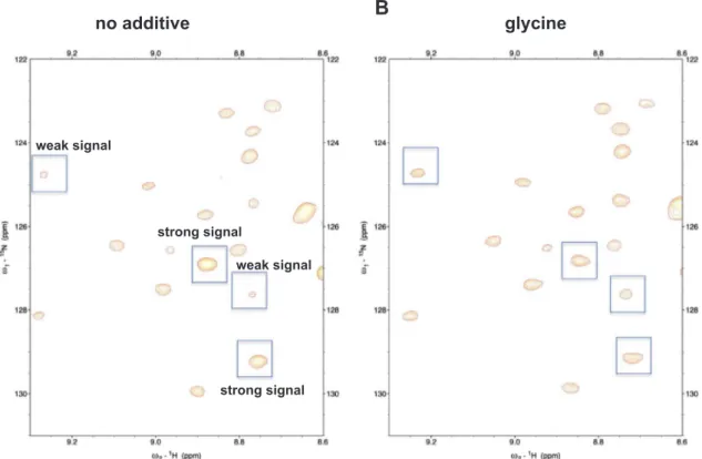
HSQC spectra of sufficient quality for further analysis were obtained in the presence of 17 different additives (Supplementary Fig. 3). Spectra collected in the presence ofβ-octylglucoside, sucrose, and NDSB 256 were not suitable for further analysis, presumably because PB2 627 was denatured or aggregated under these conditions. Spectra obtained in the presence ofL-arginineL-glutamate salt and glycine represented
ex-amples with contrastive spectral changes. L-arginine L-glutamate salt
caused a shift in position for many amide peaks (as shown in the boxes inFig. 1A) and further reduced the signal intensity of amide peaks that were already weak. As a result, many peaks in the peripheral region of the spectrum diminished or disappeared (Fig. 1A), indicating thatL -arginineL-glutamate salt increased the structural inhomogeneity of PB2
627. In contrast, glycine did not move or quench most peripheral peaks (Fig. 1B).
3.3. Quantitative analysis
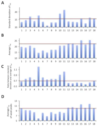
To quantitatively analyze the inhomogeneity of peak intensities, we picked 210 backbone signals that were recognized as peaks by Sparky 3. While the expected number of backbone amide signals was 215, it is quite difficult to pick a complete set of backbone amide signals due to extensive signal overlaps and the existence of numerous weak peaks that could not be clearly distinguished from noise. Thus, we anticipated that PB2 627 contains structural polymorphisms. We calculated the ratio of the signal intensities of individual resonances to the average noise, Irel, in order to measure the S/N ratios of the individual re-sonances, as well as their standard deviations to measure in-homogeneities in the signal intensities. We found that 11 additives decreased the standard deviation of Irelfor the 210 peaks compared with the no-additive condition (Fig. 2A). Furthermore, we found that the average Ireldecreased in many cases (Fig. 2B), suggesting that the decrease in standard deviation may be due to a lower average Irelvalue. We therefore calculated the ratio of the standard deviation to the average and found that the ratio increased in the presence of 10 ad-ditives, compared with the no-additive condition (Fig. 2C), suggesting that these compounds increased the inhomogeneity of Irel. The ratio increased particularly in the presence of CHAPS, trehalose, Na2SO4, (L -arginine)2SO4, and L-arginine L-glutamate salt. In contrast, neutral
amino acids and derivatives such as TMAO, betaine, L-proline, L
-α-alanine,β-alanine, and glycine reduced the ratio, suggesting that these compounds increased the homogeneity of the signal intensities. The average Irel values of the 50 weakest peaks were greater under the neutral-amino acid and derivative conditions than under the
no-additive condition (Fig. 2D). The values were markedly high under the
L-α-alanine and glycine conditions. These results suggested that the
increase in the Irelof weak signals contributed to the decreased ratio of the standard deviation to the average. The improvement observed in the quantitative analysis was confirmed qualitatively in the spectra (Fig. 3).
Next, we asked whether increasing concentration of L-α-alanine and glycine further improved the quality of the spectra (Supplementary Table 2). Average Irelfor the 210 amide peaks was greater under the condition with 0.5 M L-α-alanine than under the conditions with 0 and 1.0 M L-α-alanine. Similarly, average Irelfor the 210 peaks was the greatest under the condition with 0.5 M glycine compared with the conditions with the other glycine concentrations. In contrast, the more the concentration of the additive amino acid was, the smaller the ratio between the standard deviation and average of Irelof the amide peaks was.
3.4. NMR profiles
NMR profiling is useful for assessing the distribution of Irelvalues of proteins[26]. We profiled the Irelvalues of backbone amide protons from the1H–15N HSQC spectra (Fig. 4). Most plots in the NMR-profiles of 1,4-diaminobutane, 1,5-diaminopentane, glycerol, Na2SO4, NDSB 195, NDSB 201, and hydroxyectoine (red data points) were lower than those in the NMR-profile under the no-additive condition (blue data points). Between peak numbers ~ 15 to 210, the plots for CHAPS, trehalose, (L-arginine)2SO4, and L-arginine L-glutamate salt showed lower Irelvalues than observed with the no-additive condition, whereas the Irelvalues tended to be higher than observed with the no-additive condition between peak numbers 1 to ~ 15. With L-α-alanine and 10.0 10.0 9.5 9.5 9.0 9.0 8.5 8.5 8.0 8.0 7.5 7.5 7.0 7.0 6.5 6.5 130 130 125 125 120 120 115 115 110 110 orange: no additve green: arginine glutamate
orange: no additve blue: glycine 10.0 10.0 9.5 9.5 9.0 9.0 8.5 8.5 8.0 8.0 7.5 7.5 7.0 7.0 6.5 6.5 130 130 125 125 120 120 115 115 110 110
A
B
Fig. 1.1H-15N HSQC spectra with or without additives Overlay of PB2 627 spectra
without additives and (A) withL-arginineL-glutamate salt or (B) with glycine. The boxed peaks were markedly moved.
Fig. 2. Analysis of the homogeneity of Irelof backbone amide signals The vertical
axes indicate the standard deviation (A), average (B) and the ratio of the standard de-viation to the average (C) of Irel, respectively. The vertical axis of (D) indicates the
average Irelof the weakest 50 peaks. Lane 1: 1,4-diaminobutane; lane 2:
1,5-diamino-pentane; lane 3: CHAPS; lane 4: glycerol; lane 5: trehalose; lane 6: Na2SO4; lane 7:
NDSB195; lane 8: NDSB201; lane 9: hydroxyectoine; lane 10: (L-arginine)2SO4; lane 11:L -arginineL-glutamate salt; lane 12: TMAO; lane 13: betaine; lane 14:L-proline; lane 15:L -α-alanine; lane 16: β-alanine; lane 17: glycine; lane 18: no additive. Red bars indicate the values obtained in the absence of additives.
glycine, most plots for peak numbers between ~ 15 and 210 showed higher Irelvalues than observed with the no-additive condition, whereas the plots under these additive conditions and no-additive condition were superimposable for peak numbers 1 to ~ 15. The plots for β-alanine showed lower Irelvalues compared to the no-additive control between peak numbers 1 to ~ 15 and were superimposable between peak numbers ~ 15 to 210. Similar tendencies are observed in the cases of TMAO, betaine, andL-proline.
4. Discussion
We expressed and purified15N-labeled PB2 627, which has a nu-clear localization signal and a C-terminal RNA-binding domain. The 1H–15N HSQC data indicated that the protein was folded, and was not aggregated in solution. This observation is consistent with the previous success in crystallizing PB2 627[11–13], a process that would have required a stably folded protein. However, peaks due to backbone amides varied in intensity, suggesting that transient structural changes, structural polymorphisms, local denaturation, and/or transient, non-specific interactions occurred. Therefore, we collected spectral data in the presence of various additives that may change the structural properties of PB2 627 and improve the spectra. Consequently, we found that TMAO, betaine,L-proline, L-α-alanine, β-alanine, and glycine
im-proved the spectral quality by increasing the homogeneity of signal intensities, whereas the other compounds did not. In addition, L
-α-alanine and glycine markedly improved the average ratio of intensity of individual signals to noise levels (i.e., the Irelvalues).
A natural question is why these compounds improved the quality of the spectral data. These compounds are osmolytes, which are naturally occurring chemical chaperones [15,20,22]that enhance hydration of partially denatured proteins to prevent complete denaturation and oligomerization/aggregation [27,28]. Thus, the ability of these ad-ditives to homogenize the structural state of PB2 627 and improve the spectral quality was not surprising. The NMR profiles showed thatL-
α-alanine and glycine improved Irelvalues, except for those with small
peak numbers (i.e. those with high Irelvalues), which suggests the oc-currence of decreased structural polymorphisms and/or line broadening due to local motion over a time scale ranging from microseconds to milliseconds. In contrast, Irelvalues with small peak numbers did not change compared with those observed under the no-additive condition, suggesting that the mobility of the unstructured regions of PB2 627 was unchanged. Although most amide peaks did not move upon the addi-tion of glycine, a few peaks moved upfield, as shown inFig. 1B, which may suggest that hydrogen bonds were weakened. This, in turn, may suggest that transient interactions between PB2 627 monomers or be-tween PB2 627 and solutes decreased upon the addition of glycine, with increased hydration. Indeed, the quality of the spectrum of PB2 627 decreased when we increased the concentration of PB2 627, which suggested non-specific interaction between monomers of PB2 627 (Supplementary Table. 3). L-α-alanine and glycine have low carbon contents, each with no more than three carbon atoms, and have mul-tiple hydrophilic groups. In the cases of TMAO, betaine,L-proline, and β-alanine, the Irelvalues with small peak numbers decreased compared with those observed in the absence of additive, which suggests that these compounds decreased the mobility of the unstructured regions. Although these compounds did not improve the average Irel, it is in-triguing that they improved the ratio between the standard deviation and the average of Irelthrough an alternative mechanism to that ofL
-α-alanine and glycine. These compounds contain 3–5 carbon atoms with 1 or 2 hydrophilic group(s); thus, they are smaller than detergents and sugars. Remarkably, the effects ofL-α-alanine and β-alanine were
dis-similar, although these amino acids have identical chemical formulas. We note that these isomers differ in terms of water solubility and other characteristics, despite their high structural similarity.
No other additives tested improved the quality of the spectral data. In particular, polyols and sugars such as trehalose and sucrose resulted in extensive degradation of the spectra. This result was perhaps due to their higher molecular weight or the presence of aldehyde groups in sugars. In addition, the bulky amino acid hydroxyectoine, as well as compounds with relatively large hydrophobic groups, including
1,5-no additive
glycine
strong signal strong signal weak signal weak signalA
B
Fig. 3. Comparison of amide peaks, with or without glycine A region in the 2D1H–15N HSQC spectrum (A) without or (B) with glycine. Boxed peaks showed different intensities
diaminopentane, CHAPS,β-octylglucoside, and NDSBs, did not improve the quality of the spectra. Notably, hydrophobic molecules may either stabilize or destabilize proteins by interacting with hydrophobic pat-ches on the surface, or with the hydrophobic core, respectively[27,28]. Thus, the data may suggest that these hydrophobic additives mainly interacted with the hydrophobic core of PB2 627, presumably because hydrophobic patches on the PB2 627 surface are relatively small. In fact, there are only 2 aromatic amino acids on the surface: F76 and Y170 (Supplementary Fig. 4). Accordingly, L-arginine, which is fre-quently used to stabilize hydrophobic groups on protein surfaces[29], also degraded the spectra. In the presence ofL-arginine, the Irelof peak numbers ranging from 1 to ~ 15 increased, suggesting that the extent of mobility of the unstructured regions increased. It has been reported that
L-arginineL-glutamate salt is effective in improving NMR spectra over a
concentration range of 0.05–0.77 M [15,16]. High concentration of salts causes decreased S/N ratios with modern cryoprobes, so the pos-sibility remains that lower concentrations ofL-arginineL-glutamate and
other salts provide a different result. The inorganic salt Na2SO4also degraded the quality of spectral data, although SO42-appears early in
the Hofmeister series. This may be because SO42- is not electrically neutral, as isL-glutamate, which also did not improve the quality of the
NMR data.
Other biophysical screening methods used to optimize solution conditions, such as dynamic light scattering and the Thermofluor assay, may be easier than the present NMR-based method to check the effects of additive compounds on propensities for aggregation and thermo-stability, respectively. It is, however, quite difficult to study the prop-erties of local structural dynamics and polymorphisms by those bio-physical methods, which NMR sensitively detects.
The HSQC spectrum of PB2 627 showed well-dispersed amide sig-nals, suggesting that global folding is stable in aqueous solution, in line with its apparent crystallizability[11–13]. However, the intensities of backbone amide peaks were quite inhomogeneous, indicating that the protein structure and dynamic properties of PB2 627 are in-homogeneous. Therefore, we collected NMR data in the presence of various additives to examine whether those compounds homogenize the structural state of PB2 627. We compared the resulting spectra using the ratio between the standard deviation and average of Irelof the Fig. 4. NMR-profile of PB2 627, with or without additives The NMR profiles of the 18 conditions tested inFig. 2 are shown. The longitudinal and horizontal axes show the Irelvalues and peak
num-bers, respectively. The conditions are numbered identically as done inFig. 2. Plots for each individual additive conditions are shown with red dots, and data points obtained with the no-additive condition are shown with blue dots as a reference.
amide peaks, which represents a novel approach for assessing NMR data quality. Based on this analysis, we found that small osmolytes could improve the quality of NMR spectra by increasing homogeneity of the structural states of PB2 627. Among these compounds,L-α-alanine and
glycine remarkably improved the Irel of weak signals, which is ad-vantageous for 3-dimensional measurements. The NMR profiles in-dicated that these additives influenced the dynamic properties of PB2 627. In contrast, hydrophobic additives did not improve the quality of the spectra, presumably because such compounds perturbed the folding of PB2 627 by interacting with its hydrophobic core. These results will facilitate future structural and dynamics studies of PB2 627 in solution, as well as drug design targeting PB2 627.
It has been reported thatL-α-alanine and glycine stabilize proteins.
Matthews and Leatherbarrow reported that glycine up to 2 M con-centration increased the thermal stability of the folded structure of ly-sozyme[30]. The mechanism by whichL-α-alanine and glycine stabilize
protein structures is through hydration of protein molecules[20]. The hydration properties of these amino acids are common with other sta-bilizing chemicals such as glycerol and L-arginine L-glutamate salt.
Thus, we anticipate that these small amino acids could be generally applied to stabilize other proteins.
Our results and methodologies will also help develop NMR, che-mical, pharmacological, and quality-control techniques for analyzing protein drugs, which are now increasingly used as therapeutics. In addition, our methods will also help identify, evaluate, or develop stabilizing agents for such protein drugs, which are generally much less stable than small molecules.
Acknowledgments
Data were collected on an NMR spectrometer with an ultra-high magneticfield under the Cooperative Research Program of the Institute for Protein Research, Osaka University. This work was supported by the Japan Society for the Promotion of Science (JSPS), by a Grant-in-Aid for Scientific Research (C) 25460574 (to T.K.), and by Tokushima Bunri University.
Appendix A. Transparency document
Transparency document associated with this article can be found in the online version athttp://dx.doi.org/10.1016/j.bbrep.2017.09.003. Supplementary material. Application
Supplementary data associated with this article can be found in theonline version athttp://dx.doi.org/10.1016/j.bbrep.2017.09.003 References
[1] J.K. Taubenberger, A.H. Reid, R.M. Lourens, R. Wang, G. Jin, T.G. Fanning, Characterization of the 1918 influenza virus polymerase genes, Nature 437 (2005) 889–893.
[2] S.S. Morse, The US pandemic influenza implementation plan at six months, Nat. Med. 13 (2007) 681–684.
[3] T. Horimoto, Y. Kawaoka, Influenza: lessons from past pandemics, warnings from current incidents, Nat. Rev. Microbiol. 3 (2005) 591–600.
[4] E. De Clercq, Antiviral agents active against influenza A viruses, Nat. Rev. Drug
Discov. 5 (2006) 1015–1025.
[5] F.G. Hayden, R.L. Atmar, M. Schilling, C. Johnson, D. Poretz, D. Paar, L. Huson, P. Ward, R.G. Mills, Use of the selective oral neuraminidase inhibitor oseltamivir to prevent influenza, N. Engl. J. Med. 341 (1999) 1336–1343.
[6] P.A. Reece, Neuraminidase inhibitor resistance in influenza viruses, J. Med. Virol. 79 (2007) 1577–1586.
[7] Y. Izumi, K. Tokuda, K.A. O'dell, C.F. Zorumski, T. Narahashi, Neuroexcitatory actions of Tamiflu and its carboxylate metabolite, Neurosci. Lett. 426 (2007) 54–58. [8] A. Honda, A. Ishihama, The molecular anatomy of influenza virus RNA polymerase,
Biol. Chem. 378 (1997) 483–488.
[9] B. Crescenzo-Chaigne, S. van der Werf, N. Naffakh, Differential effect of nucleotide substitutions in the 3′ arm of the influenza A virus vRNA promoter on transcription/ replication by avian and human polymerase complexes is related to the nature of PB2 amino acid 627, Virology 303 (2002) 240–252.
[10] E.K. Subbarao, W. London, B.R. Murphy, A single amino acid in the PB2 gene of influenza A virus is a determinant of host range, J. Virol. 67 (1993) 1761–1764. [11] T. Kuzuhara, D. Kise, H. Yoshida, T. Horita, Y. Murazaki, H. Utsunomiya, H. Tsuge,
Crystallization and X-ray diffraction analysis of the RNA primer/promoter-binding domain of influenza A virus RNA-dependent RNA polymerase PB2, Acta Crystallogr. Sect. F Struct. Biol. Cryst. Commun. 65 (2009) 144–146.
[12] T. Kuzuhara, D. Kise, H. Yoshida, T. Horita, Y. Murazaki, A. Nishimura, N. Echigo, H. Utsunomiya, H. Tsuge, Structural basis of the influenza A virus RNA polymerase PB2 RNA-binding domain containing the pathogenicity-determinant lysine 627 residue, J. Biol. Chem. 284 (2009) 6855–6860.
[13] F. Tarendeau, T. Crepin, D. Guilligay, R.W. Ruigrok, S. Cusack, D.J. Hart, Host determinant residue lysine 627 lies on the surface of a discrete, folded domain of influenza virus polymerase PB2 subunit, PLoS Pathog. 4 (2008) e1000136. [14] O. Zhang, J.D. Forman-Kay, Structural characterization of folded and unfolded
states of an SH3 domain in equilibrium in aqueous buffer, Biochemistry 34 (1995) 6784–6794.
[15] S. Bagby, K.I. Tong, M. Ikura, Optimization of protein solubility and stability for protein nuclear magnetic resonance, Methods Enzymol. 339 (2001) 20–41. [16] A.P. Golovanov, G.M. Hautbergue, S.A. Wilson, L.Y. Lian, A simple method for
improving protein solubility and long-term stability, J. Am. Chem. Soc. 126 (2004) 8933–8939.
[17] L. Xiang, T. Ishii, K. Hosoda, A. Kamiya, M. Enomoto, N. Nameki, Y. Inoue, K. Kubota, T. Kohno, K. Wakamatsu, Interaction of anti-aggregation agent di-methylethylammonium propane sulfonate with acidicfibroblast growth factor, J. Magn. Reson. 194 (2008) 147–151.
[18] I. Yu, M. Nagaoka, Slowdown of water diffusion around protein in aqueous solution with ectoine, Chem. Phys. Lett. 388 (2004) 316–321.
[19] C.P. Schneider, D. Shukla, B.L. Trout, Arginine and the Hofmeister Series: the role of ion-ion interactions in protein aggregation suppression, J. Phys. Chem. B 115 (2011) 7447–7458.
[20] T. Arakawa, S.N. Timasheff, Preferential interactions of proteins with solvent components in aqueous amino acid solutions, Arch. Biochem. Biophys. 224 (1983) 169–177.
[21] M. Okanojo, K. Shiraki, M. Kudou, S. Nishikori, M. Takagi, Diamines prevent thermal aggregation and inactivation of lysozyme, J. Biosci. Bioeng. 100 (2005) 556–561.
[22] P.H. Yancey, M.E. Clark, S.C. Hand, R.D. Bowlus, G.N. Somero, Living with water stress: evolution of osmolyte systems, Science 217 (1982) 1214–1222. [23] M. Cai, Y. Huang, K. Sakaguchi, G.M. Clore, A.M. Gronenborn, R. Craigie, An
ef-ficient and cost-effective isotope labeling protocol for proteins expressed in Escherichia coli, J. Biomol. NMR 11 (1998) 97–102.
[24] F. Delaglio, S. Grzesiek, G.W. Vuister, G. Zhu, J. Pfeifer, A. Bax, NMRPipe: a mul-tidimensional spectral processing system based on UNIX pipes, J. Biomol. NMR 6 (1995) 277–293.
[25] T.D. Goddard, D.G. Kneller, SPARKY3.115〈https://www.cgl.ucsf.edu/home/ sparky/〉. (Accessed 12 April 2016), 2008.
[26] B. Pedrini, P. Serrano, B. Mohanty, M. Geralt, K. Wüthrich, NMR-profiles of protein solutions, Biopolymers 99 (2013) 825–831.
[27] T. Arakawa, R. Bhat, S.N. Timasheff, Why preferential hydration does not always stabilize the native structure of globular proteins, Biochemistry 29 (1990) 1924–1931.
[28] T. Arakawa, Mechanism of stabilization of proteins by additives in freezing, Tanpakushitsu Kakusan Koso 37 (1992) 1517–1524.
[29] D. Shukla, B.L. Trout, Interaction of arginine with proteins and the mechanism by which it inhibits aggregation, J. Phys. Chem. B 114 (2010) 13426–13438. [30] S.J. Matthews, R.J. Leatherbarrow, The use of osmolytes to facilitate protein NMR