博士学位論文
新規 NIK 阻害剤 mangiferin の
細胞死誘導効果及びその機序の解明
平成 29 年 5 月 22 日
1
序論
がんは様々なシグナル伝達を活性化させることで増殖、生存及び転移を促進
する。これらのシグナル伝達因子のうち、nuclear factor kappa B (NF-κB)は肺がん、
大腸がん、胃がんなどの様々な癌種において活性化していることが報告されて
おり [1]、この活性化ががんの発症及び進展に深く関与することが知られている。
NF-κB は p65 (RelA)、RelB、c-Rel、NF-κB1 (p50/p105)及び NF-κB2 (p52/p100)
から構成される転写因子で、通常 inhibitor kappa B (IκB)と結合し、不活性型とし
て細胞質に存在する [2, 3]。そこに、上流からの刺激が加わると、IκB は分解さ
れ、NF-κB は核内へ移行し標的遺伝子の発現を制御する。また、IκB の分解には
NF-κB-inducing kinase (NIK)や IκB kinase (IKK)が重要な役割を担う。NF-κB の活
性化は細胞の生存や増殖に関与する遺伝子群の転写を促進し、がんの進行を亢
進することが知られている [4, 5]。また、NF-κB 阻害剤は造血器腫瘍の増殖を抑
え、さらに抗がん剤に対する感受性を高めることが示されている [6]。このこと
は、NF-κB 経路ががん治療の有望なターゲット分子であり、この経路を遮断す
る薬剤の開発が求められている。
がん患者の予後を低下させる要因として転移が挙げられ、転移は多段階の複
雑な過程を経て成立する。この過程において、がん細胞における表現系の変化
が必要となり、がん細胞の遊走活性の上昇、血管内への浸潤、血管内壁への接
着、転移組織における増殖が重要である。浸潤過程では、がん細胞が発現する
タンパク分解酵素である matrix metalloproteinase (MMPs)が重要な役割を担う。
MMPs は基底膜を含む細胞外基質の分解に働く酵素で、進行性悪性腫瘍におい
て MMPs の高発現が報告されている [7]。また、接着も重要な転移過程の一つで
2
あり、very late antigens (VLAs)ががん細胞と細胞外基質の接着に関与している
[8]。そのため、MMPs 及び VLAs を抑制することが転移抑制において重要であ
ると考えられる。シグナル伝達因子の活性化も転移に関与することが知られて
おり、特に NF-κB はがんの悪性度と相関するとの報告 [9]や、MMPs 及び VLAs
の発現を制御することが示されている [10, 11, 12]。そのため、NF-κB 経路を抑
制する薬剤は転移抑制に有効であると考えられる。
Mangiferin は Mangifera indica L.などに含まれる成分で、抗炎症作用、抗酸化
作用、免疫調節作用などが報告されている [13, 14]。当研究室において、
mangiferin が造血器腫瘍に対して細胞死を誘導することを認めている [15]。しか
し、mangiferin がどのようなメカニズムで造血器腫瘍に対して細胞死を誘導して
いるかは不明である。
第 1 章では mangiferin の細胞死誘導機序について検討した。第 2 章では
mangiferin と抗がん剤併用による細胞死誘導効果の増強及びその機序の解明を
試みた。第 3 章では in vivo における mangiferin の転移抑制効果及び腫瘍増殖抑
制効果について評価した。
以下に本検討の内容を詳述する。
3
第 1 章
Mangiferin の細胞死誘導機構の解析
I.緒言
NF-κB は炎症免疫応答関連因子、アポトーシス抑制因子、細胞増殖促進因子
などの発現を誘導することで、炎症反応、免疫応答、細胞分化や増殖といった
様々な生命現象に関与している [16]。そのため、NF-κB 経路の異常は悪性腫瘍
をはじめとする炎症性疾患、自己免疫疾患などの病態の発症と維持に関与する
[17]。
NF-κB は IκB と複合体を形成することで活性が抑制されており [18]、この活
性 化 に は NIK が 重 要 な 役 割 を 果 た し て い る [19] 。 通 常 、 NIK は
cIAP/TRAF2/TRAF3 複合体によっ て分解 を受けて いる が、 lipopolysaccharide
(LPS)、CD40 ligand 等の上流からの刺激によって TRAF3 が分解され、NIK が安
定化することで下流の IKK をリン酸化する。さらに、活性化した IKK により IκB
はリン酸化を受け、プロテアソームにより分解されることで NF-κB は核に移行
し、がん細胞の生存、増殖に関与する遺伝子群の転写を誘導する [20]。そのた
め、NF-κB 経路を標的とした治療薬の開発ががん治療において有効であると考
えられる。
現在臨床応用されている NF-κB 経路を阻害する薬剤として bortezomib がある。
Bortezomib は多発性骨髄腫治療のファーストラインで使用される薬剤であり、
プロテアソームを阻害することにより、IκB の発現を安定化し NF-κB の核移行
4
を抑制する [21]。しかし、著効を示すのは多発性骨髄腫患者の 3 分の 1 であり
[22]、異なる分子機構で NF-κB 経路を阻害する薬剤の開発が重要である。
Mangiferin は天然 に 存在する ポリフ ェノ ールであ り、 Anacardiaceae 科 や
Gentianaceae 科などの植物に多く含まれる [23]。近年、mangiferin は血糖降下作
用や肝臓保護作用を有することが明らかにされており [24]、当研究室において
も、mangiferin が造血器腫瘍に対して細胞死を誘導することを認めている [15]。
しかし、mangiferin がどのようなメカニズムで造血器腫瘍に対して細胞死を誘導
しているかは不明である。そこで本章では、多発性骨髄腫細胞株における
mangiferin の細胞死誘導機序について検討した。
Ⅱ. 実験方法
1.試薬の調製
Mangiferin (Sigma, MN, USA)は dimethyl sulfoxide (DMSO)に 100 mM となるよ
うに溶解後、
0.45 μm のシリンジフィルター (Iwaki Glass, Tokyo, Japan)でろ過滅
菌したものを使用した。
2.細胞培養
ヒト多発性骨髄腫細胞株である IM9 細胞、RPMI8226 細胞及び ARH-77 細胞、
ヒト正常 B 細胞である RPMI1788 細胞を使用した。各種細胞は、25 mM
4-(2-hydroxyethyl)-1-piperazineethanesulfonic acid (HEPES; Wako, Osaka, Japan)、100
μg/mL penicillin (Gibco, CA, USA)、100 U/mL streptomycin (Gibco)、及び 10% fetal
bovine serum (FBS; Gibco)を含む RPMI1640 (Sigma)培養液にて、37℃、5% CO
25
3.Trypan blue exclusion assay
各種細胞を 96 well プレートに播種し、mangiferin を添加した。また、control
として未添加の細胞、
vehicle として 0.1% DMSO 添加の細胞を用意した。
1 日間、
3 日間、5 日間培養後の生存率を trypan blue exclusion assay により測定した。
4.Western blotting
Mangiferin を添加した IM9 細胞に cell lysis buffer を加え、遠心分離した後、そ
の上清を細胞質分画タンパクの sample とした。細胞質分画回収後のペレットを
cell lysis buffer without NP-40 で洗浄した。洗浄後のペレットに nuclear lysis buffer
を添加した。その後、遠心分離し、その上清を細胞核分画タンパクの sample と
した。タンパク定量は BCA Protein Assay (PIERCE, IL, USA)にて行った。Sample
は SDS-PAGE にて泳動し、PVDF membrane (Amersham Biosciences, IL, USA)にタ
ンパクを転写した。この membrane は 3%スキムミルクを含む Tris-buffered saline
(TBS; 10 mM Tris-HCl (pH 7.4)、150 mM NaCl)でブロッキングを行い、目的とす
るタンパクに特異的な一次抗体を反応させた。反応後、TBS で洗浄し、horseradish
peroxidase (HRP)で標識した二次抗体と反応させた。次いで、 Luminata Forte
Western HRP 基質 (Merck Millipore, MA, USA)を用いて反応させ、タンパク質を
検出した。
5. Annexin 染色
Mangiferin を添加した IM9 細胞を回収し、Muse™ Annexin V and dead cell
reagent を加えた。反応後、Muse Cell Analyzer (Merck Millipore)により Annexin 陽
性細胞数を測定した。
6
6.Caspase-3 活性の測定
Mangiferin を添加した IM9 細胞を回収し、
cell lysis buffer を添加した。
その後、
遠心分離し、上清を sample とした。Sample を Reaction Buffer と混和し、1 mM
DEVD-APC 基質を加え、インキュベートした。反応後、その蛍光強度を蛍光分
光光度計(励起光 400nm、蛍光 505nm)により測定した。
7. ミトコンドリア膜電位の測定
Mangiferin を添加した IM9 細胞を回収し、Mitopotential dye working solution を
加え、インキュベートした。その後、Muse Mito Potential 7-AAD dye を添加し、
Muse Cell Analyzer (Merck Millipore)によりミトコンドリア膜電位の低下した細
胞数を測定した。
8. Small interfering RNA (siRNA) transfection
NIK の RNA サイレンシングには、Stealth siRNA (Invitrogen, CA, USA)を用いた。
また、negative control として Stealth RNAi Negative Control Duplexes (Invitrogen)
を用いた。Stealth siRNA 及び Lipofectamine 3000 (Invitrogen)をそれぞれ無血清培
地で希釈し、インキュベートした。その後、両者を混合し、インキュベートし
たものを transfection 複合体とした。無血清培地に置き換えた IM9 細胞に、複合
体を添加し transfection を行った。Stealth siRNA の終濃度は 20 nM とした。
9.統計学的解析
上記の方法により得られた結果は平均値 ± 標準偏差で示した。また、各群の
検定には ANOVA with Dunnett により解析し、p < 0.05 のとき有意差があるとし
た。
7
Ⅲ. 結果
1. Mangiferin 添加時の細胞死誘導効果の検討
ヒト多発性骨髄腫細胞株である IM9 細胞、RPMI8226 細胞及び ARH-77 細胞と
ヒト正常 B 細胞である RPMI1788 細胞の mangiferin 添加時の生存率を測定した。
3 日後の IM9 細胞の生存率は、1 µg/mL、5 µg/mL、10 µg/mL、25 µg/mL、50 µg/mL
添加時において、それぞれ 87.0%,、66.3%、53.6%、50.9%、16.2%、5 日後の生存
率は 76.5%、46.1%、32.9%、23.5%、9.4%であった(Fig. 1A)。また、RPMI8226
細胞及び
ARH-77 細胞においても、mangiferin 添加により濃度依存的な細胞死
の誘導を認めた
(Fig. 1B and C)。しかし、RPMI1788 細胞では mangiferin 添加
による細胞死の誘導は認めなかった (Fig. 1D)。この結果より、mangiferin は多発
性骨髄腫細胞株に対して細胞死を誘導することが示唆された。
Fig. 1. Mangiferin decreases the cell viability of multiple myeloma (MM) cell lines. (A) IM9 cells were treated with 0.1% DMSO (Vehicle), mangiferin (1-50 μg/ml), or without mangiferin (Control). (B, C, D) RPMI8226, ARH-77, and RPMI1788 cells were treated with 0.1% DMSO (Vehicle), mangiferin (50-400 μg/ml), or without mangiferin (Control). Then, trypan blue exclusion assay was performed in all cells after 1, 3, and 5 days. The results are expressed as the mean ± standard deviation (SD) of three experiments performed in triplicate. * p < 0.05 compared with control.
8
2.Mangiferin 添加時の Annexin 陽性細胞数、Caspase-3 活性及びミトコンドリ
ア膜電位の検討
Mangiferin により誘導される細胞死がアポトーシスであるか明らかにするた
め、Annexin 陽性細胞数、Caspase-3 活性及びミトコンドリア膜電位について検
討した。その結果、mangiferin 投与により Annexin 陽性細胞数の増加、caspase-3
の活性化及びミトコンドリア膜電位の低下した細胞数の増加が確認された (Fig.
2)。このことから、mangiferin により誘導される細胞死はアポトーシスであるこ
とが示唆された。
Fig. 2. Mangiferin induces apoptosis, activates caspase-3, and decreases the mitochondrial membrane potential. (A) IM9 cells were treated with 0.1% DMSO (Vehicle), mangiferin (10-50 μg/ml), or without mangiferin (Control) for 2 days. The number of apoptotic cells in IM9 cells was examined using the Muse™ Annexin-V and Dead Cell Assay kit. (B) IM9 cells were treated with 0.1% DMSO (Vehicle), mangiferin (10-50 μg/ml), or without mangiferin (Control) for 36 h. The activity of caspase-3 induced by mangiferin in IM9 cells was examined using the caspase-3/CPP32 fluorometric assay kit. (C) IM9 cells were treated with 0.1% DMSO (Vehicle), mangiferin (10-50 μg/ml) or without mangiferin (Control) for 2 days. The mitochondrial membrane potential in IM9 cells was investigated using the Muse™ Mitopotential Assay kit. The results are expressed as the mean ± standard deviation (SD) of four experiments. * p < 0.05 compared with control.
9
3.Mangiferin 添加時の生存シグナルの活性動態の検討
Mangiferin により誘導される細胞死の機序を明らかにするため、細胞の生存や
増殖に関与するシグナル伝達因子の活性動態を western blotting により検討した。
その結果、NF-κB の核内での発現低下及び細胞質での発現増加が認められた
(Fig. 3)。しかし、ERK1/2、JNK1/2、mTOR の活性動態については変化を認めな
かった。このことから、mangiferin により誘導されるアポトーシスは NF-κB の核
移行阻害により引き起こされることが示唆された。
Fig. 3. Mangiferin inhibits the nuclear translocation of NF-κB. IM9 cells were treated with 0.1% DMSO (Vehicle), mangiferin (5-50 μg/ml), or without mangiferin (Control) for 2 days. The expression of NF-κB, p-ERK1/2, p-JNK1/2, and p-mTOR were detected using western blotting. The expression of ERK1/2, JNK1/2, mTOR, Lamin A/C, and β-actin were used as internal controls.
4.Mangiferin 添加時の NF-κB 経路の活性動態の検討
Mangiferin の NF-κB 核移行抑制機序を明らかにするため、NF-κB の上流シグ
ナル伝達因子である NIK、IKK 及び IκB の活性動態を western blotting により検
討した。
その結果、
NIK、
IKK、
IκB の活性低下及び IκB の発現増加を認めた (Fig.4)。
このことから、mangiferin は NIK を抑制することで、NF-κB の核移行を阻害す
ることが示唆された。
Fig. 4. Mangiferin suppresses the
phosphorylation of NIK, IKK and IκB. IM9 cells were treated with 0.1% DMSO (Vehicle), mangiferin (5-50 μg/ml), or without mangiferin (Control) for 2 days. The expression of p-NIK, p-IKK, p-IκB and IκB were detected using western blotting. The expression of total NIK, IKK and β-actin were used as an internal control.
10
5.Mangiferin 添加時のアポトーシス関連因子の発現動態の検討
NF-κB は Bcl-2 ファミリー (Bcl-xL、
Bcl-2、
Bim、
Bax)や IAP ファミリー (XIAP、
Survivin)などのアポトーシス関連因子の発現を制御することが知られている。そ
こで、mangiferin 添加時でのアポトーシス関連因子の発現動態を western blotting
により検討した。その結果、XIAP、Survivin、Bcl-xL の発現低下が認められた。
しかし、Bcl-2、Bim、Bax については変化が認められなかった (Fig. 5)。このこ
とから、mangiferin により誘導されるアポトーシスは XIAP、Survivin、Bcl-xL の
発現低下により引き起こされる可能性が示唆された。
Fig. 5. Mangiferin decreases the expression of XIAP, Survivin, and Bcl-xL protein. IM9 cells were treated with 0.1% DMSO (Vehicle), mangiferin (5-50 μg/ml), or without mangiferin (Control) for 2 days. The expression of XIAP, Survivin, Bcl-xL, Bcl-2, Bim, and Bax protein was detected using western blotting. The expression of β-actin was used as an internal control.
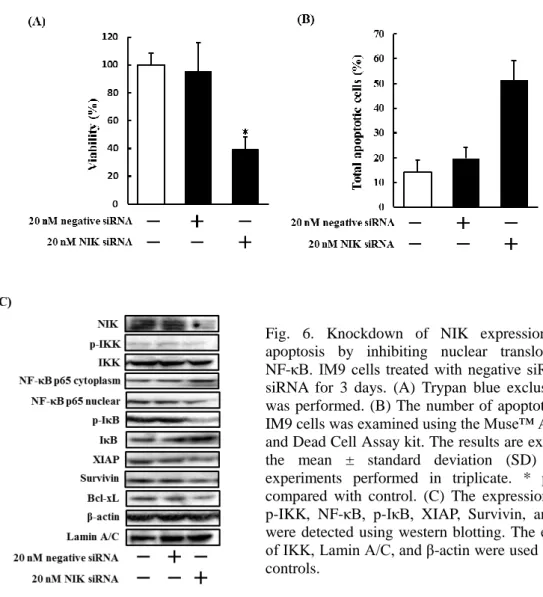
6.NIK siRNA 処理した多発性骨髄腫細胞株における細胞死誘導効果、NF-κB
経路の活性動態及びアポトーシス関連因子の発現動態の検討
上記検討結果から、mangiferin により誘導されるアポトーシスは NIK の抑制が
要因であると考えられる。そこで、NIK の抑制が多発性骨髄腫細胞株の細胞死
に重要であるか明らかにするため、NIK siRNA 処理した IM9 細胞の生存率及び
Annexin 陽性細胞数について検討した。その結果、NIK siRNA 処理により細胞生
存率の低下と Annexin 陽性細胞数の増加を認めた (Fig. 6A and B)。次に、NIK
siRNA 処理した IM9 細胞における NF-κB 経路の活性動態及びアポトーシス関連
因子の発現動態を western blotting により検討したところ、IKK、IκB の活性低下、
NF-κB の核移行阻害及び XIAP、Survivin、Bcl-xL の発現低下を認めた (Fig. 6C)。
これらの結果から、mangiferin により誘導されるアポトーシスは NIK の抑制によ
り引き起こされることが示唆された。
11
Fig. 6. Knockdown of NIK expression induces apoptosis by inhibiting nuclear translocation of NF-κB. IM9 cells treated with negative siRNA, NIK siRNA for 3 days. (A) Trypan blue exclusion assay was performed. (B) The number of apoptotic cells in IM9 cells was examined using the Muse™ Annexin-V and Dead Cell Assay kit. The results are expressed as the mean ± standard deviation (SD) of three experiments performed in triplicate. * p < 0.05 compared with control. (C) The expression of NIK, p-IKK, NF-κB, p-IκB, XIAP, Survivin, and Bcl-xL were detected using western blotting. The expression of IKK, Lamin A/C, and β-actin were used as internal controls.
Ⅳ. 考察
本章において、mangiferin は多発性骨髄腫細胞株に対して濃度依存的に細胞死
を誘導することが認められた。しかし、ヒト正常 B 細胞である RPMI1788 細胞
では mangiferin 添加による細胞死誘導は認めなかった。また、mangiferin による
細胞死はミトコンドリア膜電位の低下、Caspase-3 の活性化を介したアポトーシ
スであることを確認した。さらに、mangiferin によるアポトーシス誘導機序につ
いて検討したところ、NF-κB の核内での発現低下と細胞質中での発現増加を認
めた。しかし、ERK1/2、JNK1/2、mTOR については変化が認められなかった。
12
このことから、mangiferin は NF-κB の核移行を抑制することで、多発性骨髄腫細
胞株に対してアポトーシスを誘導することが示唆された。
IKK は IKKα、IKKβ、および IKKγ との複合体として存在し、IκB を分解する
ことで NF-κB の核移行を促進することが知られており [19, 25]、NIK は IKK 複
合体の重要な調節因子であることが報告されている。本章において、mangiferin
は NIK、IKK、IκB の活性低下及び IκB の発現増加を引き起こすことが明らかと
なった。このことから、mangiferin は NIK を阻害することにより NF-κB 経路を
抑制することが示唆された。
NF-κB は、
IAP ファミリー
(XIAP 及び Survivin)及び Bcl-2 ファミリー(Bcl-xL、
Bcl-2、
Bax、
Bim)
の発現を調節している [26]。本章において、mangiferin は Bcl-2、
Bax、Bim の発現には影響を与えず、XIAP、Survivin、Bcl-xL の発現を抑制する
ことが明らかとなった。Liang らは、NF-κB 阻害剤 berbamine が Survivin、Bcl-xL
の発現を抑制することで、多発性骨髄腫細胞株に対して細胞死を誘導すること
を示している [27]。当研究室においても、NF-κB 阻害作用を有する DMF が XIAP、
Bcl-xL の発現低下により多発性骨髄腫細胞株に対してアポトーシスを誘導する
ことを認めている [28]。このことから、mangiferin は NIK/IKK/NF-κB 経路を阻
害することで XIAP、Survivin、Bcl-xL の発現を抑制し、多発性骨髄腫細胞株に
対してアポトーシスを誘導することが示唆された。
これまでの検討結果から、mangiferin により誘導される細胞死は NIK の抑制が
要因であると考えられる。そこで、NIK siRNA 処理した多発性骨髄腫細胞株に
ついて検討したところ、細胞生存率の低下と Annexin 陽性細胞数の増加を認め
た。また、IKK、IκB、NF-κB の活性低下及び XIAP、Survivin、Bcl-xL の発現低
下を確認した。このことから、mangiferin によるアポトーシス誘導には NIK の抑
制が重要であると考えられる。
本章において、mangiferin は NIK/IKK/NF-κB 経路を抑制することで、多発性
骨髄腫細胞株に対してアポトーシスを誘導することが示唆された。現在 NIK を
標的とした治療薬は臨床応用されておらず、mangiferin は新たな作用機序を有す
る分子標的薬として期待される。
INTERNATIONAL JOURNAL OF ONCOLOGY 48: 2704-2712, 2016
2704
Abstract. Multiple myeloma (MM) is still an incurable
hema-tological malignancy with a 5-year survival rate of ~35%, despite the use of various treatment options. The nuclear factor κB (NF-κB) pathway plays a crucial role in the patho-genesis of MM. Thus, inhibition of the NF-κB pathway is a potential target for the treatment of MM. In a previous study, we showed that mangiferin suppressed the nuclear translo-cation of NF-κB. However, the treatment of MM involves a combination of two or three drugs. In this study, we examined the effect of the combination of mangiferin and conventional anticancer drugs in an MM cell line. We showed that the combination of mangiferin and an anticancer drug decreased the viability of MM cell lines in comparison with each drug used separately. The decrease in the combination of mangiferin and an anticancer drug induced cell viability was attributed to increase the expression of p53 and Noxa and decreases the expression of XIAP, survivin, and Bcl-xL proteins via inhibi-tion of NF-κB pathway. In addition, the combination treatment caused the induction of apoptosis, activation of caspase-3 and
the accumulation of the cells in the sub-G1 phase of the cell cycle. Our findings suggest that the combination of mangiferin and an anticancer drug could be used as a new regime for the treatment of MM.
Introduction
Multiple myeloma (MM) is a B cell malignancy characterized by clonal proliferation of plasma cells in the bone marrow (1). MM is the most common hematological malignancy, second only to non-Hodgkin's lymphoma and accounts for 10% of all hematological malignancies and 1% of all cancers (2). MM treatment comprises vincristine/adriamycin/dexamethasone or melphalan/prednisolone chemotherapy and novel agents such as thalidomide, lenalidomide, pomalidomide, and bortezomib (3,4). However, MM remains an incurable disease with a 5-year survival rate of ~35% (5). In addition, most of the drugs used for MM treatment have side effects that limit their utility. Thus, there remains an unmet need for novel therapies for MM treatment.
The nuclear factor κB (NF-κB) pathway plays a crucial role in the survival, growth, and drug resistance of different types of cancers, including MM (6,7). Constitutive NF-κB activity is present in human MM cell lines and cells of the MM patients (8). The NF-κB family includes RelA (p65), RelB, c-Rel, p50 (NF-κB1), and p52 (NF-κB2) proteins (9). NF-κB is typi-cally a heterodimer composed of p50 and p65 subunits and is constitutively present in the cytosol and the nucleus. In the cytosol, NF-κB is inactivated by its association with inhibitor of NF-κB (IκB) (10). Upon stimulation, IκB is phosphorylated by IκB kinases marking it for proteasomal degradation and thereby allowing nuclear translocation of NF-κB (5,11). Then, NF-κB binds to specific DNA sequences and promotes the transcription of its target genes (12). The NF-κB pathway regulates the gene expression of cell cycle regulators (c-Myc, cyclin D, cyclin E, p21, and p27) and anti-apoptotic molecules [B cell leukemia 2 (Bcl-2), B cell leukemia-xL (Bcl-xL), x-linked inhibitor of apoptosis (XIAP), and c-IAP] (13,14).
Mangiferin enhances the sensitivity of human multiple
myeloma cells to anticancer drugs through suppression
of the nuclear factor
κ
B pathway
TOmOyA TAkEDA1, MAsANOBU TsUBAkI1, TOsHIkI kINO1, AYAkO kAWAMURA1, sHOTA IsOYAMA1, TATsUkI ITOH4, MOTOHIRO IMANO5, GENzOH TANABE2, OsAMU MURAOkA2, HiDEAki mATsuDA3, TAkAO sATOU6 and sHOzO NisHiDA1
1Division of Pharmacotherapy, 2Laboratory of Pharmaceutical Organic Chemistry, 3Department of Natural Drugs Resources,
kinki University school of Pharmacy, kowakae, Higashi-Osaka; 4Department of Food science and Nutrition, kinki university school of Agriculture, Nara; Departments of 5surgery and 6Pathology,
kinki University school of Medicine, Osakasayama, Osaka, Japan Received November 19, 2015; Accepted March 15, 2016
DOi: 10.3892/ijo.2016.3470
Correspondence to: Dr shozo Nishida, Division of Pharmacotherapy, kinki University school of Pharmacy, kowakae, Higashi-Osaka 577-8502, Japan
E-mail: [email protected]
Abbreviations: AML, acute myeloid leukemia; Bax, B cell leukemia-2 associated X; Bcl-2, B cell leukemia-2; Bcl-xL, B cell leukemia-xL; ERk1/2, extracellular signal-regulated kinase 1/2; IAP, inhibitors of apoptosis; IκB, inhibitor of κB; JNk1/2, c-Jun N-terminal protein kinase 1/2; MM, multiple myeloma; NF-κB, nuclear factor κB; PUMA, p53 upregulated modulator of apoptosis; XIAP, X-linked inhibitor of apoptosis protein
Key words: multiple myeloma, mangiferin, NF-κB, anticancer drug, p53, Noxa, X-linked inhibitor of apoptosis protein, survivin, Bcl-xL
TAkEDA et al: MANGIFERIN ENHANCEs THE sENsITIVITY OF MULTIPLE MYELOMA CELLs 2705
Recent studies have reported that NF-κB inhibitors induced apoptosis in hematopoietic tumor cells through downregula-tion of anti-apoptotic proteins (15). Therefore, the inhibidownregula-tion of NF-κB signaling is a potential target for the treatment of MM.
Recently, plant and plant-derived drugs have been recognized as one of the most attractive approaches for cancer therapy (16). In addition, many drugs derived from plants have been shown to be useful and effective in sensi-tizing tumors to conventional agents, prolonging survival time, and preventing the side effects of chemotherapy (17,18). Mangiferin, 1,3,6,7-tetrahydroxyxanthone-C2-β -D-glucoside, is a compound extracted from plants belonging to the Anacardiaceae and Gentianaceae families, including
Mangifera indica L. (19). Mangiferin has been reported to
have various bioactivities, such as anti-oxidant, antitumor, antidiabetic, anti-inflammatory, and immunomodulatory activities (19). Previous studies have revealed that mangiferin has anticancer effects in acute myeloid leukemia (AML) cell lines (20). In addition, we showed that mangiferin induced apoptosis by inhibiting the nuclear translocation of NF-κB. However, the treatment of MM involves a combination of two or three drugs, including adriamycin, vincristine, and melphalan. In this study, we examined the effect of the combi-nation of mangiferin and conventional anticancer drugs in MM cell lines.
Materials and methods
Materials. Mangiferin (C19H18O11) and melphalan were purchased from sigma (st. Paul, MN, UsA), and dissolved in dimethyl sulfoxide. These reagents were dissolved in phosphate-buffered saline (PBs) and filtered through 0.45-µm syringe filters (iwaki Glass, Tokyo, Japan) before use in the experiments described below.
Adriamycin and vincristine were purchased from sigma. These reagents were dissolved in PBs and used for the various assays described below.
Cell culture. IM9 cells and RPMI8226 were obtained from
Health science Research Resources Bank (Osaka, Japan). IM9 cells were cultured in RPMI-1640 medium (sigma) containing 10% fetal bovine serum (Gibco, Carlsbad, CA, UsA), 100 µg/ml penicillin (Gibco), 100 u/ml streptomycin (Gibco), and 25 mM 4-(2-hydroxyethyl)-1-piperazine ethanesulfonic acid (Wako, Osaka, Japan). All cell lines were maintained at 37˚C in an atmosphere containing 5% CO2.
Trypan blue exclusion assay. The cells were plated in 96-well
plates at 2x10⁴ cells/ml and treated with mangiferin, anticancer drugs, a combination of both, or without mangiferin (control). After incubation, the cells were stained with trypan blue and the number of stained cells was counted at days one, three, and five.
Western blotting
Preparation of nuclear extracts for NF-κB. The cells treated
with mangiferin, anticancer drugs, a combination of both, or without mangiferin (control) were washed with cold PBs and lysed using a lysis buffer containing 100 mM Tris-HCl
(pH 7.4), 1 mm EDTA, 0.5% NP-40, 1 µm pepstatin, 1 µm leupeptin, 2 mm sodium orthovanadate, 1 µm calpain inhibitor, phosphatase inhibitor cocktail I/II, and 1 mM phenylmethylsulfonyl fluoride (PMsF). The lysates were centrifuged at 14,000 rpm for 5 min, and the supernatant, which contained the cytoplasmic extracts, was stored at -80˚C. The nuclear pellet was resuspended in cold nuclear extraction buffer for 30 min. The extract was centrifuged at 14,000 rpm for 5 min, and the supernatant containing the nuclear extract was obtained. The proteins were measured using the BCA protein assay kit (Pierce, Rockford, IL, UsA). Total cellular proteins (30 µg of protein) from the cytoplasmic or the nuclear extract were separated using 10% sodium dodecyl sulfate (sDs) polyacrylamide gels. The proteins were transferred to polyvinyl difluoride (PVDF) membranes (Amersham, Arlington Heights, IL, UsA). The membranes were blocked with 5% skim milk and incubated overnight at 4˚C with rabbit anti-human NF-κB p65 (Cell signaling Technology, Beverly, MA, UsA) and rabbit anti-human lamin A/C antibodies (santa Cruz Biotechnologies, santa Cruz, CA, UsA). After binding to an appropriate horseradish peroxidase-conjugated secondary antibody, the proteins were visualized using Luminata Forte Western HRP substrate (Millipore, MA, UsA) according to the manufacturer's instructions.
Preparation of whole cell lysates. The cells treated with
mangiferin, anticancer drugs, a combination of both, or without mangiferin (control) were washed with cold PBs and lysed with a lysis buffer containing 100 mM Tris-HCl (pH 7.4), 1 mm EDTA, 0.5% NP-40, 1 µm pepstatin, 1 µm leupeptin, 2 mm sodium orthovanadate, 1 µm calpain inhibitor, phos-phatase inhibitor cocktail I/II, and 1 mM PMsF. The proteins were measured using the BCA protein assay kit (Pierce). The extracts (30 µg of protein) were separated using 10% sDs poly-acrylamide gels. The proteins were then transferred to PVDF membranes (Amersham). The membranes were blocked with 5% skim milk and incubated overnight at 4˚C with each of the following antibodies: rabbit anti-human phospho-p44/42 mitogen-activated protein kinase (MAPk, ERk1/2), rabbit anti-human p44/42 MAPk (ERk1/2), mouse anti-human phospho-IκB, rabbit anti-human IκB, rabbit anti-human phospho-stress-activated protein kinase/c-jun N-terminal kinase (JNk1/2), rabbit anti-human JNk1/2, rabbit anti-human cyclin D, rabbit anti-human cyclin E, rabbit anti-human XiAP, rabbit anti-human survivin (Cell signaling Technology), mouse anti-human β-actin (sigma), rabbit anti-human p53, rabbit anti-human p21, rabbit anti-human p27, rabbit anti-human PUMA, rabbit anti-human NOXA, rabbit anti-human Bcl-xL, rabbit human Bax, rabbit human Bim, and rabbit anti-human Bcl-2 (santa Cruz Biotechnologies). After binding of an appropriate horseradish peroxidase-conjugated secondary antibody, the proteins were visualized using Luminata Forte Western HRP substrate (Millipore) according to the manufac-turer's instructions.
Flow cytometry. The cells treated with mangiferin, anticancer
drugs, a combination of both, or without mangiferin (control) were washed with cold PBs, and fixed in 70% ethanol. The cells were resuspended in PBs and 50 µg/ml propidium iodide was added. Then, the samples were measured on a BD-LsR
INTERNATIONAL JOURNAL OF ONCOLOGY 48: 2704-2712, 2016
2706
flow cytometer (BD Biosciences, CA, usA). Cell cycles were analyzed on Cell Quest software (BD Biosciences).
Analysis of apoptosis by flow cytometry. Measurement of
cells undergoing apoptosis was performed with the Muse™ Annexin V and Dead Cell Assay kit (merck millipore, Darmstadt, Germany), according to the manufacturer's instruc-tions. The cells were treated with mangiferin, anticancer drugs, a combination of both, or without mangiferin (control) for 48 h. Then, Muse Annexin V and dead cell reagent was added. After incubation for 20 min at room temperature, apoptotic cells were applied to a Muse Cell Analyzer (Merck Millipore).
Measurement of the proteolytic activity of caspase-3. The
activity of caspase-3 was determined using the caspase-3/ CPP32 fluorometric assay kit (BioVision mountain View, CA, UsA) according to the manufacturer's instructions. The cells were treated with mangiferin, anticancer drugs, a combination of both, or without mangiferin (control) for 36 h. Then, the cells were washed in PBs and lysed using the lysis buffer provided in the kit. The cell lysates were centrifuged at 14,000 rpm for
5 min, and the reaction buffer containing 1 mM Asp-Glu-Val-Asp-7-amino-4-trifluoromethylcoumarin was added to the supernatants and incubated at 37˚C for 2 h. subsequently, the absorbance was measured using a fluorescence spectropho-tometer (Hitachi, Tokyo, Japan) at an emission wavelength of 505 nm and an excitation wavelength of 400 nm.
Statistical analysis. All results are expressed as means ±
stan-dard deviation of several independent experiments. Multiple comparisons of the data were performed using analysis of variance with Dunnett's test. P-values <5% were considered significant.
Results
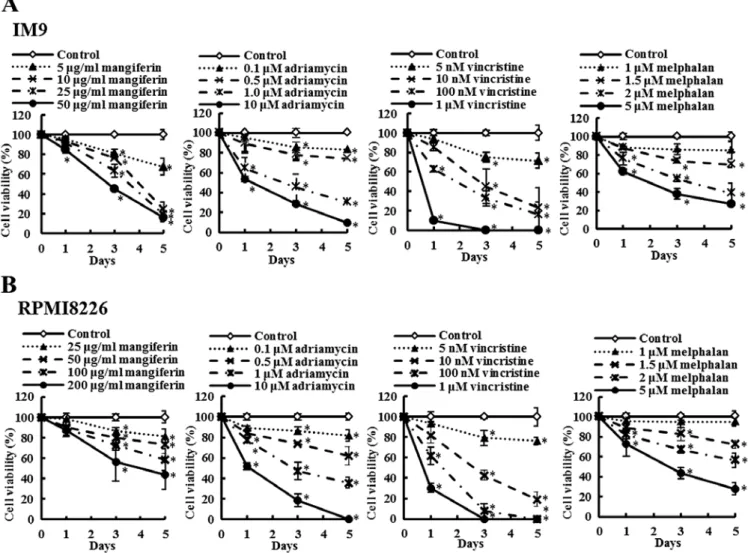
Mangiferin, adriamycin, vincristine, and melphalan decrease the viability of the MM cell line. The effects of mangiferin
and the three anticancer drugs (adriamycin, vincristine, and melphalan) on the viability of the MM cell lines (IM9 and RPMI8226) as determined by the trypan blue exclusion assay are shown in Fig. 1. IM9 cells were treated in the absence
Figure 1. Mangiferin, adriamycin, vincristine, and melphalan decrease the viability of IM9 cells and RPMI8226 cells. (A) IM9 cells were treated with mangiferin (5-50 µg/ml), adriamycin (0.1-10 µm), vincristine (5 nm-1 µm), and melphalan (1-5 µm). (B) RPmi8226 cells were treated with mangiferin (25-200 µg/ml), adriamycin (0.1-10 µm), vincristine (5 nm-1 µm), and melphalan (1-5 µm). Then, trypan blue exclusion assay was performed in im9 cells and RPmi8266 cells after one, three, and five days. The results are expressed as the mean ± standard deviation (sD) of three experiments performed in triplicate. *p<0.05 compared
TAkEDA et al: MANGIFERIN ENHANCEs THE sENsITIVITY OF MULTIPLE MYELOMA CELLs 2707
(control) or presence of mangiferin (5-50 µg/ml), adriamycin (0.1-10 µm), vincristine (5 nm-1 µm), or melphalan (1-5 µm). After three days, the viability of IM9 cells treated with 5, 10, 25, and 50 µm mangiferin was 80.5, 76.3, 63.3 and 45.3%, respectively, whereas that after five days was 67.2, 24.5, 19.3 and 16.3%, respectively. After three days, the viability of IM9 cells treated with 5, 10, 25, and 50 µm adriamycin was 85.5, 78.0, 45.4 and 28.9%, respectively, whereas that after five days was 83.1, 74.3, 30.2 and 9.7%, respectively. Vincristine and melphalan showed results similar to those observed with adriamycin. In addition, RPMI8226 cells also showed results similar to those observed with IM9 cells (Fig. 1B). These results indicated that mangiferin, adriamycin, vincristine, and melphalan decreased the viability of MM cell lines in a concentration-dependent manner.
Mangiferin enhances the sensitivity of human multiple myeloma cells to anticancer drugs. The effects of mangiferin,
the three anticancer drugs, and the combination of each anti-cancer drug and mangiferin on the viability of the MM cell lines (IM9 and RPMI8226) were determined by trypan blue exclusion assay and are shown in Fig. 2. IM9 cells were treated with either mangiferin (5 µg/ml), adriamycin (0.5 µm), vincristine (5 nm), melphalan (1.5 µm), or combination of mangiferin with an anticancer drug. After three days, the viability of im9 cells treated with 5 µg/ml mangiferin, 0.5 µm adriamycin, or combination of both was 72.0, 69.8 and 45.0%, respectively. The combination of mangiferin and vincristine or melphalan showed results similar to those observed with adriamycin. In addition, RPMI8226 cells also showed results similar to those observed with IM9
Figure 2. mangiferin enhances the effect of anticancer drugs on mm cell lines. (A) im9 cells were treated with mangiferin (5 µg/ml) or adriamycin (0.5 µm), vincristine (5 nm), and melphalan (1.5 µm), or a mixture of mangiferin with an anticancer drug. (B) RPmi8226 cells were treated with mangiferin (25 µg/ml) or adriamycin (0.5 µm), vincristine (5 nm), and melphalan (1.5 µm), or a mixture of mangiferin with an anticancer drug. Then, trypan blue exclusion assay was performed in im9 cells and RPmi8226 cells after one, two, and three days. The results are expressed as the mean ± standard deviation (sD) of three experiments performed in triplicate. *p<0.05 compared with control.
INTERNATIONAL JOURNAL OF ONCOLOGY 48: 2704-2712, 2016
2708
cells (Fig. 2B). These results show that the combination of mangiferin and an anticancer drug significantly reduced the viability of the MM cell line in comparison to the use of each of these drugs separately.
The combination of mangiferin and an anticancer drug suppresses the nuclear translocation of NF-κB. In a previous
study, we showed that mangiferin inhibits the nuclear translo-cation of NF-κB in AML cell lines (20). However, mangiferin did not affect the levels of ERk1/2, Akt, and p38MAPk phos-phorylation. To clarify the molecular mechanisms underlying the effects of combination of mangiferin and other anticancer drugs, we investigated the nuclear translocation of NF-κB and expression of phosphorylated IκB and IκB proteins by using western blotting. Our results showed that the combination of mangiferin and each of the other anticancer drugs signifi-cantly suppressed the nuclear translocation of NF-κB (Fig. 3). Further, we observed no changes in the levels of ERk1/2 and JNk1/2 phosphorylation (Fig. 4). These results indicated that the decrease in the combination of mangiferin and an anti-cancer drug induced cell viability was attributed to inhibition of the NF-κB pathway.
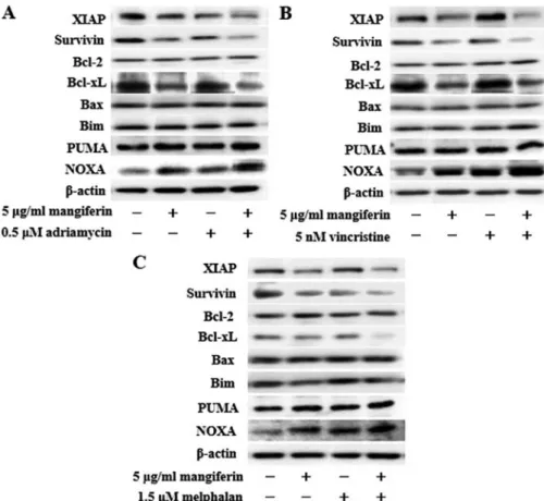
The combination of mangiferin and an anticancer drug increases the expression of p53 and Noxa and decreases the
expression of XIAP, survivin, and Bcl-xL proteins. NF-κB is a nuclear factor known to activate the expression of genes involved in cell proliferation and cell survival (anti-apoptotic proteins and pro-apoptotic proteins). Therefore, we examined the expression of proteins involved in cell proliferation and cell survival by using western blotting. Our results showed that the combination of mangiferin and an anticancer drug upregu-lated the expression of p53 and Noxa and downreguupregu-lated that of XIAP, survivin, and Bcl-xL proteins in comparison with mangiferin alone (Figs. 5 and 6). However, we observed no changes in the expression of cyclin D, cyclin E, p27, p21, Bcl-2, Bax, Bim, and PUMA proteins.
The combination of mangiferin and an anticancer drug causes the accumulation of cells in the sub-G1 phase of the cell cycle. p53 plays important roles in various phases of the
cell cycle. Thus, we examined cell cycle regulation in IM9 cells treated with a combination of mangiferin and each of the other anticancer drugs by flow cytometry. Our results showed that the combined treatment increased the accumulation of cell population in the sub-G1 phase (Fig. 7). These results are indicative of apoptosis.
The combination of mangiferin and an anticancer drug induces apoptosis by activating caspase-3. We measured
Figure 3. The combination of mangiferin and an anticancer drug suppresses the nuclear translocation of NF-κB. (A) IM9 cells were treated with mangif-erin (5 µg/ml) or adriamycin (0.5 µm), vincristine (5 nm), and melphalan (1.5 µm), or a mixture of mangifmangif-erin with an anticancer drug for three days. (B) RPmi8226 cells were treated with mangiferin (25 µg/ml) or adriamycin (0.5 µm), vincristine (5 nm), and melphalan (1.5 µm), or a mixture of mangiferin with an anticancer drug for three days. The expression of NF-κB, and, phospho-IκB was detected using western blotting. The expression of IκB, Lamin A/C, and β-actin were used as internal controls.
TAkEDA et al: MANGIFERIN ENHANCEs THE sENsITIVITY OF MULTIPLE MYELOMA CELLs 2709
apoptotic cells using the muse™ Annexin V and Dead Cell Assay kit. IM9 cells were treated with a combination of mangiferin and each of the other anticancer drugs for two days. Our results showed that mangiferin increased the number of apoptotic cells in a concentration-dependent manner (Fig. 8A). Apoptosis is induced by an interaction between various initi-ator and effector caspases. Caspase-3 is a crucial effector of
the apoptosis pathway. We investigated caspase-3 activation in IM9 cells treated with a combination of mangiferin and each of the other anticancer drugs by using the caspase-3/CPP32 fluorometric assay kit. The combination of mangiferin and an anticancer drug activated caspase-3 (Fig. 8B). These results showed that the combined treatment induced apoptosis by activating caspase-3.
Figure 4. The combination of mangiferin and an anticancer drug do not change the levels of ERk1/2 and JNk1/2 phosphorylation. IM9 cells were treated with mangiferin (5 µg/ml) or (A) adriamycin (0.5 µm), (B) vincristine (5 nm), and (C) melphalan (1.5 µm), or a mixture of mangiferin with an anticancer drug for three days. The expression of phospho-ERk1/2, and phospho-JNk1/2 was detected using western blotting. The expression of ERk1/2, JNk1/2, and β-actin were used as internal controls.
Figure 5. The combination of mangiferin and an anticancer drug increases the expression of p53. im9 cells were treated with mangiferin (5 µg/ml) or (A) adria-mycin (0.5 µm), (B) vincristine (5 nm), and (C) melphalan (1.5 µm), or a mixture of mangiferin with an anticancer drug for three days. The expression of cyclin D, cyclin E, p53, p27, and p21 were detected using western blotting. The expression of β-actin was used as internal control.
INTERNATIONAL JOURNAL OF ONCOLOGY 48: 2704-2712, 2016
2710
Figure 6. The combination of mangiferin and an anticancer drug increase the expression of Noxa, and decrease the expression of XIAP, survivin, and Bcl-xL proteins. im9 cells were treated with mangiferin (5 µg/ml) or (A) adriamycin (0.5 µm), (B) vincristine (5 nm), and (C) melphalan (1.5 µm), or a mixture of mangiferin with an anticancer drug for three days. The expression of XIAP, survivin, Bcl-2, Bcl-xL, Bax, Bim, PUMA, and Noxa were detected using western blotting. The expression of β-actin was used as internal control.
Figure 7. The combination of mangiferin and an anticancer drug causes the accumulation of the cells in the sub-G1 phase of the cell cycle. IM9 cells were treated with mangiferin (5 µg/ml) or adriamycin (0.5 µm), vincristine (5 nm), and melphalan (1.5 µm), or a mixture of mangiferin with an anticancer drug for three days. Cell cycle distribution was analyzed at an hour after PI staining.
TAkEDA et al: MANGIFERIN ENHANCEs THE sENsITIVITY OF MULTIPLE MYELOMA CELLs 2711
Discussion
Despite the development of increasingly effective therapies, MM remains an incurable disease with an average survival of 3-5 years following diagnosis. In addition, most of the compounds used for MM treatment have side effects that limit their utility. Presumably, the side effects of these compounds could be decreased by reducing their dose and using them in combination with another drug (21). The nuclear factor κB (NF-κB) pathway plays a crucial role in the pathogenesis of MM (22,23). Thus, inhibition of the NF-κB pathway is a potential target for the treatment of MM. We have previously shown that mangiferin induced apoptosis in AML cell lines via inhibition of the NF-κB pathway (20). Additionally, it
was reported that mangiferin in combination with oxaliplatin counteract the development of resistance to oxaliplatin in colon cancer cells by reducing active NF-κB (24). However, the effect of the combination of mangiferin and conventional anticancer drugs in mm cell lines remain to be clarified. in particular, the molecular mechanism has not been elucidated thus far. In this study, we examined the effect of the combina-tion of mangiferin and convencombina-tional anticancer drugs in MM cell lines.
We showed that mangiferin, adriamycin, vincristine, and melphalan decrease the viability of MM cell lines. The combination of mangiferin and each of the above-mentioned anticancer drugs significantly reduced the viability of the MM cell line in comparison with each of these drugs used alone. Furthermore, our results showed that the combination treatment significantly suppressed the nuclear translocation of NF-κB. However, we observed no changes in the levels of ERk1/2 and JNk1/2 phosphorylation. In agreement with previous reports, constitutive activation of NF-κB promoted multiple myeloma cell growth and survival, and the NF-κB inhibitor dimethyl fumarate induced apoptosis in MM cell lines (15,25). In addition, other studies showed that celastrol induces chemosensitization through downregulation of NF-κB in MM cell lines (17). These results indicate that the combi-nation of mangiferin and other anticancer drugs exert their effects on MM through downregulation of NF-κB pathway.
NF-κB initiates the transcriptional activation of prosur-vival genes and proliferation-promoting genes (26). We observed that the combination of mangiferin and each of the other anticancer drugs significantly increased the expression of p53 and Noxa and decreased the expression of XIAP, survivin, and Bcl-xL, proteins. In addition, the combination treatment caused the induction of apoptosis, activation of caspase-3 and the accumulation of the cells in the sub-G1 phase of the cell cycle. The tumor suppressor p53 induces apoptosis by transactivation of its downstream apoptotic regulators such as Noxa (27). survivin, a member of the inhibitor of apoptosis protein family, protects cells from caspase-dependent apop-totic pathways. survivin overexpression has been reported in various hematopoietic and solid cancers (28-30). XIAP is the most potent endogenous direct inhibitor of caspases and is thus considered a key physiological regulator of cell death. MM cells express high levels of XIAP regulated by the NF-κB pathway (31). The Bcl-2 family member Bcl-xL is an anti-apoptotic protein; Bcl-xL overexpression has been reported in MM cell lines (32,33). These results suggest that the combina-tion of mangiferin and an anticancer drug induces apoptosis by increasing the expression of p53 and Noxa and decreasing that of XIAP, survivin, and Bcl-xL proteins via inhibition of the NF-κB pathway.
In conclusion, our results showed that the combination of mangiferin and an anticancer drug decreased the viability of MM cell lines in comparison with each of these drugs used separately. The decrease in the combination of mangiferin and an anticancer drug induced cell viability was attributed to the induction of apoptosis, activation of caspase-3, and the accumulation of the cells in the sub-G1 phase of the cell cycle via inhibition of nuclear translocation of NF-κB. Importantly, 40% of multiple myeloma patients show constitutive activation of the NF-κB pathway. Our findings showed that the
combina-Figure 8. The combination of mangiferin and an anticancer drug induces apoptosis by activating caspase-3. (A) IM9 cells were treated with mangif-erin (5 µg/ml) or adriamycin (0.5 µm), vincristine (5 nm), and melphalan (1.5 µm), or a mixture of mangiferin with an anticancer drug for two days. The number of apoptotic cells in IM9 cells was examined using the Muse Annexin V and Dead Cell Assay kit. (B) im9 cells were treated with man-giferin (5 µg/ml) or adriamycin (0.5 µm), vincristine (5 nm), and melphalan (1.5 µm), or a mixture of mangiferin with an anticancer drug for 36 h. The activity of caspase-3 induced by mangiferin in IM9 cells was examined using the caspase-3/CPP32 fluorometric assay kit. The results are expressed as the mean ± standard deviation (sD) of four experiments. *p<0.05 compared with
INTERNATIONAL JOURNAL OF ONCOLOGY 48: 2704-2712, 2016
2712
tion of mangiferin and an anticancer drug selectively inhibited the NF-κB pathway without inhibiting other signaling factors. In addition, we found that mangiferin enhanced the effect of conventional anticancer drugs (adriamycin, vincristine, and melphalan) commonly used in multiple myeloma treatment. Our results provided evidence of the potential of the combina-tion of mangiferin and an anticancer drug as a new regime for the treatment of MM.
Acknowledgements
This study was supported in part by a Grant-in-Aid for scientific Research (C) from the Japan society for the Promotion of science (JsPs) and by Ministry of Education, Culture, sports, science, and Technology (MEXT)-supported Program for the strategic Research Foundation at Private Universities, 2014-2018.
References
1. Raab Ms, Podar k, Breitkreutz I, Richardson PG and Anderson kC: Multiple myeloma. Lancet 374: 324-339, 2009. 2. Bladé J, Cibeira MT and Rosiñol L: Novel drugs for the treatment
of multiple myeloma. Haematologica 95: 702-704, 2010. 3. Rajkumar sV: Treatment of multiple myeloma. Nat Rev Clin
Oncol 8: 479-491, 2011.
4. Wang X, zhang M, Wang M, He P, Liu X, Chen L, Xi J, Wang M, Li J, Liu H, et al: Arsenic trioxide combined with VCmP or VAD chemotherapy in patients with refractory or relapsed multiple myeloma in a single institution of China. Indian J Hematol Blood Transfus 30: 259-264, 2014.
5. Paramore A and Frantz s: Bortezomib. Nat Rev Drug Discov 2: 611-612, 2003.
6. sethi G, sung B and Aggarwal BB: Nuclear factor-kappaB acti-vation: From bench to bedside. Exp Biol Med (Maywood) 233: 21-31, 2008.
7. Tsubaki M, Takeda T, Ogawa N, sakamoto k, shimaoka H, Fujita A, itoh T, imano m, ishizaka T, satou T, et al: Over-expression of survivin via activation of ERk1/2, Akt, and NF-κB plays a central role in vincristine resistance in multiple myeloma cells. Leuk Res 39: 445-452, 2015.
8. Cormier F, monjanel H, Fabre C, Billot k, sapharikas E, Chereau F, Bordereaux D, molina TJ, Avet-Loiseau H and Baud V: Frequent engagement of RelB activation is critical for cell survival in multiple myeloma. PLos One 8: e59127, 2013. 9. karin m and Greten FR: NF-kappaB: Linking inflammation
and immunity to cancer development and progression. Nat Rev Immunol 5: 749-759, 2005.
10. Ma C, zuo W, Wang X, Wei L, Guo Q and song X: Lapatinib inhibits the activation of NF-κB through reducing phosphoryla-tion of IκB-α in breast cancer cells. Oncol Rep 29: 812-818, 2013.
11. Hideshima T, ikeda H, Chauhan D, Okawa y, Raje N, Podar k, mitsiades C, munshi NC, Richardson PG, Carrasco RD, et al: Bortezomib induces canonical nuclear factor-kappaB activation in multiple myeloma cells. Blood 114: 1046-1052, 2009.
12. Hayden Ms and Ghosh s: shared principles in NF-kappaB signaling. Cell 132: 344-362, 2008.
13. Panwalkar A, Verstovsek s and Giles F: Nuclear factor-kappaB modulation as a therapeutic approach in hematologic malignan-cies. Cancer 100: 1578-1589, 2004.
14. Joyce D, Albanese C, steer J, Fu m, Bouzahzah B and Pestell RG: NF-kappaB and cell-cycle regulation: The cyclin connection. Cytokine Growth Factor Rev 12: 73-90, 2001.
15. Tsubaki M, Ogawa N, Takeda T, sakamoto k, shimaoka H, Fujita A, itoh T, imano m, satou T and Nishida s: Dimethyl fumarate induces apoptosis of hematopoietic tumor cells via inhibition of NF-κB nuclear translocation and down-regulation of Bcl-xL and XIAP. Biomed Pharmacother 68: 999-1005, 2014.
16. Olaku O and White JD: Herbal therapy use by cancer patients: A literature review on case reports. Eur J Cancer 47: 508-514, 2011.
17. Jeong sJ, koh W, kim B and kim sH: Are there new therapeutic options for treating lung cancer based on herbal medicines and their metabolites? J Ethnopharmacol 138: 652-661, 2011. 18. Wang LH, Li Y, Yang sN, Wang FY, Hou Y, Cui W, Chen k,
Cao Q, Wang s, zhang TY, et al: Gambogic acid synergistically potentiates cisplatin-induced apoptosis in non-small-cell lung cancer through suppressing NF-κB and MAPk/HO-1 signalling. Br J Cancer 110: 341-352, 2014.
19. Luo F, Lv Q, zhao Y, Hu G, Huang G, zhang J, sun C, Li X and Chen k: Quantification and purification of mangiferin from Chinese Mango (Mangifera indica L.) cultivars and its protective effect on human umbilical vein endothelial cells under H(2)O(2)-induced stress. Int J Mol sci 13: 11260-11274, 2012.
20. shoji k, Tsubaki m, yamazoe y, satou T, itoh T, kidera y, Tanimori Y, Yanae M, Matsuda H, Taga A, et al: Mangiferin induces apoptosis by suppressing Bcl-xL and XIAP expressions and nuclear entry of NF-κB in HL-60 cells. Arch Pharm Res 34: 469-475, 2011.
21. slawinska-Brych A, zdzisinska B, mizerska-Dudka m and kandefer-szerszen M: Induction of apoptosis in multiple myeloma cells by a statin-thalidomide combination can be enhanced by p38 MAPk inhibition. Leuk Res 37: 586-594, 2013.
22. kannaiyan R, Hay Hs, Rajendran P, Li F, shanmugam mk, Vali s, Abbasi T, kapoor s, sharma A, kumar AP, et al: Celastrol inhibits proliferation and induces chemosensitization through down-regulation of NF-κB and sTAT3 regulated gene products in multiple myeloma cells. Br J Pharmacol 164: 1506-1521, 2011.
23. Tsubaki M, komai M, Itoh T, Imano M, sakamoto k, shimaoka H, Ogawa N, mashimo k, Fujiwara D, Takeda T, et al: Inhibition of the tumour necrosis factor-alpha autocrine loop enhances the sensitivity of multiple myeloma cells to anticancer drugs. Eur J Cancer 49: 3708-3717, 2013.
24. du Plessis-stoman D, du Preez J and van de Venter m: Combination treatment with oxaliplatin and mangiferin causes increased apoptosis and downregulation of NFκB in cancer cell lines. Afr J Tradit Complement Altern Med 8: 177-184, 2011. 25. Trocoli A and Djavaheri-mergny m: The complex interplay
between autophagy and NF-κB signaling pathways in cancer cells. Am J Cancer Res 1: 629-649, 2011.
26. DiDonato JA, mercurio F and karin m: NF-κB and the link between inflammation and cancer. immunol Rev 246: 379-400, 2012.
27. zhu HJ, Liu L, Fan L, zhang LN, Fang C, zou zJ, Li JY and Xu W: The BH3-only protein Puma plays an essential role in p53-mediated apoptosis of chronic lymphocytic leukemia cells. Leuk Lymphoma 54: 2712-2719, 2013.
28. Tsubaki M, satou T, Itoh T, Imano M, komai M, Nishinobo M, Yamashita M, Yanae M, Yamazoe Y and Nishida s: Over-expression of mDR1 and survivin, and decreased Bim Over-expression mediate multidrug-resistance in multiple myeloma cells. Leuk Res 36: 1315-1322, 2012.
29. Tsubaki M, komai M, Itoh T, Imano M, sakamoto k, shimaoka H, Takeda T, Ogawa N, mashimo k, Fujiwara D, et al: By inhibiting src, verapamil and dasatinib overcome multidrug resistance via increased expression of Bim and decreased expressions of mDR1 and survivin in human multidrug-resistant myeloma cells. Leuk Res 38: 121-130, 2014.
30. markovic O, marisavljevic D, Cemerikic-martinovic V, martinovic T, Filipovic B, stanisavljevic D, zivković R, Hajder J, stanisavljevic N and mihaljevic B: survivin expres-sion in patients with newly diagnosed nodal diffuse large B cell lymphoma (DLBCL). med Oncol 29: 3515-3521, 2012.
31. Desplanques G, Giuliani N, Delsignore R, Rizzoli V, Bataille R and Barillé-Nion s: Impact of XIAP protein levels on the survival of myeloma cells. Haematologica 94: 87-93, 2009.
32. Lin J, Wu y, yang D and zhao y: induction of apoptosis and antitumor effects of a small molecule inhibitor of Bcl-2 and Bcl-xl, gossypol acetate, in multiple myeloma in vitro and in vivo. Oncol Rep 30: 731-738, 2013.
33. Xu L, yang D, Wang s, Tang W, Liu m, Davis m, Chen J, Rae Jm, Lawrence T and Lippman ME: (-)-Gossypol enhances response to radiation therapy and results in tumor regression of human prostate cancer. Mol Cancer Ther 4: 197-205, 2005.
24
第 3 章
新規 NIK 阻害剤 mangiferin による
転移抑制効果及び腫瘍増殖抑制効果
I.緒言
悪性腫瘍は多くの効果的な薬剤や治療法の出現にも関わらず未だ根治するこ
とが困難な疾患で、その原因として転移が挙げられる。そのため、転移を抑制
することががん患者の予後改善につながると考えられるが、現在転移を抑制す
る治療法は国内外において開発されていない。
がんの転移は、原発巣からの遊離、血管内への侵入、血管内壁への接着、組
織への浸潤という多段階の過程を経て成立するため、治療には分子標的を基盤
としたアプローチが重要である。このうち浸潤過程は、MMPs が重要な役割を
果たす [38]。MMPs は宿主の基底膜や extracellular matrix (ECM)などの組織構築
を破壊するタンパク分解酵素で、特に分泌型の MMP-1、MMP-2、MMP-9 や膜
結合型の MMP-14 ががんの進行や浸潤に関与することが示されている [39]。こ
れらは乳癌や肺癌など種々の悪性腫瘍において確認され、がん細胞の浸潤に深
く関与することが報告されている [40]。また接着過程は VLAs が重要であり、
特に VLA-4、VLA-5、VLA-6 は腫瘍細胞の転移及び接着に関与することが知ら
れている [41]。そのため、MMPs 及び VLAs を抑制することが転移抑制におい
て重要であると考えられる。
25
転移には様々な細胞内シグナル伝達因子が関与し、特に NF-κB は乳癌や前立
腺癌、悪性黒色腫などにおいて高発現していることが知られている [42, 43]。
NF-κB は核内に移行することで遺伝子の転写活性を促進し、Bcl-2、Bcl-xL のよ
うな抗アポトーシス因子を発現することにより細胞の生存に関与している [44]。
さらに、NF-κB が恒常的に活性化しているがん細胞では MMPs が高発現してお
り、がん細胞の浸潤能を増加させることが知られている [45]。このことから、
NF-κB は転移抑制および腫瘍増殖抑制のターゲット分子として有用であると考
えられる。
第 1 章において、mangiferin は NIK を抑制することで NF-κB 経路を阻害する
ことが示された。そこで、本章ではマウス由来の悪性黒色腫細胞株である B16
melanoma BL6 (B16BL6)細胞を用いて、in vivo における mangiferin の転移抑制効
果及び腫瘍増殖抑制効果について検討した。
Ⅱ. 実験方法
1. 試薬の調製
Mangiferin (Sigma)は DMSO に溶解後、0.45 μm のシリンジフィルター (Iwaki
Glass)でろ過滅菌したものを使用した。
2. 使用動物及び使用細胞株
使用動物には 6 週齢の C57BL/6 系雄性マウスを用いた。また、使用細胞株は
マウス由来の悪性黒色腫細胞株 B16BL6 細胞を用いた。B16BL6 細胞は、25 mM
HEPES (Wako)、100 μg/mL penicillin (Gibco)、100 U/mL streptomycin (Gibco)、及
26
び 10% FBS (Gibco)を含む RPMI1640 (Sigma)培養液にて、37℃、5% CO
2の条件
下で培養した。
3. Mangiferin 投与による転移抑制効果
B16BL6 細胞をマウスの footpad に皮下注射した。3 週間後、原発巣を切除し、
mangiferin を 1 日 1 回経口投与した。なお、control として 0.1% DMSO を経口投
与した。経口投与は原発巣切除翌日から 3 週間継続して行い、その後マウスを
解剖し、肺の転移結節数を算定した。
4. Mangiferin 投与による腫瘍増殖抑制効果
B16BL6 細胞をマウスの footpad に皮下注射し、翌日から mangiferin を 1 日 1
回連日経口投与した。なお、control として 0.1% DMSO を経口投与した。腫瘍体
積は 17 日間測定し、その後原発巣を切除し、sample として回収した。
5.Western blotting
実験方法 4. で切除した原発巣に lysis buffer を添加した。その後、遠心分離し、
その上清を細胞質分画タンパクの sample とした。細胞質分画回収後のペレット
を lysis buffer without NP-40 で洗浄した。洗浄後のペレットに nuclear lysis buffer
を添加した。その後、遠心分離し、その上清を細胞核分画タンパクの sample と
した。タンパク定量は BCA Protein Assay (PIERCE)にて行い、これを sample とし
た。Sample は SDS-PAGE にて泳動し、PVDF membrane (Amersham Biosciences)
にタンパクを転写した。この membrane は 3%スキムミルクを含む TBS (10 mM
Tris-HCl (pH 7.4)、150 mM NaCl)でブロッキングを行い、目的とするタンパクに
特異的な一次抗体を反応させた。反応後、TBS で洗浄し、HRP で標識した二次
27
抗体と反応させた。次いで、Luminata Forte Western HRP 基質 (Merck Millipore)
を用いて反応させ、タンパク質を検出した。
6. Total RNA extraction and quantitative real-time PCR
実験方法 4. で切除した原発巣に TRIzol reagent (Invitrogen)を添加し、total RNA
を抽出した。次に、抽出した total RNA は SuperScript First-Strand Synthesis System
(Invitrogen)により cDNA を合成した。抽出した cDNA、SYBR Premix Ex Taq
(Takara Biomedical, Siga, Japan)、VLAs、MMPs に特異的にデザインされたプライ
マーを用いて、ABI PRISM 7000 sequence detection system (Applied Biosystems, CA,
USA)により MMPs 及び VLAs mRNA 発現を定量した。なお、MMPs 及び VLAs
mRNA の発現は GAPDH mRNA 発現により標準化した。
7. Collagenase 活性
実験方法 4. で切除した原発巣から回収した sample に FITC でラベルした基質
を加え、インキュベートした。反応後、氷冷した酵素反応停止剤を加え、蛍光
強度を蛍光分光光度計 (励起光 495 nm、蛍光 520 nm)にて測定した。
8. Mangiferin 投与による生存期間への影響
B16BL6 細胞をマウスの尾静脈から注射し、翌日から mangiferin を 1 日 1 回連
日経口投与した。なお、control として 0.1% DMSO を経口投与した。経口投与は
尾静脈注射翌日からマウスが死亡するまで継続して行った。
9. 統計的解析
上記の方法により得られた結果は平均値 ± 標準誤差で示した。各群の検定に
28
は ANOVA with Dunnett、生存曲線の検定にはカプラン・マイヤー法を用い、p <
0.05 のとき有意差があるとした。
Ⅲ. 結果
1. Mangiferin 投与による転移抑制効果及び腫瘍増殖抑制効果
Mangiferin 投与により、B16BL6 細胞の肺転移を抑制するか自然転移モデルに
より検討した。その結果、mangiferin 投与群では肺転移結節数の減少が認められ
た (Fig. 1A)。次に、mangiferin 投与により腫瘍増殖を抑制するか検討した。その
結果、control と比較して腫瘍増殖の抑制が確認された (Fig. 1B)。
Fig. 1. Mangiferin inhibits spontaneous metastasis and tumor growth. Mice footpads were inoculated with B16BL6 cells. (A) Treatment with 0.1% DMSO (Control) or mangiferin (50, 100, and 200 mg/kg) began 1 day after removal of the footpad tumor. The animals were sacrificed at 21 days after tumor removal, and metastasis nodules in the lungs were counted. The results are expressed as the mean ± standard deviation (SD) of 10 mice. * p < 0.05 compared to control. (B) Treatment with 0.1% DMSO (Control) or mangiferin (50, 100, and 200 mg/kg) began 1 day after inoculation. The results are expressed as the mean ± standard deviation (SD) of 10 mice. * p < 0.05 compared to control.
2. Mangiferin 投与による生存シグナルの活性動態の検討
29
た。そこで、マウスの原発巣から回収した sample を用いて、NF-κB 経路の活性
動態を western blotting により検出した。その結果、NIK、IKK、IκB 及び NF-κB
の活性低下が認められた (Fig. 2A)。また、mangiferin が NF-κB 経路以外の生存
シグナルに影響するか検討したところ、Akt、p38、ERK1/2、mTOR の活性動態
については変化が認められなかった (Fig. 2B)。このことから、mangiferin 投与に
よる転移及び腫瘍増殖の抑制には NIK/IKK/NF-κB 経路阻害が要因となることが
示唆された。
Fig. 2. Mangiferin specifically suppresses the NF-κB pathway. Mice footpads were inoculated with B16BL6 cells. Treatment with 0.1% DMSO (Control) or mangiferin (50, 100, and 200 mg/kg) began 1 day after inoculation. After 21 days, the primary tumors were harvested. (A, B) The expression of phosphorylated and total NIK, IKK, IκB, NF-κB, Akt, p38, ERK, and mTOR were detected by western blotting. The expression of β-actin and Lamin A/C were used as internal controls.
3. Mangiferin 投与による MMPs 及び VLAs の発現動態
MMPs や VLAs は、がん細胞の浸潤、運動、接着に関与することが知られてい
る。また、NF-κB の活性化は MMPs 及び VLAs の発現を誘導することも報告さ
れている [47]。そこで、マウスの原発巣から回収した sample を用いて、MMPs
と VLAs の発現動態について検討した。その結果、control と比較して mangiferin
投与群において MMP-1、MMP-2、MMP-9 および MMP-14 mRNA 発現低下が認
30
められた (Fig. 3A-D)。さらに、Type I 型コラゲナーゼ活性と Type IV 型コラゲナ
ーゼ活性について検討したところ、mangiferin 投与群において Type I 型コラゲナ
ーゼ活性及び Type IV 型コラゲナーゼ活性の低下を確認した。また、MMP-14 タ
ンパクの発現低下も認められた (Fig. 3E-G)。
VLAs の発現動態について検討したところ、control と比較して mangiferin 投与
により VLA-4、VLA-5、VLA-6 mRNA 及びタンパクの発現低下が認められた (Fig.
4)。このことから、mangiferin 投与による転移抑制には MMPs と VLAs の発現低
下が関与する可能性が示唆された。
Fig. 3.Mangiferin inhibits the expression and activity of MMPs. Mice footpads were inoculated with B16BL6 cells. Treatment with 0.1% DMSO (Control) or mangiferin (50, 100, and 200mg/kg) began 1 day after inoculation. After 21 days, the primary tumors were harvested. (A-D) Total RNA was extracted, and the (A)MMP-1, (B)MMP-2, (C)MMP-9, and (D)MMP-14 mRNA levels were determined by real-time PCR. The results are expressed as ratios of test: control after normalization to the GAPDH mRNA levels. The results are representative of 5 independent experiments. * p < 0.05 compared to control. (E, F) Activity levels of (E) type I collagenase (MMP-1) and (F) the type IV collagenases (MMP-2 and MMP-9) in the primary tumors. The results are representative of 3 independent experiments. * p < 0.05 compared to control. (G) The expression of MMP-14 was detected by western blotting. The expression of β-actin was used as internal controls.
31
Fig. 4. Mangiferin inhibits the expression of VLAs. Mice footpads were inoculated with B16BL6 cells. Treatment with 0.1% DMSO (Control) or mangiferin (50, 100, and 200 mg/kg) began 1 day after inoculation. After 21 days, the primary tumors were harvested. (A-C) Total RNA was extracted, and the (A) VLA-4 mRNA, (B) VLA-5 mRNA, and (C) VLA-6 mRNA levels were determined by real-time PCR. The results are expressed as ratios of test: tumor control after normalization to the GAPDH mRNA levels. The results are representative of 5 independent experiments. * p < 0.05 compared to control. (D) The expression of VLA-4, VLA-5, and VLA-6 protein were detected by western blotting. The expression of β-actin was used as internal controls.