Morphological Analysis of Nautilus pompilius
著者
TANABE Kazushige, HAYASAKA Shozo, TSUKAHARA
Junzo
journal or
publication title
南方海域調査研究報告=Occasional Papers
volume
4
page range
38-49
URL
http://hdl.handle.net/10232/15858
Kagoshima Univ. Res. Center S. Pac..Occasional Papers, No. 4. p. 38-49, 1985
3.
Morphological Analysis of Nautilus pompilius
byKazushige Tanabe1', Shozo Hayasaka2' and Junzo Tsukahara31
Abstract
Based on the two population samples from the Suva and the Pacific Harbour areas, Viti Levu Island, Fiji Islands, morphological variation and relative growth of soft and hard tissues of Nautilus pompilius were analyzed. In both samples development of gonad begins at the stage of more than 130 mm in shell diameter. Soft tissue, gonad and shell of mature males are usually heavier and larger than those of mature females. Shell form ratios are slightly different between sexes, and most mature males have broader and higher whorl apertural area than females. Morphological comparison of the two samples with that from the Philippines shows a fairly large geographical variation not only in size and weight of the mature animals but also in the nepionic size between the two separated habitats.
Introduction
Following the description of trapping experiments, results of laboratory work on the morphologic variation and allometric growth of softand hard tissues in the samples from the Suva and the Pacific Harbour areas are given below with special reference to the sexual dimorphism. Moreover, the measurement data on the Fijian samples were compared with those on the
Philippine ones (Hayasaka et al, 1982) to discuss the geographical variation of shell size in
nepionic and mature stages.
Materials and Methods
Materials
Measurement data of all specimens captured (Record of Trapping Experiment, tables 3-4 in this volume) were analyzed for understanding the variation of fundamental shell morphology and the allometric relationship between the weights of soft and hard tissues. Shells of selected 23 specimens captured from the Suva area in this project (SV 5-3. 12-1, 13-1-6, 13-8-14, 13-16, 13-18-19, 14-1-5) and of five additional specimens (four males (F 35, 37-38, 42) and one female (F 42)} captured by Hayasaka and Shinomiya in 1981 (Hayasaka and Shinomiya, 1982, fig. 1) from
1) Department of Earth Sciences, Faculty of Science, Ehime University. Matsuyama 790, japan.
2) Institute of Earth Sciences, Faculty of Science, Kagoshima University, Kagoshima 890, Japan.
almost the same area as the one mentioned above were used for individual relative growth analysis
of radius vector in median section. Aside from the seven immature specimens (SV 13-4, 5, 11, 12,
16, 14-2 and F 42), shells of the remaining 21 specimens were also examined for the ontogenetic
change of shell form ratios in cross section. Weight data of sexual organs in selected specimens
given by Tsukahara (1985, table 1) are cited in this paper.
In addition to the specimens from Fiji Islands, shells of nine specimens collected by
Hayasaka and others from Tanon Strait, the Philippines in 1981 (B 3-6, 18, 23, 25, 41 and 52;
see Hayasaka et al., 1982, fig. 3 and table 10 for their detailed locations and biological data)
were also used in this paper.
Methods
Field methods have already described in the earlier chapter. For the analysis of individual
relative growth and variation of shell form radius vector of a spiral (R), nepionic size (= shell
diameter at the end of the nepionic constriction) and a last-formed septal thickness were measured on the median-sectioned specimen with an aid of a profile projector (NIKON, V-12) attached to a digital micrometer (accuracy 1p.m). The former character was measured at intervals of 45°. In
this paper an adaptical end of caecum was adopted as an origin of thecoordinate axes for growth
analysis (the reason is to be described in the text). After having measured the above characters, most median-sectioned shells were further cut perpendicular along the 0°-180° line.
Shell diameter (D), whorl breadth (B) and height (H), half length of umbilicus (C) and flank
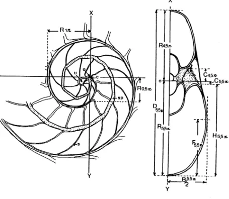
Fig. 1. Basic morphology, orientation and measurements ofthe shell of N. pompilius in median
Kagoshima Univ. Res. Center S. Pac, Occasional Papers. No.4, 1985 40
position (F) were measured on the "half" cross section at intervals of 180 by means of a profile
projector. Based on these measurements, the following four parameters proposed by Raup (1966)
and Chamberlain (1976) were calculated ; thus (1) Whorl expansion rate (W) (=(Rn/Rn-l)2],
(2)Distance ofthe whorls to the coiling axis (D) (=C/R), (3) Relative whorl thickness (S) (= B/
H), and (4) Flank position (FR) (= F/D). Among these parameters, W reflects whorl inflation,
while Dmeans the relative size ofumbilicus. Sand F characterize the fundamental whorl shape.
Basic morphology and measurements in median and cross section are diagrammatically
illustrated in Fig. 1. Biometrical analysis of the measurements was made on a desk computer
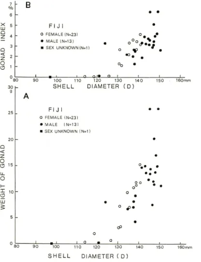
(NEC PC-9801 F). 7 % 6 B • • 5 FIJ I • 4 O FEMALE (N«23) °»- •• 3 • MALE (N-13) • SEX UNKNOWN (N-1) • o if> • 2 1 0 1 «__l 1 O OJ*. Q 1 1 1 1 a < z: O cs 15 I 10 CD 100 110 SHELL 120 130 140 DIAMETER (D) FIJ I O FEMALE (N=23) • MALE (N=13) • SEX UNKNOWN (N=1) J _ 90 100 110 120 130 140 SHELL DIAMETER ( D) -L- _U • • • o f . • cP
Fig. 2. Scatter plots of gonad weight (A) or gonad index (B) versus shell diameter for selected
specimens captured from the Suva and Pacific Harbour areas, Viti Levu Island, Fiji
(August-September, 1983). Gonad index is the ratio oftestis or ovary weight to total soft tissue weight.
Sexual Dimorphism
As we could capture various-sized Nautilus from the areas studied, allometric relationships of a shell and soft tissue were further examined in relation to sexual maturity.
The average relative growth of wet gonad (ovary or testis) weight versus shell diameter in the selected specimens from Suva and Pacific Harbour areas (Fig. 2 A) shows that in females ovary weight increases exponentially as shell diameter exceeds 120mm and attains to a maximum at the stage around 140mm in shell diameter. In contrast to this, full development of testis in males is prolonged to the stage between 140-150 mm in shell diameter. Therefore, at the stages between 120-140 mm in shell diameter gonad weight in females is slightly larger than that of the males of similar shell size. Most specimens at the stages larger than 140mm in shell diameter are males. Testis in full-grown males appears to be much heavier than ovary in mature females.
Scatter plots of gonad index (ovary or testis weight/soft tissue weight) versus shell diameter (Fig. 2 B) also suggest that gonad in both males and females increases exponentially through the stages larger than 130-140 mm in shell diameter. If a provisional standard of sexual maturity is
i o ID 111 D CO CO 500 g 400
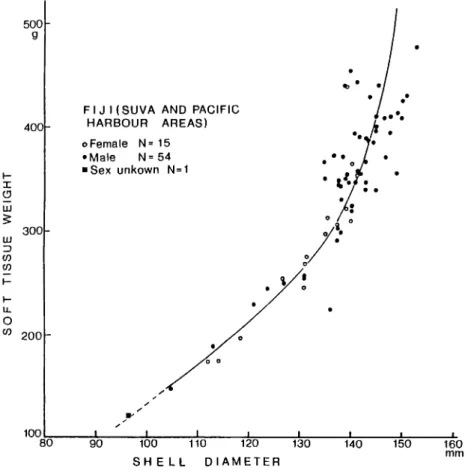
FIJ I (SUVA AND PACIFIC HARBOUR AREAS) o Female N= 15 •Male N=54 • Sex unkown N=1 300 o CO 200 100 _1_ J_ J L 80 90 100 110 120 130 SHELL DIAMETER _1_ 140 150 160 m m
Fig. 3. Scatter plots of soft tissue weight versus shell diameter for selected specimens from the Suva and Pacific Harbour areas. Curve fits by eye.
Kagoshima Univ. Res. Center S. Pac. Occasional Papers, No. 4, 1985 42
regarded to be 3.0 on gonad index, mature males are mostly larger than mature females. Also, in the specimens larger than 140mm in shell diameter, males usually possess a heavier soft tissue than females (Fig. 3). Such a sexual dimorphism expressed by the shell size or body weight has already been confirmed by us in the samples of Nautilus pompilius from Tafion Strait, the Philippines (Hayasaka et al. 1982) and N. cf. pompilius [ = N. belauensis Saunders, 1981) from Palau (Saunders and Spinosa, 1978).
Biometrics of Shells
Ontogenetic allometry
It has already been demonstrated that the growth of a spiral of many chambered cephalopod shells can be approximated by the following equation :
R = aeKH
where R and ® are a radius vector and a total rotation angle from the coiling axis respectively, and a is a constant (Moseley, 1838; Thompson, 1917; Huxley, 1932; OBATA,
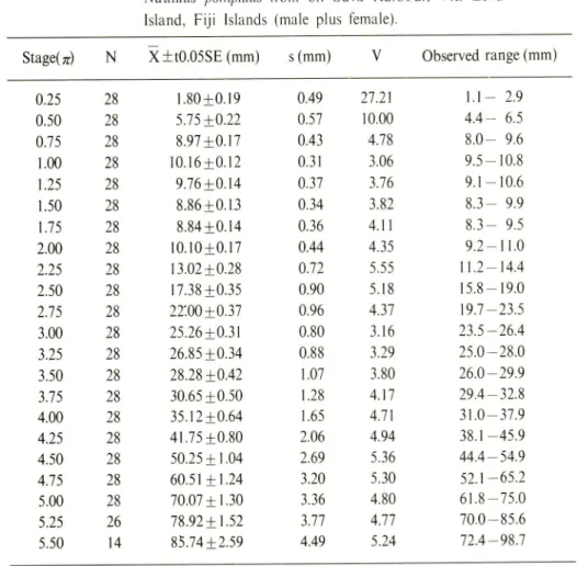
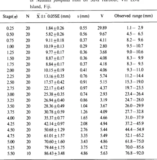
1959,1960).Hi-Table I. Statistical data for the radius vector in the sample of
Nautilus pompilius from off Suva Harbour. Viti Levu
Island, Fiji Islands (male plus female).
Staged)
N
X+t0.05SE (mm)
s(mm)
V
Observed range (mm)
0.25 28 1.80+0.19 0.50 28 5.75+0.22 0.75 28 8.97+0.17 1.00 28 10.16+0.12 1.25 28 9.76+0.14 1.50 28 8.86+0.13 1.75 28 8.84 + 0.14 2.00 28 10.10+0.17 2.25 28 13.02+0.28 2.50 28 17.38+0.35 2.75 28 22'.00 + 0.37 3.00 28 25.26+0.31 3.25 28 26.85+0.34 3.50 28 28.28+0.42 3.75 28 30.65+0.50 4.00 28 35.12+0.64 4.25 28 41.75+0.80 4.50 28 50.25 + 1.04 4.75 28 60.51 ±1.24 5.00 28 70.07 + 1.30 5.25 26 78.92+1.52 5.50 14 85.74 + 2.59 0.49 27.21 0.57 10.00 0.43 4.78 0.31 3.06 0.37 3.76 0.34 3.82 0.36 4.11 0.44 4.35 0.72 5.55 0.90 5.18 0.96 4.37 0.80 3.16 0.88 3.29 1.07 3.80 1.28 4.17 1.65 4.71 2.06 4.94 2.69 5.36 3.20 5.30 3.36 4.80 3.77 4.77 4.49 5.24 1.1-- 2.9 4.4-- 6.5 8.0-- 9.6 9.5- 10.8 9.1--10.6 8.3-- 9.9 8.3-- 9.5 9.2--11.0 11.2--14.4 15.8--19.0 19.7--23.5 23.5--26.4 25.0--28.0 26.0--29.9 29.4--32.8 31.0--37.9 38.1 -45.9 44.4--54.9 52.1--65.2 61.8 -75.0 70.0--85.6 72.4--98.7
rano (1977), Hirano and Obata (1979) and Hirano et al. (1980) applied the above equation
to express the growth of a radius vector in Nautilus pompilius and N. macromphalus. These
authors set a coiling axis at a center of the inscribed circle in the umbilical perforation. The
umbilical perforation in Nautilus is, however, not an initial portion of shell growth unlike a
protoconch of ammonites and gastropods, but is formed after the completion of the first whorl.
For this reason, we used an adaptical end of a caecum as an initial part of whorl growth (Fig. 1).
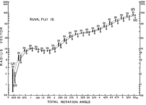
Statistical data for growth of radius vector in the specimens examined are listed in Tables 1-3. Coefficients of variation are relatively small in the specimens examined, other than the case in the
earliest stage. As shown in Fig. 4, the growth pattern of radius vector can not be expressed by a
single allometric equation. A conspicuous decrease of radius vector near the end of the first
whorl is observed, and this probably owes to a decline of whorl growth associated with a
formation of a nepionic constriction. In the stage after the second whorl the slope of allometric
shell growth in males seems to be slightly larger than that of females, but the means of radius
vector of males and females at the same total rotation angle do not show any statistically
significant difference.
Ontogenetic changes in shell form
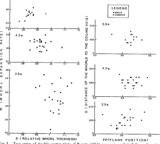
The results of this analysis may be concisely summarized in the S -W and D -FP scatter diagrams in Fig. 5. The two series of scatter diagrams show that the variation of shell form gradually decreases with growth. At the stages of 2.5 n and 4.5 n, males and females do not show
a significant difference in shell form, but at 5.5 n stage (probably mature or almost mature stage)
males have larger W and S values than females. This fact correlates well with the growth of soft
tissue weight (Fig. 2), and both data postulate that in mature or almost mature stage body chamber
Q25 Q5 0.75 1 1.25 1.5 1.75 2 2.25 2.5 2.75 3 3.25 3.5 3.75 TOTAL ROTATION ANGLE
4.25 4.5 4.75 5.25 5.5^
Fig. 4. Semi-logarithmic diagram showing the ontogenetic growth of radius vector versus total
rotation angle for selected twenty-eight specimens from the Suva area. Horizontal line and black or white rectangles indicate the arithmetic mean and twice of standard deviation.
Kagoshima Univ. Res. Center S. Pac, Occasional Papers, No. 4, 1985
volume or weight of males is usually greater than those of females.
44
Growth of the last septum thickness
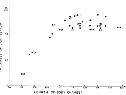
The relationship between the last septum thickness and the body chamber length was examined in the twenty-eight median-sectioned specimens from the Suva area. Except for ri\Q young specimens, all specimens have already attained to secret a final septum, judging from closing together of the last two or three septa. As shown in Fig. 6, in the specimens of less than 110 mm shell diameter the last septum gradually thickens with the increase of body chamber length. The septum attains to a full thickness (ca. 1.2-1.3 mm) when body chamber reaches to 115 in spiral angle. Fig. 6 also suggests that the growth of body chamber (= length of outer shell wall and volume of soft tissue) has been continued after the completion of the last septum. In this figure amounts of cameral liquid within the last chamber are also shown near the plots of selected thirteen specimens (ml unit). The relationship between the relative thickness of the last septum and the amount of liquid in the last chamber for the eleven specimens are shown in Fig. 7. In the specimen, SV 12-3 with a very thin, incomplete last septum (0.06 mm in thickness; about 9 % of the thickness of the preceeding septum), the newest chamber is completely filled with liquid. In
5.5 * < CC 2 o co 2 < Q_ X 111 CC O I 3.0 2.5 0.7 4.8 • 4.5 0.8 4.5* 2.5*: .* I 0.9 0.8 0.9 1 1.1 12
S ( RELATIVE WHORL THICKNESS)
LEGEND •MALE o FEMALE X < 0.10 5.5*
COILING
JO o o • • o • • • • o III I <* • • P o 1 , CO _J CC O ^ 0.10 HI X 4.5^ 0.3 0.4 0.5 2.5* 0.10 • • • & o o • 0.05 • • • • • o • fp • • • 0 1 0.3 0.4 0.5 FP(FLANK p o s i t i o n )Fig. 5. Two series of double scatter plots of Raup's (1966) parameters of shell forms for selected
specimens of N. pompilius from the Suva area, showing the ontogenetic changes of shell
the specimens, SV 10-6, 13-4 and 13-18 with an intermediate last septal thickness (ca. 20-70% of the total thickness of the preceeding septum), about 20-30% of the final chamber has already been emptied. After having attained to a maximum septal thickness, several specimens still possess small amounts of liquid (about 5 -25 %of a chamber volume) within their last chamber. However, in the specimens with a body chamber longer than 120°, the last chamber has almost been emptied. These observations provide the following conclusions: (1) first emptying occurs long before the completion of the last septum, (2) growth of body chamber and a gradual pumping of cameral liquid have been continued after the last septum has reached its maximum thickness. Fig. 7 also strongly suggests that the emptying rates gradually decrease with growth of body chamber .
Ward et al. (1981, fig. 5) and Ward and Chamberlain (1983, fig. 3) described the relation ship between cameral liquid volume and last septum thickness in N. macromphalus and TV.
pompilius on the basis of long-term radiographic observations of living animals in captivity.
According to their observations, in both species first emptying occurs when the last septum reaches to 15 to 60 %of the expected final thickness. Our data apparently differ from those of Ward et
al. (1981) and Ward and Chamberlain (1983) in the thinner septum thickness when first
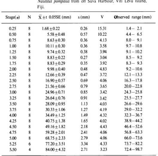
emptying occurs. The reason for this appears to be the difference in emptying patterns of animals Table 2. Statistical data for the radius vector in the male samplesof
of Nautilus pompilius from off Suva Harbour, Viti Levu Island, Fiji.
StageU) N X±t 0.05SE(mm) s(mm) V Observed range (mm)
1.1- 2.9 4.5- 6.5 8.2- 9.6 9.5-10.7 9.0-10.6 8.3- 9.9 8.3- 9.5 9.3-11.0 11.2-14.4 15.3-19.0 19.7-23.5 23.4-26.4 24.7-28.0 26.0-29.9 27.7-32.8 31.0-37.9 37.2-45.9 44.4-54.9 52.1-65.2 61.8-75.0 70.0-85.6 76.8-92.0 0.25 20 1.84+0.26 0.55 29.89 0.50 20 5.82+0.26 0.56 9.67 0.75 20 9.11+0.18 0.37 4.11 1.00 20 10.19+0.13 0.29 2.80 1.25 20 9.77+0.17 0.36 3.68 1.50 20 8.87+0.17 0.36 4.08 1.75 20 8.84+0.17 0.37 4.18 2.00 20 10.15+0.19 0.41 4.06 2.25 20 13.16+0.35 0.76 5.74 2.50 20 17.57+0.42 0.91 5.15 2.75 20 22.17+0.45 0.97 4.37 3.00 20 25.38+0.35 0.74 2.93 3.25 20 26.94 ±0.40 0.86 3.19 3.50 20 28.36+0.49 1.04 3.67 3.75 20 30.78 ±0.59 1.26 4.09 4.00 20 35.37+0.77 1.65 4.66 4.25 20 42.14 ±0.97 2.08 4.94 4.50 20 50.68 + 1.29 2.76 5.44 4.75 20 61.01 + 1.57 3.35 5.49 5.00 20 70.60 ±1.60 3.43 4.86 5.25 20 79.44 ±1.75 3.75 4.72 5.50 10 86.43 ±3.48 4.86 5.63
Kagoshima Univ. Res. Center S. Pac, Occasional Papers, No.4, 1985 46
under natural and artificial conditions. Many workers have suggested that the cameral liquid in
living Nautilus has a primary function to support calcifying septa and siphuncle against implosion
by ambient hydrostatic pressure, for the gas pressure within the chambers never exceeds
atomos-pheric pressure regardless of depth (Denton and Gilpin-Brown, 1966; Collins et al., 1980;
Ward et al., 1980, 1981 ; Ward and Chamberlain, 1983). Therefore, drainage of cameral
liquid must occur after the septum attains a sufficient thickness to withstand the high hydrostatic
pressure in the habitat. From our observation, it has been realized that even a incomplete septum
with only 20%of the final thickness can withstand the pressure of more than 40 atm.
Geographic Variation of Shell Morphology
It is well known that Nautilus pompilius is widely distributed in the tropical South Pacific.
The Philippines and Fiji Islands are situated in the northwestern and southeastern margins of the
wide habitat realm of this species (Hamada, 1977; Saunders, 1981 b). Based on the two samples from Tanon Strait, the Philippines and from the Suva area, Fiji Islands (see Hayasaka
Table 3. Statistical data for the radius vector in the female sample of
Nautilus pompilius from off Suva Harbour, Viti Levu Island,
Fiji.
Staged) N X+t 0.05SE (mm) s(mm) V Observed range (mm)
1.4- 2.1 4.4- 6.5 8.0- 9.1 9.7-10.8 9.1-10.2 8.5- 9.2 8.3- 9.3 9.2-10.6 12.1-13.3 16.3-17.8 20.0-22.8 24.3-25.8 25.5-27.7 26.6-29.6 29.0-32.2 32.3-36.7 38.9.-44.2 46.4-52.6 56.8-63.3 66.0-73.6 73.7-82.2 72.4-98.7 0.25 8 1.68 ±0.22 0.26 15.31 0.50 8 5.58 ±0.48 0.57 10.22 0.75 8 8.63 ±0.30 0.36 4.13 1.00 8 10.11 ±0.30 0.36 3.58 1.25 8 9.74 ±0.32 0.38 3.94 1.50 8 8.83 ±0.22 0.27 3.04 1.75 8 8.83 ±0.29 0.35 3.92 2.00 8 9.98 ±0.40 0.48 4.83 2.25 8 12.66 ±0.39 0.47 3.72 2.50 8 16.90 ±0.57 0.69 4.06 2.75 8 21.56 ±0.66 0.79 3.65 3.00 8 24.96 ±0.71 0.85 3.42 3.25 8 26.64 ±0.76 0.91 3.42 3.50 8 28.09 ±0.95 1.13 4.03 3.75 8 30.33 ±1.06 1.27 4.19 4.00 8 34.49 ±1.25 1.49 4.32 4.25 8 40.75 ±1.38 1.65 4.02 4.50 8 49.16±1.82 2.18 4.43 4.75 8 59.28 ±2.01 2.41 4.06 5.00 8 68.75 ±2.33 2.79 4.06 5.25 6 77.20 ±3.51 3.34 4.33 5.50 4 84.00 ±4.32 2.71 3.23
D I-CL W CO < CO CO UJ z • • ' 100 105 110 115 120
LENGTH OF BODY CHAMBER
125 130
Fig. 6. Plots of last septal thicknenss versus body chamber length for selected twenty-eight specimens of N. pompilius from the Suva area. The number beside some plots indicates cameral liquid volume (ml) in the last chamber.
100%- • • 80 -♦ • 60 -40 • l • i 20 < ♦IMMATURE • MATURE OR ALMOST MATURE 0 • •
AMOUNT OF LIQUID IN CHAMBER
(PERCENT FILLED)
Fig. 7. Plots of relative thickness of the last septum versus amounts of liquid volume within the last chamber (percent chamber volume) for selected specimens from the Suva area. Relative thickness of the last septum is expressed as percentage of total thickness of the preceeding septum for immature shells or that of the expected thickness (1.3 mm) of the full-grown final septum for almost mature shells.
Kagoshima Univ. Res. Center S. Pac, Occasional Papers, No.4, 1985 48
et al., 1982 and Hayasaka and Shinomiya, 1982 for detailed records), Tanabe et al. (1983) discussed the geographic variation of several morphological characters of N. pompilius between the two isolated areas. They concluded that the two samples show a statistically significant difference in the size and weight distributions in the mature stage. Definition of mature stage in our previous papers (Hayasaka et al., 1982 ; Tanabe et al, 1983) was, however, relied on the characteristic features of the shell such as a presence of a blacked and thickened aperture, closing together of the final two or three septa and a thickening of the last septum. In this study, we also examined a development of gonad in some specimens. As a result, it was realized that the mature females and males with a well-developed gonad have the shells of about 140 mm and 150 mm in diameter respectively (Fig. 2, B). These average shell sizes are about 20 mm smaller than those (Hayasaka et at, 1982, fig. 19; Tanabe et al, 1983, table 3) of mature males and females from
Tahon Strait, the Philippines. Thus, this paper provides more concrete data for the geographic
variation of this species in the mature stage.
Table 4. Statistical data on the nepionic shell size for the samples of N.
pompilius from the Fiji Islands (Suva area) and the Philippines
(Tafion Strait and Bohol Strait near Siquijor Islands).
Sample N X ±t 0.05SE (mm) s(mm) V Observed range (mm)
Suva 28 31.03+1.10 2.84 9.16 24.9-35.4
Tafion 6 25.72+0.95 0.90 3.50 24.7-27.1
Bohol i 22.95 + 2.25
0.25 1.09 22.7-23.2
In addition to the difference in mature stage, the present species shows a fairly large intraspecific variation in the nepionic shell size between Fiji and the Philippines. As shown in Table 4, the size ranges from 24.9 mm to 35.4 mm in the specimens from the Fiji Islands (Suva and Pacific Harbour areas); and the mean nepionic shell size (31.0 mm ; N= 28) is much larger than those of the samples from Tafion and Bohol Straits, the Philippines. From the oxygen isotopic analysis of the shells Cochran et al. (1981) and Taylor and Ward (1983) suggested that the nepionic constriction in Nautilus is probably marked at the time of hatching. If this opinion is correct, the above difference in the nepionic shell size among the three population samples
indicates the geographic variation in the size of post-embryonic young between Fiji and the
Philippines.
References
Chamberlain, J. A. Jr., 1976: Flow patterns and drag coefficients of chambered cephalopod shells. Palaeontology, 19, 539-563.
Cochran, J. K., Rye, D. M. and Landman, N. H., 1981 : Growth rate and habitat of Nautilus
pompilius inferred from radioactive and stable isotope studies. Paleobiology, 1, 469-480.
Collins, D. H., Ward, P. D. and Westermann, G. E. G., 1980 : Function of cameral water in
Nautilus. Paleobiology, 6, 168-172.
Biol. Assoc. U. K., 46, 723-759.
Hamada, T.,1977: Distribution and some ecological barriers on the habitat condition of
Nautilus and its application to the rearing of Nautilus macromphalus. Sci. Paps. Coll. Gen. Educ, Univ. Tokyo, 27, 89-102.
Hayasaka, S. and Shinomiya, A., 1982 : Marine ecological study on the habitat of Nautilus in the environs of Viti Levu Island, Fiji Islands. Sci. Res. Rep. Oceania, (NA V '81) Kagoshima
Univ. Res. Center S. Pac, I. 69-73 (in Japanese).
, Saisho, T., Kakinuma, Y., Shinomiya, A., Oki, K., Hamada, T.Janabe, K., Kanie, Y., Hattori, M., Vande Vusse, F., Alcala, U Cordero, P. A. Jr., Cabrera, J. J.
and Garcia, R. G., 1982: Field study on the habitat of Nautilus in the environs of Cebu and Negros Islands, the Philippines. Mem. Kagoshima Univ., Res. Center S. Pac, 3(1).
67-115.
Hirano, H., 1977: Biometrical characteristics of Nautilus pompilius. Gakujutsu Kenkyu (The
Sci. Res.), Ser. Biol. Geol., School of Educ, Waseda Univ., 26, 13-23.
and Obata, I., 1979 : Shell morphology of Nautilus pompilius and A^. macromphalus.
Bull. Natl. Sci. Mus., Ser. C (Geol. & Paleont.), 5, 113-130.
, and Tanabe, K. 1980: Biometric characteristics (In: Hamada, T., Obata, I. and Okutani, T. eds., 1980) p. 34-43. Tokai Univ. Press, Tokyo.
Huxley. J. S., 1932: Problems of Relative Growth. 312 p. Dover Publ., New York.
Moseley, H., 1838: On the geometrical forms of turbinated and discoid shells. Phil. Trans. Royal Soc. Lond. in 1838, 351-370.
Obata, I., 1959 : Croissance sur quelques Especes des Desmoceratidae. Mem. Fac. Sci., Kyushu
Univ., Ser. D (Geol.), 9, 33-45.
, 1960: Spirale de quelques ammonites. Ibid., 9, 151-163.
Raup, D. M., 1966: Geometric analysis of shell coiling: general problems. /. Paleont., 40,
1178-1190.
Saunders, W. B., 1981 a : A new species of Nautilus from Palau. The Veliger, 24, 1-7.
, 1981 b : The species of living Nautilus and their distribution. Ibid., 24, 8-17. Tanabe, K., Hayasaka, S., Saisho, T., Shinomiya, A. and Aoki, K. 1983: Morphologic
variation of Nautilus pompilius from the Philippines and Fiji Islands (In : Hayasaka, S. ed, 1983.). Kagoshima Univ., Res. Center S. Pac, Occasional Papers., 1, 9-21.
Taylor, B. E. and Ward, P. D., 1983: Stable isotopic studies of Nautilus macromphalus Sowerby (New Caledonia) and Nautilus pompilius L. (Fiji). Palaeogeogr., Palaeoclimatol,
Palaeoecol., 41, 1-16.
Thompson, D., 1917: On growth and Form. 1116 p. Cambridge Univ. Press, London.
Tsukahara, J., 1985: Histological and histochemical studies of gonads of Naultilus pompilius from Fiji (In: Hayasaka, S. ed., 1985). Mem. Kagoshima Univ., Res., Center S. Pac, Occasional Papers, 4, 50-60.
Ward, P. D. and Chamberlain, J. A. Jr., 1983: Radiographic observation of chamber forma tion cycle in Nautilus pompilius. Nature, 303, 57-59.
, Greenwald, L. and Greenwald, O., 1980: The buoyancy of the chambered
Nautilus. Sci. Amer., 243, 190-203.
, and Magnier, Y., 1981 : The chamber formation cycle in Nautilus
Figs. 1-3. Median sections of the shells of Nautilus pompilius from Suva area, Fiji Islands.
1. Early whorls of specimen SV 13-2 (male). Scale bar indicates 5 mm. Arrow points
to a primary constriction.
2. Specimen SV 15-3 (female). Scale bar represents 1cm. 3. Specimen SV 14-3 (male). Scale bar represents 1cm.
Tanabe et al.: Morphological Analysis f
i
>
/
( v
/
¥*
' X\
j ^ iT!l|
Plate 7I
XFigs. 1-6. Dorsal views of the mature or almost mature shells of Nautilus pompilius from the
Suva area, Fiji Islands. All X2/3. 1. Specimen SV 13 - 3 (mature female). 2. Specimen SV 5 - 3 (mature female). 3. Specimen SV 12-1 (almost mature female). 4. Specimen SV 14-5 (almost mature female). 5. Specimen SV 14-2 (almost mature female). 6. Specimen SV 14-3 (mature female).
Tanabe et al.: Morphological Analysis Plate
lift
HHnHHP 4 H illlllv:
P 1
>.••:.•.:.•::;•Figs. 1-4. Nautilus pompilius from Suva area, Fiji Islands. Lateral (a) and dorsal (b) views of each specimen. All X 0.5.
1. Specimen SV 10-4 (immature male). 2. Specimen SV 10-3 (immature female). 3. Specimen SV 10-2 (immature male). 4. Specimen SV 10-1 (immature male).
Tanabe et al.: Morphological Analysis Plate 9