IRUCAA@TDC : Loss of heterozygosity and microsatellite instability on chromosome 2q in human oral squamous cell carcinoma
10
0
0
全文
(2) 17. Bull Tokyo Dent Coll (2005) 46 (1–2): 17–25. Original Article. Loss of Heterozygosity and Microsatellite Instability on Chromosome 2q in Human Oral Squamous Cell Carcinoma Hideyuki Numasawa, Nobuharu Yamamoto, Akira Katakura and Takahiko Shibahara Department of Oral and Maxillofacial Surgery, Tokyo Dental College, 1-2-2 Masago, Mihama-ku, Chiba 261-8502, Japan. Received 1 April, 2005/Accepted for Publication 27 July, 2005. Abstract Allelic imbalance or loss of heterozygosity (LOH) and microsatellite instability (MSI) have been used to identify regions on chromosomes that may contain putative tumor suppressor genes. To obtain a detailed understanding of genetic alterations in oral cancer, 10 highly polymorphic markers mapped on chromosome 2 were used to examine 25 cases of oral squamous cell carcinoma (SCC). With these, we analyzed chromosome 2q for LOH in 25 primary oral SCCs and constructed a deletion map for this arm of the chromosome. LOH was detected in 16 (64%) of the 25 informative samples at one or more of the loci examined. MSI was observed in 5 (20%) of the 25 cases. Among the loci examined, LOHs were restricted to D2S1328 and D2S206 on chromosomes 2q14-21 and 2q36, respectively, with the former locus showing a rate of 5 (20.8%) and the latter a rate of 6 (25%) of the 24 informative cases. These observations taken in conjunction with data from 40 former cases analyzed at our laboratory suggest that the high incidence of LOH at chromosome 2q is associated with carcinogenesis of oral SCC. The regions that comprise the D2S1328 and D2S206 loci may play an important role in the development of oral SCC, perhaps containing sites that harbor a putative tumor suppressor gene. Key words:. Chromosome 2— Oral squamous cell carcinoma— Loss of heterozygosity (LOH)— Microsatellite instability (MSI)— Tumor suppressor gene. Introduction. is known about the molecular basis of oral SCC compared to other human malignancies. Inactivation of the tumor suppressor gene (TSG) is considered to be associated with carcinogenesis, and alterations in TSGs are widely accepted to be critical events in the. Oral squamous cell carcinoma (SCC) is the most common malignant disease of the oral and maxillofacial region, and the sixth most frequent cancer worldwide12). However, little. This paper is a thesis submitted by Dr. H. Numasawa to the Graduate School of Tokyo Dental College.. 17.
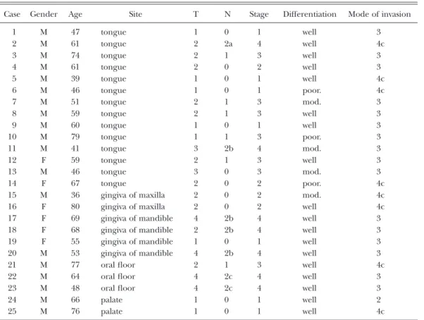
(3) 18. Numasawa H et al.. Table 1 Summary of clinicopathologic features in 25 oral SCCs Case. Gender. Age. Site. T. N. Stage. Differentiation. Mode of invasion. 1 2 3 4 5 6 7 8 9 10 11 12 13 14 15 16 17 18 19 20 21 22 23 24 25. M M M M M M M M M M M F M F M F F F F M M M M M M. 47 61 74 61 39 46 51 59 60 79 41 59 46 67 36 80 69 68 55 53 77 64 48 66 76. tongue tongue tongue tongue tongue tongue tongue tongue tongue tongue tongue tongue tongue tongue gingiva of maxilla gingiva of maxilla gingiva of mandible gingiva of mandible gingiva of mandible gingiva of mandible oral floor oral floor oral floor palate palate. 1 2 2 2 1 1 2 2 1 1 3 2 3 2 2 2 4 2 1 4 2 4 4 1 1. 0 2a 1 0 0 0 1 1 0 1 2b 1 0 0 0 0 2b 2b 0 2b 1 2c 2c 0 0. 1 4 3 2 1 1 3 3 1 3 4 3 3 2 2 2 4 4 1 4 3 4 4 1 1. well well well well well poor. mod. well well poor. mod. well mod. poor. mod. well well well well well well well well well well. 3 4c 3 3 4c 4c 3 3 3 3 3 3 3 4c 4c 4c 3 3 3 3 4c 3 3 2 4c. M: Male, F: Female. well: well differentiated, mod.: moderately differentiated, poor.: poorly differentiated. multi-step process leading toward the development of cancer. It is generally accepted that the transformation of normal tissue into malignant tissue follows an accumulation of genetic changes in the oncogenes and TSGs1). Frequent allelic losses at specific chromosomal loci in several types of human cancer have provided strong evidence to suggest the existence of TSGs on each chromosome where a deletion was detected. Cytogenetic and molecular biologic studies of human cancers have identified chromosomal abnormalities, including partial deletions of specific loci15). In particular, loss of heterozygosity (LOH) and microsatellite instability (MSI) on human chromosome 2 is known to be involved in various types of human cancers, such as neuroblastoma16), thyroid cancer 25), lung cancer11), stomach can-. cer7), head and neck cancer14), and granulosa cell tumor 24). Based on these observations, 2 common loss sites have been identified in the 2q14-21 and 2q36 regions, suggesting the presence of possible TSGs at these regions. In oral cancer, cytogenetic analysis has revealed consistent chromosomal alterations involving chromosomes 3p13,18), 4q20), 5q22), 7q21), 8p10), 9p6), 10q30), 11q19), 13q8), 18q23), 20q2), 21q29) and 22q5). Our previous allelotyping study of oral 40 SCCs revealed high frequencies of LOH on the long arm of chromosome 227,28). In the present study, we examined 25 oral tumors with 10 high polymorphic microsatellite markers as a preliminary step toward isolating putative TSGs associated with oral SCC on human chromosome 2, and constructed a detailed deletion map of this chromosome..
(4) 19. Allelic Loss on Chromosome 2q in Oral Cancer. Table 2 Allelic imbalances (LOH and MSI) at 10 microsatellite loci on chromosome 2q in 25 oral SCCs Locus symbol. Chromosomal location. D2S436 D2S1328 D2S111 D2S202 D2S116 D2S1327 D2S155 D2S164 D2S133 D2S206. 2q11.1-14 2q14-21 2q23-33 2q32 2q32-35 2q33 2q35 2q35 2q36 2q36. Frequency of LOH: % (LOH/informative cases) 10.0 20.8 16.0 17.4 8.3 9.1 4.1 13.6 4.5 25.0. Materials and Methods Samples were obtained from 25 Japanese patients with oral SCC who visited the Tokyo Dental College Chiba Hospital over a 7-year period between 1997 and 2003 (Table 1). Informed consent was obtained from all patients and also from their families. This study was also approved by the Institutional Ethics Committee of Tokyo Dental College. The samples consisted of 25 primary tumors and the same number of normal tissues corresponding to those primary tumors taken from the same patients. Three of these cases showed metastatic tumors of the lymph node. In these cases, further samples were taken of the metastasized tumors. They were collected either at the time of surgical resection (no chemotherapy or radiotherapy was given before surgical resection in any of the cases) or at the time of biopsy. Clinicopathologic staging was analyzed using the UICC TNM staging system17). 1. Tissue samples The obtained tissues were divided into two segments: one was frozen immediately after careful removal from the surrounding normal tissue and stored at ⳮ80°C until extraction of DNA, and the other was fixed in 10% formalin for pathologic diagnosis. Histopathologic diagnosis was performed according to the. (2/20) (5/24) (4/25) (4/23) (2/24) (2/22) (1/24) (3/22) (1/22) (6/24). Frequency of MSI: % (MSI/informative cases) 5.0 4.5 0.0 0.0 0.0 4.5 0.0 9.1 0.0 4.2. (1/20) (1/24) (0/25) (0/23) (0/24) (1/22) (0/24) (2/22) (0/22) (1/24). International Classification of Tumors26). 2. DNA extraction All cases had histologically confirmed SCC of the oral cavity, and the tumor samples for DNA extraction were inspected to make sure that they consisted of more than 80% tumor. DNA extraction from all tumor and most of the normal tissue samples was performed from the fresh frozen specimens that had been preserved in liquid nitrogen immediately after resection. First, the tissues were powdered in liquid nitrogen. Next, they were centrifuged to obtain deposition. After that, 1,000 l of TNES buffer (10 mM Tris-HCl (pH8.0), 150 mM NaCl, 10 mM EDTA, 0.1% SDS) and 30 l Proteinase K (100 g/ml) were added to the powder and incubated in a hot water-bath overnight at 50°C. Genomic DNAs were extracted by phenol-chloroform extraction and refined, and then they were washed and precipitated with ethanol4). A spectrophotometric method was used to estimate the concentrations of the extracted DNA and the DNA extracts were kept frozen at ⳮ80°C. 3. PCR and microsatellite analysis We used 10 microsatellite markers located on chromosome 2q. All primers were obtained from Research Genetics (Huntsville, AL, Table 2). PCR amplification was performed at.
(5) 20. Numasawa H et al.. Fig. 1 Deletion mapping of chromosome 2q in 25 primary tumors The case number is shown above each column, and the 10 microsatellite markers are on the left.. a total reaction volume of 20 l, as described previously3). Each PCR reaction mixture contained 250 ng of sample DNA, 20 pmol of each primer, 10 mM Tris-HCl (pH8.3), 50 mM KCl, 3.0 mM MgCl2, 2 mM dNTP, and 0.5 units of Taq DNA polymerase (PerkinElmer Cetus, Norwalk, CT). Thirty-five cycles consisting of denaturation at 94°C for 1 min, annealing at 55°C for 1 min, and extension at 72°C for 1 min were performed with a DNA Thermal Cycler (Perkin-Elmer Cetus, Norwalk, CT). After dilution with an adequate volume of formamide-dye mixture (95% formamide, 20 mM EDTA, 0.05% bromophenol blue, and 0.05% xylene cyanol), the PCR products were heat-denatured (98°C, 5 min), chilled on ice, and electrophoresed on 6% urea-formamide-polyacrylamide gel at 3W for 2 to 3 hours, depending on the fragment size. Silver staining of the gels was performed using a DNA Silver Staining Kit (Amersham Pharmacia Biotech AB, Sweden). To ensure reproducibility for each instance of LOH or MSI, all tests were performed under the same conditions.. 4. Assessment of LOH and microsatellite instability (MSI) We assessed LOH by scanning a stained gel using a densitometer, followed by analysis with National Institute of Health (NIH) software (Image version 1.62, Dr. W. Rasband, NIH, Bethesda, MD, USA). We compared the intensities of the signals in tumor DNA with those of the corresponding normal DNA. A reduction in signal intensity of more than 50% in a tumor sample compared with that in a normal counterpart was required as the criterion for the presence of LOH. Commonly deleted regions were defined as those loci most frequently showing LOH together with multiple interstitial deletions27). MSI in the DNA samples was assessed as positive in those cases which had additional bands in the tumor sample that were not observable in the corresponding normal samples and in those cases which showed a band shift in the tumor sample that contrasted with the pattern of the corresponding normal bands. 5. Statistical analysis A Fisher’s exact test was performed to evaluate the significance of the correlation.
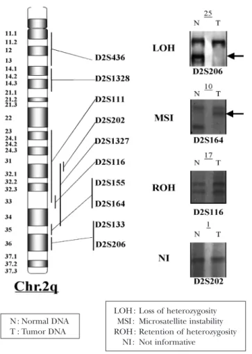
(6) Allelic Loss on Chromosome 2q in Oral Cancer. N : Normal DNA T : Tumor DNA. LOH : MSI : ROH : NI :. 21. Loss of heterozygosity Microsatellite instability Retention of heterozygosity Not informative. Fig. 2 Typical examples of allelic imbalances (LOH and MSI) on 2q Microsatellite polymorphism analysis in oral SCC specimens. Case numbers are shown above, and locus symbols are indicated below. Paired normal (N) and tumor (T) samples demonstrating loss of the upper allele (LOH, case no. 25), microsatellite instability (MSI, case no. 10), retain of heterozygosity (ROH, case no. 17), and uninformative (NI, case no. 1).. between LOH and the clinicopathological parameters. The accepted level was p⬍0.05.. Results Ten microsatellite markers on the long arm of chromosome 2 were analyzed in 25 oral SCCs with various grades of differentiation and clinical stages. At all the loci examined on this chromosome in normal tissues, 20 or more cases were heterozygotes. Sixteen out of twenty-five cases (64%) of the oral SCCs showed a loss of heterozygosity in one or. more of the microsatellite markers on chromosome 2. The locations of the markers and frequencies of LOH and MSI are summarized in Table 2 and Fig. 1. A high frequency of LOH was observed at the D2S1328 locus (20.8%) on 2q14-21, and the D2S206 locus (25%) on 2q36. MSI in chromosome 2q was present in 5 out of 25 cases (20%) of tumors (Table 2, Fig. 1). Typical examples of LOH, MSI, retention of heterozygosity case (ROH) and not informative case (NI) on 2q are shown in Fig. 2. The frequencies of LOH in the lymph node metastases revealed that 2 out of the 3.
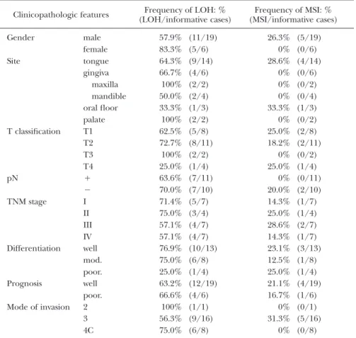
(7) 22. Numasawa H et al.. Table 3 Summary of clinicopathologic features in 25 oral SCCs Clinicopathologic features Gender Site. T classification. pN TNM stage. Differentiation. Prognosis Mode of invasion. male female tongue gingiva maxilla mandible oral floor palate T1 T2 T3 T4 Ⳮ ⳮ I II III IV well mod. poor. well poor. 2 3 4C. Frequency of LOH: % (LOH/informative cases) 57.9% 83.3% 64.3% 66.7% 100% 50.0% 33.3% 100% 62.5% 72.7% 100% 25.0% 63.6% 70.0% 71.4% 75.0% 57.1% 57.1% 76.9% 75.0% 25.0% 63.2% 66.6% 100% 56.3% 75.0%. (66.7%) showed LOH at D2S436. In our previous studies on 7 cases of lymph node metastases, LOH was also found at the same locus in 1 out of the 3 informative cases examined. However, among the total 7 cases, LOH was additionally found at D2S1327, D2S155, and D2S164, yielding frequencies of 33.3 (2/6), 100 (3/3), 57.1 (4/7) and 33.3%28). We compared our results with the clinicopathological features of each tumor (Table 3). A number of sites displaying LOH at 2q could be detected in early stage lesions, and the frequencies of LOH tended to be higher in the later clinical stages, although no statistical difference was observed. No significant correlation between incidence of LOH and grade differentiation was noted. MSI was present at 4 loci in the chromosome, but again there was. (11/19) (5/6) (9/14) (4/6) (2/2) (2/4) (1/3) (2/2) (5/8) (8/11) (2/2) (1/4) (7/11) (7/10) (5/7) (3/4) (4/7) (4/7) (10/13) (6/8) (1/4) (12/19) (4/6) (1/1) (9/16) (6/8). Frequency of MSI: % (MSI/informative cases) 26.3% 0% 28.6% 0% 0% 0% 33.3% 0% 25.0% 18.2% 0% 25.0% 0% 20.0% 14.3% 25.0% 28.6% 14.3% 23.1% 12.5% 25.0% 21.1% 16.7% 0% 31.3% 0%. (5/19) (0/6) (4/14) (0/6) (0/2) (0/4) (1/3) (0/2) (2/8) (2/11) (0/2) (1/4) (0/11) (2/10) (1/7) (1/4) (2/7) (1/7) (3/13) (1/8) (1/4) (4/19) (1/6) (0/1) (5/16) (0/8). no apparent association between the incidence of MSI and histopathological grading or clinical status.. Discussion In order to isolate putative TSGs via positional cloning, a number of detailed deletion maps have been constructed for each chromosome in relation to a range of cancers in human. In particular, chromosome 2 is wellknown to frequently alter in patients with any one of several types of malignant tumor, such as neuroblastoma16), thyroid cancer 25), lung cancer11), stomach cancer 7), head and neck cancer14), or granulosa cell tumor 24). Two of our recent allelotyping studies on oral SCC.
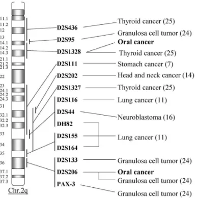
(8) Allelic Loss on Chromosome 2q in Oral Cancer. 23. Fig. 3 Commonly deleted regions on 2q in human cancers Comparisons of reported examples in other cancers on chromosome 2q with the oral cancers in the microsatellite analysis reported herein. Malignant tumors of cancers in thyroid, stomach, lung, head and neck, and granulose cell tumor have been already analyzed on 2q, but no report on oral cancers has been made except by us. The numbers in the parentheses indicate the reference number.. also revealed high frequencies of LOH on the long arm of chromosome 227,28). Therefore, in the present study, in order to confirm the findings of those studies, we further examined the same 10 microsatellite markers on chromosome 2q by using new samples of SCCs. In our earlier studies on 40 cases of oral SCCs, we found that the incidence of LOH in 10 microsatellite markers on chromosome 2 was 67.5% (27/40) in primary tumors. High frequencies of LOH at these loci (65%) were also found in this study. Among those found in this study, we found 2 distinct putative tumor suppressor loci, D2S206 and D2S1328. High frequencies of LOH at these loci were also found in the results of our earlier studies. Frequent allelic loss of the D2S206 locus located on band 2q36 was also reported in granulosa cell tumor10). This finding suggests. that a putative suppressor gene located at this region plays a role in both granulosa cell tumor and oral cancer. The second region, including the D2S1328 locus at chromosome 2q14-21, was found to have frequent allelic loss in thyroid cancer 25). This finding suggests that a putative suppressor gene located at this region plays an important role in both thyroid and oral cancers (Fig. 3). However, among these high-frequency LOHs, oral cancer was found to be the most prevalent. Therefore, this suggests that LOH at this location includes an important TSG in the development of oral cancers. A more detailed deletion map needs to be compiled in order to further clarify this finding. In 7 metastatic tumors of our previous studies on 40 cases of oral SCC, we found higher frequencies of LOH in at D2S436 (33.3%), D2S1327 (33.3%), D2S155 (100%),.
(9) 24. Numasawa H et al.. and D2S164 (57.1%) compared with LOH in primary tumors28). Deletion of one of these loci D2S436 was also found in 2 of the 3 metastatic cases of this study. These findings suggest that these loci contribute to the development of metastase. However, this may also be attributed either to a sampling bias (types of tumor studied, patient population differences) or to differences in technical and interpretive approaches. Taking into account the LOHs of primary tumors, it would appear that alterations at several regions on chromosome 2q play an important role in the genesis of oral SCC. In this study, no statistically significant correlation was found between presence of LOH, TNM stage, and histological grade in the patients. These results indicate that inactivation of the TSG is an early event in oral SCC, because low-stage tumors have a similar frequency of LOH to more advanced cases. Our findings showed a high incidence of MSI at multiple loci in oral SCC in 20% of the cases examined (5/25). Another important finding of the present study was the higher incidence of MSI in tumors located in the tongue compared with at other anatomic locations in the oral cavity9). This suggests that carcinogenesis in tongue tissue may be influenced by different TSGs to that at other sites in the oral cavity, such as the gingiva or the palate. However, more specimens are necessary to confirm this result because our sample size was small. In conclusion, our present data taken together with the results of our previous studies27,28), suggest that putative TSGs are located in the 2q14-21 and 2q36 regions and that inactivation may occur at an early stage in the progression of oral tumors.. 3). 4). 5). 6). 7). 8). 9). 10). 11). 12) 13). References 1) Fearon ER, Vogelstein B (1990) A genetic model for colorectal tumorigenesis. Cell 61:759–767. 2) Imai FL, Uzawa K, Miyakawa A, Shiiba M, Tanzawa H (2001) A detailed deletion map of. 14). chromosome 20 in human oral squamous cell carcinoma. Int J Molecular Medicine 7:43–47. Komiya A, Suzuki H, Aida S, Yatani R, Shimazaki J (1995) Mutational analysis of CDKN2 (CDK4I/MTS1) gene in tissues and cell lines of human prostate cancer. Jpn J Cancer Res 86:622–625. Maniatis T, Fritsch EF, Sambrook J (1982) Molecular Cloning; A Laboratory Manual, 1st ed., pp.280–281, Cold Spring Harbor Laboratory Press, Cold Spring Harbor, New York. Miyakawa A, Wang XL, Nakanishi H, Imai FL, Shiiba M, Miya T, Imai Y, Tanzawa H (1998) Allelic loss on chromosome 22 in oral cancer: Possibility of the existence of a tumor suppressor gene on 22q13. Int J Oncol 13:705–709. Nakanishi H, Wang XL, Imai FL, Kato J, Shiiba M, Miya T, Imai Y, Tanzawa H (1999) Localization of a novel tumor suppressor gene loci on chromosome 9p21-22 in oral cancer. Anticancer Res 19:29–34. Nishizuka S, Tamura G, Terashima M, Satodate R (1998) Loss of heterozygosity during the development and progression of differentiated adenocarcinoma of the stomach. J Pathol 185:38–43. Ogawara K, Miyakawa A, Shiiba M, Uzawa K, Watanabe T, Wang XL, Sato T, Kubosawa H, Kondo Y, Tanzawa H (1998) Allelic loss of chromosome 13q14.3 in human oral cancer: Correlation with lymph node metastasis. Int J Cancer 79:312–317. Ogawara K, Uzawa K, Nakanishi H, Yokoe H, Wang XL, Tanzawa H, Sato K (1997) Frequent microsatellite instability in oral cancer. Oncol Rep 4:161–165. Ono K, Miyakawa A, Fukuda M, Shiiba M, Uzawa K, Watanabe T, Miya T, Yokoe H, Imai Y, Tanzawa H (1999) Allelic loss on the short arm of chromosome 8 in oral squamous cell carcinoma. Oncol Rep 6:785–789. Otsuka, T, Kohno T, Mori M, Noguchi M, Hirohashi S, Yokota J (1996) Deletion mapping of chromosome 2 in human lung carcinoma. Genes Chromosomes Cancer 16: 113–119. Parkin DM, Pisani P, Ferlay J (1993) Estimates of the worldwide incidence of eighteen major cancers in 1985. Int J Cancer 54:594–606. Partridge M, Emilion G, Langdon JD (1996) LOH at 3p correlates with a poor survival in oral squamous cell carcinoma. Br J Cancer 73:366–371. Ransom DT, Barnett TC, Bot J, de Boer B, Metcalf C, Davidson JA, Turbett GR (1998) Loss of heterozygosity on chromosome 2q: possibly a poor prognostic factor in head and neck cancer. Head and Neck 20:404–410..
(10) Allelic Loss on Chromosome 2q in Oral Cancer. 15) Suzuki H, Emi M, Komiya A, Fujiwara Y, Yatani R, Nakamura Y, Shimazaki J (1995) Localization of a tumor suppressor gene associated with progression of human prostate cancer within a 1.2Mb region of 8p22-21.3. Genes Chromosomes Cancer 13:168–174. 16) Takita J, Hayashi, Y, Kohno T, Shikeki M, Yamaguchi N, Hanada R, Yamamoto K, Yokota J (1995) Allelotype of neuroblastoma. Oncogene 11:1829–1834. 17) UICC (1987) TNM Classification of Malignant Tumours, 4th ed., pp.16–18, Springer, Berlin. 18) Uzawa N, Akanuma D, Negishi A, Amagasa T, Yoshida M (2000) Loss of heterozygosity on the short arm of chromosome 3 in oral squamous cell carcinomas: Relationship between loss of heterozygosity on 3p25-pter region and clinical and histological features. Jpn J Oral Maxillofac Surg 46:455–461. (in Japanese) 19) Uzawa K, Suzuki H, Komiya A, Nakanishi H, Ogawara K, Tanzawa H, Sato K (1996) Evidence for two distinct tumor-suppressor gene loci on the long arm of chromosome 11 in human oral cancer. Int J Cancer 67:510–514. 20) Wang XL, Uzawa K, Imai FL, Tanzawa H (1999) Localization of a novel tumor suppressor gene associated with human oral cancer on chromosome 4q25. Oncogene 18:823–825. 21) Wang XL, Uzawa K, Miyakawa A, Shiiba M, Watanabe T, Sato T, Miya T, Yokoe H, Tanzawa H (1998) Localization of a tumour-suppressor gene associated with human oral cancer on chromosome 7q31.1. Int J Cancer 75:671–674. 22) Wang XL, Uzawa K, Nakanishi H, Tanzawa H, Sato K (1997) Allelic imbalance on the long arm of chromosome 5 in oral squamous cell carcinoma. Int J Oncol 10:535–538. 23) Watanabe T, Wang XL, Miyakawa A, Shiiba M, Imai Y, Sato T, Tanzawa H (1997) Mutational state of tumor suppressor genes (DCC, DPC4) and alteration on chromosome 18q21 in human oral cancer. Int J Oncol 11:1287–1290. 24) Watson RH, Roy Jr WJ, Davis M, Hitchcock A, Campbell IG (1997) Loss of heterozygosity at the ␣-inhibin locus on chromosome 2q is not. 25). 26). 27). 28). 29). 30). 25. a feature of human granulosa cell tumors. Gynecol Oncol 65:387–390. Williams ST, Douglas WS, Zahid K, Doris JT, Samuel Jr AW, Paul JG (1997) Allelotype of follicular thyroid carcinomas reveals genetic instability consistent with frequent nondisjunctional chromosomal loss. Genes Chromosomes Cancer 19:43–51. World Health Organization (1971) International Histological Classification of Tumours; Histological Typing of Oral and Oropharyngeal Tumours, No. 4, WHO, Geneva. Yamamoto N, Mizoe J, Numasawa H, Tsujii H, Shibahara T, Noma H (2003) Allelic loss on chromosomes 2q, 3p and 21q: possibly a poor prognostic factor in oral squamous cell carcinoma. Oral Oncol 39:796–805. Yamamoto N, Mizoe J, Numasawa H, Yokoe H, Uzawa K, Shibahara T, Tsujii H, Noma H, Tanzawa H (2003) Allelic loss of chromosome 2 in human oral squamous cell carcinoma: correlation with lymph node metastasis. Oral Oncol 39:64–68. Yamamoto N, Noma H, Shibahara T (2001) Allelic imbalance on the long arm of chromosome 21 in human oral squamous cell carcinoma: relationship between allelic imbalances (LOH and MSI) and clinicopathologic features. Bull Tokyo Dent Coll 42:211–223. Yamashita Y, Miyakawa A, Mochida Y, Aisaki K, Yama M, Shiiba M, Watanabe T, Yokoe H, Uzawa K, Imai Y, Tanzawa H (2002) Genetic aberration on chromosome 10 in human oral squamous cell carcinoma. Int J Oncol 20: 595–598.. Reprint requests to: Dr. Takahiko Shibahara Department of Oral and Maxillofacial Surgery, Tokyo Dental College, 1-2-2 Masago, Mihama-ku, Chiba 261-8502, Japan E-mail: [email protected].
(11)
図


+3
関連したドキュメント
(24) Similarly, T26 inhibited both Pim-3 kinase activity in a cell-free system and in vitro cell proliferation of human pancreatic cancer cell lines at micromo- lar
NELL1 (a) and NELL2 (b) mRNA expression levels in renal cell carcinoma cell lines OS-RC-2, VMRC-RCW, and TUHR14TKB and control HEK293T cells were analyzed using quantitative
Abbreviations: DSBs, DNA double-strand breaks; ESCC, esophageal squamous cell carcinoma; γ H2AX, H2AX phospho rylation; HDACs, histone deacetylases; HR, homologous
To examine the expression of cell competition markers at the interface between normal and transformed epithelial cells, we focused on studying the p53 signature of the human
As it is involved in cell growth, IER3 expression has been examined in several human tumors, including pancreatic carcinoma, ovarian carcinoma, breast cancer, and
The objectives of this study were to evaluate the formation of lymphvascular niches in lymph nodes of patients with oral squamous cell carcinoma (OSCC), and investigate the roles
学位授与番号 学位授与年月日 氏名
Recently, we reported that the CSC markers epithelial cell adhesion molecule (EpCAM) and CD90 are expressed independently in primary HCCs and cell lines, and CD90 + cells share
