岡崎女子短期大学研究紀要45号 抜粋
平成24年3月25日
Measurement of Nocturnal Physical Activity Using a
Three-Dimensional Accelerometer in Children with Cerebral Palsy
Jun Shiragaki
Nobuaki Iwasaki
Introduction
The circadian rhythm is a biological rhythm that has been shown to be related to mental health (Fukuda & Ishihara, 2001). Therefore, sleep should be considered an important element of any individual's physical and mental health (Hayashi & Katada, 2002).
It has been suggested that the sleep of patients with cerebral palsy (CP) is often disturbed (Prechtl et al., 1973 ; Hori & Kodama, 1975), and sleep problems in children with severe CP may be attributed to obstructive sleep apnea (Magardino & Tom, 1999) caused by their inability to change body position (Kotagal et al., 1994).
Various methods have been used to measure physical activity (Laport et al., 1985). However, those former methods were complicated and costly ; hence, they have not been established. In that regard, a 3D accelerometer is useful for assessing daily physical activity, in that it can produce accurate data, is easy and convenient, does not inhibit daily activities of subjects, and has good test-retest reliability (Sugimoto, 1995 ; Sugimoto et al., 1997).
A 3D accelerometer is also useful in measuring the physical activity of children with CP (Shiragaki et al., 2000) ; Shiragaki et al. (2000) reported for the first time on
measurement of physical activity by applying a 3D accelerometer method to children with CP in free-living conditions. Moreover, the data generated by the 3D accelerometer method is useful to demonstrate patterns of nocturnal physical activity in children with CP (Shiragaki et al., 2000).
The purpose of this study was to reveal the nocturnal physical activity of children with CP using a 3D accelerometer and to make a comparative study of nocturnal physical activity patterns in children with CP and in normal children.
Subjects and methods Subjects
Thirty-one boys (mean 8.27, SD 3.08 years) with spastic cerebral palsy who had no major athetoid or ataxic characteristics participated in this study (CP group). All subjects with CP have intellectual disabilities as accompanying symptoms. The control subjects were 12 normal boys (mean 6.92, SD 2.46 years) without neurological abnormalities (control group). No significant differences were observed in age between the CP group and control group. The CP group was divided into four subgroups according to gross motor function (Johnson et al., 1951) : 8 children were bed-ridden (bed-ridden
**Department of Preschool Education, Okazaki Women's Junior College ** Department of Pediatrics, Ibaraki Prefectural University of Health Sciences
【研究論文】
Measurement of Nocturnal Physical Activity Using a
Three-Dimensional Accelerometer in Children with Cerebral Palsy
Jun Shiragaki*
Nobuaki Iwasaki**
Abstract
The purpose of this study was to research nocturnal physical activity in children with cerebral palsy (CP) by a 3D accelerometer and to compare nocturnal physical activity patterns in children with CP with those in normal children. It was considered that 3D accelerometer could show patterns of nocturnal physical activity in CP subjects objectively, and could be useful in estimating the effort of changing body position and in investigating the quality of physical activity.
group), 10 children were capable of rolling (rolling group), 8 children were capable of crawling (crawling group), and 5 children were capable of standing and walking (standing / walking group). There were significant differences between the age of the five groups including the control group (4/38, F=4.205, p<0.01). Multiple comparisons by the LSD method had the results : control group < standing / walking group, bed-ridden group < standing / walking group, rolling group < standing / walking group, crawling group < standing / walking group.
In the CP group, 21 children were being treated with an anticonvulsant (monotherapy), 8 children with more than two anticonvulsants (polytherapy), and 2 children received no antiepileptic drugs. Informed consent was obtained in documents signed by parents.
Methods
In this study, the Actigraph (model Mini-motionlogger, Ambulatory Monitoring Inc., NY, USA) was used for measuring physical activity over 24 hours. The Actigraph is a three-dimensional (3D) accelerometer that utilizes a piezo-electric transducer and a mechanical and electronic monitor to measure physical activity. The 3D accelerometer was attached to the subjects' non-dominant wrist or the wrist of lower motor age. The 3D accelerometer can translate body movement into an electric signal and interfaces with an personal computer (model DYNABOOK EZ-486, Toshiba, Tokyo, Japan) for programming and for downloading the data. The data is continually sampled by microprocessor and then stored in 16K digital memory. The 3D accelerometer is housed in a 4.4L x 3.3W x 0.95H cm aluminum case that weighs approximately 5.6g. The 3D accerelometer was set so that the epoch time was 1 minute and the sensitivity was 18 (standard sensitivity). This setting counted all directional movement more than 0.01 G. Sugimoto et al. (1997) discovered that there was a relationship between the intensity of each action and the Actigraph
output, using 10 healthy subjects under free-living conditions. This study followed the setting of Sugimoto et al. (1997). Moreover, periodicity in nocturnal physical activity was calculated using the formula below :
D=0.00001 (404A-4+598A-3+326A-2
+441A-1+1408A0+508A+1+350A+2)
where D<1=sleep, D≧1=wake, A0, Ai-1, Ai+1, etc. = activity scores for the present minute, the previous minute, the following minute, etc,
Measurement was performed uniformly within the July-August period, in order to reduce the error range of physical activity by seasons. In addition, the parents of the subjects reported on the subjects' states of life such as sleep, waking, meal time and bath time during the measurement period.
All statistical analyses were carried out using the Statview software program for Macintosh. The Kruskal-Wallis analysis of variance (ANOVA) method was used for between-group comparisons. A level of p<0.05 was considered statistically significant. Values were expressed as means±SEM.
Results
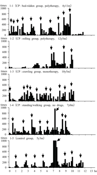
Fig. 1 shows a representative example of the results of measuring nocturnal physical activity by 3D accelerometer in one subject each from the bed-ridden, rolling, crawling, and standing / walking groups. Values were measured on settings of a 1-minute epoch time added every 10 minutes in order to show the periodicity of nocturnal physical activity. The value in the vertical axis shows the number of times acceleration more than 0.01 G every 10 minutes while the value in horizontal axis shows time. Arrow signs in the figures show awakenings according to the formula of Cole et al. (1992). In normal children, high activity every 90 minutes as well as periodicity of REM sleep and non-REM sleep was recognized (1-5). In children with CP, there were some cases in which periodicity at regular intervals was recognized
(1-4), while other cases showed different results such as a case with short intervals, a case whose periodicity disappeared just before awakening (1-1), and a case without periodicity for a period of time after falling a sleep.
Fig. 2 shows calculated sleeping time using the formula of Cole et al. (1992) based on the measurements by the 3D accelerometer. There were no significant differences in sleeping hours between the CP group and the control group (t=0.51, df=41, ns). Sleeping hours by level of motor function were as follows : bed-ridden group, mean 9.30 ± 2.14 hours, rolling group, 9.81 ± 1.65 hours ; crawling group, 9.32 ± 1.94 hours ; and standing / walking group, 9.46 ± 1.35 hours ; there were no significant differences (df=4/38, F=0.19, ns). Sleeping hours in each level by the numbers of kinds of anti-convulsants
were as follows : no drugs group, 10.06 ± 0.25 hours ; monotherapy group, 9.24 ± 1.90 hours ; and polytherapy group, 10.03 ± 1.49 hours ; there were no significant differences in sleeping hours among the three groups (df=3/39, F=0.60, ns).
In the CP group, the mean and SD between sleeping hours based on the questionnaire survey of subjects' parents and sleeping hours measured by 3D accelerometer were 1.06 ± 0.67 hours.
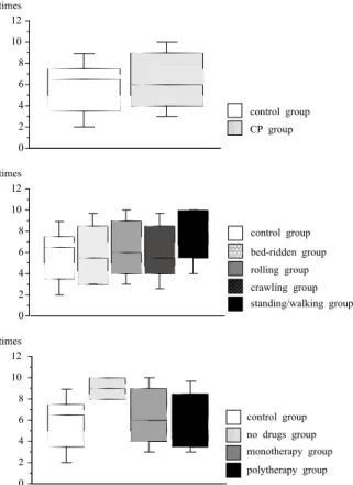
Fig. 3 shows the sleep periodicity investigated by the output of the 3D accelerometer. The means and SD of the sleep periodic time were 5.92 ± 2.71 times for the control group and 6.32 ± 2.71 times for the CP group. There were no significant differences in sleep periodic time between the CP group and the control group (t=0.44, df=41, ns). The means and SDs of the sleep periodic time by motor function level were 5.88 ± 2.85 times for the bed-ridden group, 6.30 ± 2.83 times for the rolling group, 6.00 ± 2.78 times for the crawling group, and 7.60 ± 2.61 times for the standing / walking group. There
1-4(CP : standing/walking group, no drugs, 7y0m)
0 200 400 600 800 1000 times
1-2(CP : rolling group, polytherapy, 12y5m)
0 200 400 600 800 1000 times
1-5(control group, 5y3m)
0 1 2 3 4 5 6 7 8 9 10 11 12 13 hours 0 200 400 600 800 1000 times
1-1(CP : bed-ridden group, polytherapy, 4y11m)
0 200 400 600 800 1000 times
1-3(CP : crawling group, monotherapy, 10y5m)
0 200 400 600 800 1000 times
Fig.1 The representative example of the measuring results of nocturnal physical activity by the 3D accelerometer in typical subjects. Each value shows the 3D accelerometer date per 10 minutes. The arrow signs shows the awakenings (Cole et al., 1992).
CP group control group 0 2 4 6 8 10 12 14 hours 16 standing/walking group crawling group rolling group bed-ridden group control group 0 2 4 6 8 10 12 14 hours 16 monotherapy group control group polytherapy group no drugs group 0 2 4 6 8 10 12 14 hours 16
were no significant differences in sleep periodic time between each level of motor function (df=4/38, F=0.39, ns). And the means and SDs of the sleep periodic time of each group by number of kinds of anti-convulsant were 9.00 ± 1.41 times for the no drugs group, 6.24 ± 2.74 for the monotherapy group, and 5.88 ± 2.75 for the polytherapy group. The result of the one-way ANOVA between the four groups including control showed no significant differences in sleep periodic time between groups according to the numbers of kinds of anti-convulsant (df=3/39, F=0.80, ns).
Fig. 4 shows the waking time after sleep onset investigated from the output of the 3D accelerometer. The means and SDs of the awakenings after sleep onset were 1.54 ± 0.71 hours for the control group and 1.73 ± 0.71 hours for the CP group. There were no significant differences in awakening time after sleep onset between the CP group and the control group (t=-0.79, df=41, ns). The means and SDs of awakening time after sleep onset by
level of motor function were 1.87 ± 0.96 hours for the bed-ridden group, 1.61 ± 0.67 hours for the rolling group, 1.72 ± 0.70 hours for the crawling group, and 1.79 ± 0.45 hours for the standing / walking group ; there were no significant differences in awakening time after sleep onset by level of motor function (df=4/38, F=0.29, ns). Finally, the means and SD of the awakening time after sleep onset by level of numbers of kinds of anti-convulsants were 2.07 ± 0.05 times hours for the no drugs group, 1.64 ± 0.64 for the monotherapy group, and 1.89 ± 1.00 for the polytherapy group. The result of one-way ANOVA between the four groups including the control showed no significant differences in awakening time after sleep onset between groups according to numbers of kinds of anti-convulsants (df=3/39, F=0.59, ns).
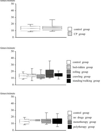
Fig. 5 shows the nocturnal physical activity measured by 3D accelerometer. The means and SDs of the nocturnal physical activity were 13.58 ± 4.28 times / minute for the control group and 15.40 ± 7.01 times / minute for the CP group.
CP group control group 0 2 4 6 8 10 12 times 0 2 4 6 8 10 12 times standing/walking group crawling group rolling group bed-ridden group control group 0 2 4 6 8 10 12 times monotherapy group control group polytherapy group no drugs group
Fig. 3 Sleep periodic time led by 3D accelerometer
CP group control group hours standing/walking group crawling group rolling group bed-ridden group control group monotherapy group control group polytherapy group no drugs group
Fig. 4 Awakenings after sleep onset 0 1 2 3 4 hours 0 1 2 3 4 hours 0 1 2 3 4
There were no significant differences in nocturnal physical activity between the CP group and control group (t=-0.84, df=41, ns). The means and SDs of nocturnal physical activity by level of motor function were 14.86 ± 6.57 times / minute for the bed-ridden group, 13.54 ± 5.50 times / minute for the rolling group, 18.51 ± 10.03 times / minutes for the crawling group, and 14.98 ± 4.39 times / minutes for the standing / walking group ; there were no significant differences in nocturnal physical activity by level of motor function (df=4/38, F=0.88, ns). Finally, the means and SDs of nocturnal physical activity by numbers of kinds of anti-convulsants were 15.20 ± 1.56 times / minutes for the no drugs group, 15.84 ± 7.42 times / minutes for the monotherapy group, and 14.29 ± 7.13 times / minutes for the polytherapy group. The result of one-way ANOVA between the four groups including the control showed no significant differences in nocturnal physical activity between groups according to numbers of kinds of anti-convulsants (df=3/39, F=0. 34, ns).
Discussion
Physical activity is defined as all movements of the body by the muscular system consuming energy ; it is distinct from exercise, which is planned, structured and repetitious physical activity aimed at maintaining and improving the physique (Casperson and Poweel, 1985). Many different attempts to assess physical activity have been reported up to this day (Laport et al., 1985). Physical activity differs in the quantity of energy consumed and required physical strength. Also, the methods to assess physical activity vary, and typically assessed subjects vary by method. Methods for assessing physical activity can be classified as direct and indirect. As direct methods, there are (a) self-administered quantitative histories or physical activities recall survey, (b) observations using video camera, and (c) mechanical and electronic monitors ; as indirect methods, there are (d) methods using energy metabolism and (e) methods using food intake. Self-administered quantitative histories or physical activities recall surveys are convenient and inexpensive, and can supply detailed information of physical activity about a large number of people (Melanson and Freedson, 1996). However, this method has inherent drawbacks in that the subjectivity of the subject influences the results, which may not reflect physical activity accurately. And it is not a feasible method for physically handicapped children or subjects who have intellectual disabilities and communication disorders as accompanying symptoms, who are unable to report for themselves. Observation using video camera is objective, has good test-retest reliability and can record physical activity patterns (Melanson and Freedson, 1996). But it is only a measurement of physical activity under experimentation, which makes it almost impossible to measure the physical activity of a subject all day. On the other hand, indirect methods such as methods using energy metabolism and methods using food intake are objective, pertinent and useful, but they are expensive and may possibly measure different things from real physical activity ; therefore,
CP group control group standing/walking group crawling group rolling group bed-ridden group control group monotherapy group control group polytherapy group no drugs group
Fig. 5 Nocturnal physical activity 0 10 20 30 40 50 times/minute 0 10 20 30 40 50 times/minute 0 10 20 30 40 50 times/minute
their usefulness is limited.
This study is the first report on measurement of nocturnal physical activity which applies the 3D accelerometer method to children with CP in free-living conditions. The 3D accelerometer is useful for assessing daily physical activity : it can produce accurate data, is easy and convenient, does not inhibit the daily activities of subjects, and has good test-retest reliability (Sugimoto, 1995 ; Sugimoto et al., 1997). Recently it has been used not only on adults but also on children. Studies have been done on changes of body movement accompanied by neonatal development (Guilleminault et al., 1996), sleep disorders (Acebo et al., 1999 ; Franck et al., 1999) and ADHD (Inoue et al., 1998) ; however, investigations of children with CP are rare. It was proved that the 3D accelerometer is useful at measuring physical activity by counting a certain value of acceleration in a certain amount of time. Bach et al. (1994) and Reddihough et al. (1990) made an attempt to measure the movement of the upper limbs of CP subjects by 3D accelerometer, analyzed the frequency, which is different from the way we used it in this study, and compared the results with clinical assessments of function of the upper limbs. They reported that assessment of function of upper limbs using the 3D accelerometer did not always reflect gross motor assessment. But previous reports (Acebo et al., 1999 ; Inoue et al., 1998 ; Sugimoto et al., 1997) inducated that this method is reliable and established it as a quantitative index of physical activity.
In this study, we quantified and investigated nocturnal physical activity using a 3D accelerometer. Numerous studies have investigated prolongation of REM sleep latency (Hori & Kodama, 1975 ; Hayashi et al., 1990), presence of spike and slow-wave activity (Zucconi & Bruni, 2001), gross body movements (Fukumoto et al., 1981 ; Hayashi et al., 1990) and twitch body movements (Hiura et al., 1984 ; Hayashi et al., 1990) using electromyography while studies which investigated nocturnal physical activity in free condition during sleeping
have been rare.
In this study, the periodicity of nocturnal physical activity in CP was recognized (Fig. 1), and the 3D accelerometer proved useful in measuring patterns of nocturnal physical activity. Sleep problems are common in handicapped children. Several studies have shown sleep problems associated with intellectual disabilities (Quine, 1991 ; Poindexter & Bihm, 1994 ; Wiggs & Stores, 1996 ; Pillar et al. 1998 ; Didden et al., 2002 ; Hayashi & Katada, 2002) and in cerebral palsy (Shibagaki et al., 1985 ; Kotagal et al., 1994; Zucconi & Bruni, 2001). In previous studies, irregular sleep-wake rhythms (Hayashi & Katada, 2002), increased arousal time (Prechtl et al., 1973 ; Hori & Kodama, 1975) and short sleep patterns (Shibagaki et al., 1985; Poindexter & Bihm, 1994) were indicated, but there were no significant differences in sleep periodic time (Fig. 3) and in number of awakenings OR awakening time after sleep onset (Fig. 4) in this study. As Fig.1 shows, there were some cases whose periodic time showed high, low, and irregular. In previous studies of sleep fragmentation or disruption (Kotagal et al., 1994 ; Hayashi & Katada, 2002), irregular sleep-wake rhythms (Hayashi & Katada, 2002) were indicated, and analysis of the periodicity of nocturnal physical activity, with detailed information of each case might be necessary.
Awakening and sleep could be distinguished using the formula of Cole et al. (1992). There was a gap between the parents' reports and the actual sleep time as investigated by 3D accelerometer, and delayed sleep onset and early morning arousal were recognized, as the previous study indicated. Although delayed sleep onset and early morning arousal were recognized, there were no significant differences between actual sleep times in CP and the control group (Fig. 2). The lives of CP patients almost totally rely on their parents' care, and their regular habits in everyday life are determined by the parents ; thus, they go to bed early and get up early compared to the control group. But the actual sleep time of CP subjects
was almost same as that of control group, even considering delayed sleep onset and sleep fragmentation.
There were no significant differences in nocturnal physical activity between the CP group and control group, and there were no significant differences between each level by motor function or between groups by numbers of kinds of anti-convulsants (Fig.5). Several studies have shown a relationship between sleep problems and daytime problem behaviors (Quine, 1991 ; Wiggs & Stores, 1996 ; Richdale et al., 1999). Previous studies indicated spasticity, which leads to respiratory malfunction in patients with CP (Zucconi & Bruni, 2001), and the increased tone of spastic muscles, which also perform during sleep (Gamstorp, 1985). Treatment for sleep disturbance occurring by brain damage is important in view of the improvement of increased muscle tone (Zucconi & Bruni, 2001).
Hypoactivity is most common in CP children. The ratio of total daily energy expenditure to resting metabolic rate (Bandini et al., 1991) and daily physical activity measured by 3D accelerometer (Shiragaki et al., 2000) in CP was significantly lower than those in healthy control subjects. The authors investigated physical activity by motor function level, as in this study, and the result showed that the higher motor function the subject has, the higher physical activity showed. The possible factors as to why there were significant differences in daily physical activity and no significant differences in nocturnal physical activity are several: (a) involuntary movements (b) physical activity caused by changes in body position (c) epileptic spasm (d) nocturnal sleep fragmentation owing to low energy consumption in the daytime.
As a solution to the problem of measuring of physical activity, 3D accelerometer is limited to measuring the quantity of physical activity, and is not for measuring the detailed movement of the whole body or each part of the body. It will be a subject from now on to investigate the reliability of its detailed information about nocturnal movements and the quality of each
movement, such as involuntary movements and physical activity caused by changes in body position, using video camera at the same time. It will be useful to investigate physical activity in each state such as when resting / sleeping, when epileptic spasms occur, when they change in body position so that the output from 3D accelerometer might be distinguished. In this study, we attached a 3D accelerometer to the non-dominant wrist or the wrist of lower motor age following the method of the previous study (Sugimoto et al., 1997). This is considered to be an appropriate method, including the part of body to which it is attached, enabling measurement at each level of motor function. The physical activity which was derived might differ according to which part of the body it is attached to, for in CP, the paralyzed part varies by individual, e.g., hemiplegia and diplegia, and many times the range of motion is limited by deformity and contracture. Hence, it might be necessary to investigate physical activity with the accelerometer attached to various parts of body. But if attached to the lumbus, the subject might have a pain when rolling, and if attached to the ankle joint the subject might feel uncomfortable, and it might get turned off by crossing of the lower limbs, characteristic of spastic CP, or it may restrict free motion. Therefore, it is still necessary to investigate which part of the body is best for attaching the 3D accelerometer in CP subjects.
References
Acebo C., Sedeh A., Seifer R., Tzischinsky O., Wolfson A. R., Hafer A. & Carskadon M. A. (1999) Estimating sleep patterns with activity monitoring in children and adolescents : how many nights are necessary for reliable measures? Sleep, 22, 95-103.
Bach T. M., Reddihough D. S., Burgess G., Johnson L. M. & Byrt T. A. (1994) Comparison of subjective and objective measures of movement performance in children with cerebral palsy. Developmental Medicine and Child Neurology, 36, 974-979.
Bandini L. G., Schoeller D. A., Fukagawa N. K., Wykes L. J. & Dietz W. H. (1991) Body composition and energy expenditure in a d o l e s c e n t s w i t h c e r e b r a l p a l s y o r myelodysplasia. Pediatric Research, 29, 70-77. Casperson C. J. & Powell K. E. (1985) Physical activity, exercise, physical fitness : Definitions and distinctions for health-related research. Public Health Reports, 100, 126. Cole R. J., Kripke D. F., Gruen W., Mullaney D.
J. & Gillin J. C. (1992) Automatic sleep / wake identification from wrist activity. Sleep, 15 (5), 461-469.
Didden R., Korzilius H., van Aperlo B., van Overloop C. & de Vries M. (2002) Sleep problems and daytime problem behaviours in children with intellectual disability. Journal of Intellectual Disability Research, 46(7), 537-547.
Fukuda K. & Ishihara K. (2001) Age-related changes of sleeping pattern during adolescence. Psychiatry and Clinical Neurosciences, 55, 231-232.
Franck L. S., Johnson L. M., Lee K., Hepner C., Lambert L., Passeri M., Manio E., Dorenbaum A. & Wara D. (1999) Sleep disturbances in children with human immunodeficiency virus infection. Pediatrics, 104 (5), e62.
Fukumoto M., Mochizuki N., Takeishi M., Nomura Y. & Segawa M. (1981) Studies of body movements during night sleep in infancy. Brain and Development, 3(1), 37-43. Gamstorp I. (1985) Paediatric neurology. London :
Butterworths, 274-293.
Guilleminault C., Ieger D., Pelayo R., Gould S., Hayes B. & Miles L. (1996) Development of circadian rhythmicity of temperature in full-term normal infants. Neurophysiologie Clinique, 26 (1), 21-29.
Hayashi E. & Katada A. (2002) Sleep in person with intellectual disabilities : a questionnaire survy. Japanese Journal of Special Education, 39 (6), 91-101.
Hayashi M., Inoue Y., Iwakawa Y. & Sasaki H. (1990) REM abnormalities in severe athetoid
cerebral palsy. Brain and Development, 12, 494-497.
Hiura K., Hashimoto T., Fukuda K., Endo S., Tayama M., Mori A., Fujino K. & Miyao M. (1984) Polysomnography on lissencephaly. No To Hattatsu, 16, 359-364.
Hori T. & Kodama M. (1975) Polygraphical study of sleep in cerebral palsied adults. Rinsho Noha, 17, 168-172. (in Japanese)
Inoue K., Nadaoka T., Oiji A., Morioka Y., Totsuka S., Kanbayashi Y. & Hukui T. (1998) Clinical evaluation of attention-deficit hyperactivity disorder by objective quantitative measures. Child Psychiatry and Human Development, 28 (3), 179-188.
Jan M. M. (2000) Melatonin for the treatment of handicapped children with severe sleep disorders. Pediatric Neurology, 23, 229-232. Johnson M. K., Zuck F. N. & Wingate K. (1951)
Measurement of motor handicaps in children with neuromuscular disorder as cerebral palsy. The Journal of Bone and Joint Surgery, 33, 698-707.
Kotagal S., Gibbons V. & Stith J. (1994) Sleep abnormalities in patients with severe cerebral palsy. Developmental Medicine and Child Neurology, 36, 304-311.
Laport R. E., Montoye H. J. & Casperson C. J. (1985) Assessment of physical activity in epidemiologic research : Problems and prospects. Public Health Reports, 100, 131-146.
Magardino T. M. & Tom L. W. C. (1999) Surgical Management of obstructive sleep apnea in children with cerebral palsy. Laryngoscopme, 109, 1611-1615.
Melanson E. J. & Freedson P. S. (1996) Physical activity assessment : A review of methods. Critical Reviews in Food Science and Nutrition, 36 (5), 385-396.
Pillar G., Etzioni A., Shahar E. & Lavie P. (1998) Melatonin treatment in an institutionalized child with psychomotor retardation and an irregular sleep-wake pattern. Archives of disease in childhood, 79, 63-64.
of short-sleep patterns in institutionalized individuals with profound mental retardation. American Journal on Mental Retardation, 98 (6), 776-780.
Prechtl H. F. R., Theorell K. & Blair A. W. (1973) Behavioral state cycles in abnormal infants. Developmental Medicine and Child Neurology, 15, 606-615.
Quine L. (1991) Sleep problems in children with severe mental handicap. Journal of Mental Deficiency Research, 35, 269-290.
Reddihough D., Bach T., Burgess G., Oke L. & Hudson I. (1990) Objective test of the quality of motor function of children with cerebral palsy: Preliminary study. Developmental Medicine and Child Neurology, 32, 902-909. Richdale A., Cotton S. & Hibbit K. (1999)
Sleep and behaviour disturbance in Prader-Willi syndrome: a questionnaire study. Journal of Intellectual Disability Research, 43, 380-392.
Shibagaki M., Kiyono S. & Takeuchi T. (1985) Nocturnal sleep in mentally retarded infants
with cerebral palsy. Electroencephalography and Clinical Neurophysiology, 61, 465-471. Shiragaki J., Iwasaki N. & Fujita K. (2000)
Measurement of physical activity using Actigraph method in children with cerebral palsy. Shinshin Syougaigaku Kenkyuu, 24, 1-8. (In Japanese)
Sugimoto A. (1995) Measurement of daily physical activity by the motion sensor. Tokyo Jikeikai Medical Journal, 110, 163-173. Suigimoto A., Hara Y., Findley T. W. &
Yonemoto K. (1997) A useful method for measuring daily physical activity by a three-derection monitor. Scandinavian Journal of Rehabilitation Medicine, 29, 37-42.
Wiggs L. & Stores G. (1996) Severe sleep disturbance and daytime challenging behaviour in children with severe learning disabilities. Journal of Intellectual Disability Research, 40, 518-528.
Zucconi M. & Bruni O. (2001) Sleep disorders in children with neurologic diseases. Seminars in Pediatric Neurology, 8 (4), 258-275.