R E V I E W
Open Access
Oral environment and cancer
Yasusei Kudo
1*, Hidesuke Tada
1,3, Natsumi Fujiwara
2, Yoshiko Tada
3, Takaaki Tsunematsu
1, Yoichiro Miyake
4and Naozumi Ishimaru
1Abstract
Cancer is now the leading cause of death in Japan. A rapid increase in cancer mortality is expected as Japan is
facing a super-aged society. Many causes of cancer are known to be closely linked to life style factors, such as
smoking, drinking, and diet. The oral environment is known to be involved in the pathogenesis and development
of various diseases such as bronchitis, pneumonia, diabetes, heart disease, and dementia. Because the oral cavity
acts as the bodily entrance for air and food, it is constantly exposed to foreign substances, including bacteria and
viruses. A large number of bacteria are endemic to the oral cavity, and indigenous oral flora act to prevent the
settlement of foreign bacteria. The oral environment is influenced by local factors, including dental plaque, tartar,
teeth alignment, occlusion, an incompatible prosthesis, and bad lifestyle habits, and systemic factors, including
smoking, consumption of alcohol, irregular lifestyle and eating habits, obesity, stress, hormones, and heredity. It has
recently been revealed that the oral environment is associated with cancer. In particular, commensal bacteria in the
oral cavity are involved in the development of cancer. Moreover, Candida, human papilloma virus and Epstein-Barr
virus as well as commensal bacteria have been reported to be associated with the pathogenesis of cancer. In this
review, we introduce recent findings of the correlation between the oral environment and cancer.
Keywords: Cancer, Oral environment, Bacteria, Candida, Human papilloma virus, Epstein-Barr virus
Background
During 2012, approximately 14.1 million new cases of
human cancer were reported globally [1]. In Japan, the
estimated number of cancer deaths during 2014 was
approximately 367,000 [2]. Regarding the age-specific
causes of death, during 2013, cancer was the leading
cause of death among 40–89 years in Japan [2]. Tobacco
use accounts for approximately 22 % of cancer deaths
[3], followed by obesity, a poor diet, lack of physical
activity, and consumption of alcohol, which collectively
account for 10 % of cancer deaths [3, 4]. Other factors
include infections, exposure to ionizing radiation, and
environmental pollutants [5]. Thus, various causes of
cancer are known to be closely involved in lifestyle
choices, such as smoking, drinking, and diet.
The human body is inhabited by over 100 trillion
microbial cells living in symbiosis with the host. Many
bacteria are endemic to the oral cavity. More than 700
bacterial species inhabit the oral cavity, including at least
11 bacterial phyla and 70 genera. Individuals that
prac-tice oral hygiene have 1,000–100,000 bacteria living on
each tooth surface, whereas those who do not regularly
practice dental hygiene can have between 100 million
and 1 billion bacteria on each tooth surface [6].
Al-though some bacteria of the oral cavity are harmful and
can cause serious disease, many of the oral bacteria are
in fact beneficial in preventing diseases. Thus, the oral
cavity is inhabited by complex multispecies bacterial
communities that usually exist in a balanced
immunoin-flammatory state with the host [7]. It is now established
that many chronic inflammatory conditions are caused
by an imbalance between host-microbiota interactions,
resulting in a dysbiotic community, deregulated immune
responses, and eventually disease outcomes. Oral
com-mensal bacteria play a critical role in the development of
oral diseases, including periodontal disease and tooth
loss, and maintenance of a normal oral physiological
environment [8, 9]. In addition, oral commensal bacteria
are involved in the pathogenesis of cancer [10]. Thus,
the oral environment including oral commensal bacteria
is known to be involved in the pathogenesis and
devel-opment of various diseases, such as bronchitis and
* Correspondence:[email protected]
1Department of Oral Molecular Pathology, Institute of Biomedical Sciences,
Tokushima University Graduate School, Tokushima, Japan Full list of author information is available at the end of the article
© 2016 Kudo et al. Open Access This article is distributed under the terms of the Creative Commons Attribution 4.0 International License (http://creativecommons.org/licenses/by/4.0/), which permits unrestricted use, distribution, and reproduction in any medium, provided you give appropriate credit to the original author(s) and the source, provide a link to the Creative Commons license, and indicate if changes were made. The Creative Commons Public Domain Dedication waiver (http://creativecommons.org/publicdomain/zero/1.0/) applies to the data made available in this article, unless otherwise stated.
pneumonia, diabetes, heart disease, and dementia. The
oral environment is influenced by local factors, including
dental plaque, tartar, teeth alignment, occlusion, an
incompatible prosthesis, and bad lifestyle habits, and
systemic factors, including smoking, consumption of
al-cohol, an irregular lifestyle, eating habits, obesity, stress,
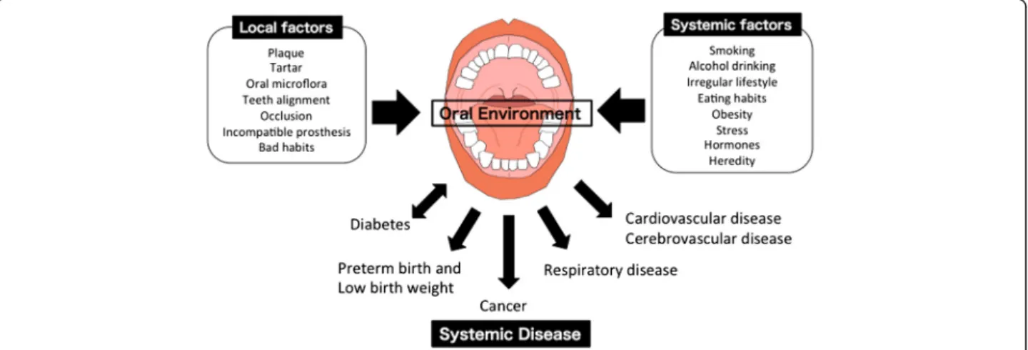
hormones, and heredity factors (Fig. 1). Majority of the
causes of cancer are thought to be related to tobacco use
and heavy alcohol consumption. In the oral
environ-ment, poor oral hygiene and viral and Candida infections
can be risk factors for cancer.
Oral bacteria and cancer
The link between oral infections and adverse systemic
conditions has attracted a great deal of attention in the
dental and medical fields. A major consequence of the
systemic spread of oral commensals and pathogens to
distant body sites has been the disruption of immune
surveillance and homeostasis, resulting in the promotion
or acceleration of pathogenic processes. Oral bacteria
are involved in systemic infections and inflammation
such as cardiovascular disease, adverse pregnancy
out-comes, rheumatoid arthritis, aspiration pneumonia,
in-flammatory bowel disease, organ inflammations, and
cancer (Fig. 1). Cumulative studies have revealed that
oral bacteria are involved in the initiation or progression
of certain cancers. Chronic or dysregulated inflammation
contributes to tumor development, partly through the
modulation of the tumor microenvironment [11].
Epi-demiological studies have shown that the risk of cancer
is increased in people with periodontal disease or tooth
loss caused by oral bacteria [12]. Oral diseases, including
periodontitis, have been related to the risk of oral and
gastrointestinal cancers, such as oral, esophageal, gastric
and pancreatic cancers [13, 14]. Although the underlying
mechanism for the associations between oral health
status and cancers are not completely understood, it
has been reported that Fusobacterium nucleatum and
Porphyromonas
gingivalis
are
involved
in
cancer
development.
F.
nucleatum,
an
anaerobic
Gram-negative
oral
commensal, is associated with periodontitis, adverse
pregnancy outcomes, cardiovascular disease, rheumatoid
arthritis, inflammatory bowel disease, and colon cancer
[15]. This bacterial species is a key component of
peri-odontal plaque because of its abundance and ability to
coaggregate with other species in the oral cavity [16, 17].
Elevated F. nucleatum levels were significantly detected
in people with colon cancers compared with that in
people with normal colon tissue [18, 19]. The bacteria
genera of Fusobacterium, Porphyromonas,
Peptostrepto-coccus, and Mogibacterium were found to be enriched in
the mucosa-adherent microbiota of people with colon
cancer [20]. Moreover, the overabundance of
Fusobacter-ium
in people with colon cancer was positively
associ-ated with lymph node metastasis [18]. In cases of colon
cancer, a positive correlation between mRNA levels for
several local cytokines and Fusobacterium species has
been observed [21]. Although the mechanism by which
F. nucleatum
might contribute to the pathogenesis of
such a diverse clinical spectrum is unknown, recent
studies have demonstrated the mechanisms of F.
nuclea-tum
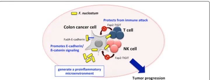
in the involvement of colon cancer as follows: i) F.
nucleatum
induces oncogenic and inflammatory
re-sponses to stimulate growth of colon cancer cells
through FadA adhesin via binding with E-cadherin and
ensuing activation of ß-catenin signaling [22], ii) F.
nucleatum
generate a proinflammatory
microenviron-ment for the progression of colon cancer through the
recruitment of tumor-infiltrating immune cells [23], and
Fig. 1 Influence of Oral Environment on Systemic Disease. The oral environment is influenced by local factors, including dental plaque, tartar, teeth alignment, occlusion, an incompatible prosthesis, and bad lifestyle habits, and systemic factors, including smoking, consumption of alcohol, an irregular lifestyle, eating habits, obesity, stress, hormones, and heredity factors. The oral environment is involved in systematic diseases, such as diabetes, preterm birth and low birth weight, cancer, respiratory disease, cardiovascular disease and cerebrovascular disease
iii) F. nucleatum interferes with the host immunity by
engaging its bacterial protein Fap2 with the inhibitory
immunoreceptor TIGIT on NK and T cells [24] (Fig. 2).
Accumulated evidence has shown strong
correla-tions between a number of chronic periodontal
bacteria containing Prevotella, Porphyromonas and
Streptococcus
spp. and orodigestive cancer, including
oral squamous cell carcinoma and pancreatic cancer
[25–31]. P. gingivalis have an abundance of virulence
factors and can be present in periodontal pockets. P.
gingivalis
has a major disruptive effect on local
im-mune responses in the periodontal area [7]. Moreover,
it can survive within the oral epithelial tissues and
can evade the host immune response [32–37]. In vitro
studies have demonstrated that i) P. gingivalis strains
can induce the expression of the B7-H1 and B7-DC
receptors in oral cancer cells, which might facilitate
immune evasion by oral cancers [38] and ii) P.
gingi-valis
activates the ERK1/2-Ets1, p38/HSP27, and
PAR2/NF-kB pathways to induce proMMP9
expres-sion, after which the proenzyme is activated by
gingi-pains to promote cellular invasion of oral cancer cells
[39]. In primary cultures of gingival epithelial cells, P.
gingivalis
is strongly antiapoptotic and can suppress
chemically induced apoptosis via activation of the
Jak1/Akt/Stat3 signaling pathway [40, 41], suggesting
that the antiapoptotic effect of P. gingivalis may be
involved in carcinogenesis. Thus, both P. gingivalis
and F. nucleatum are involved in cancer progression
via activation of cell signaling and/or facilitating
immune evasion. However, the possible link between
oral bacterium and cancer development remains to be
investigated in molecular detail.
Viral and Candida infection and cancer
Tobacco and alcohol are considered as major risk factors
for cancer; however, viral and candidal infection is
increasingly being identified to play significant roles in
cancer
development [42].
Human
papillomaviruses
(HPVs) are small, double-stranded DNA viruses that
induce hyperproliferative lesions in epithelial tissues
[43, 44]. More than 100 types of HPV have been
identi-fied. Among them, high-risk HPV types, including HPV
type 16 (HPV-16), HPV-18, HPV-31, HPV-33, and
HPV-42, induce lesions in the genital tract that can
pro-gress to malignancy [44–46]. HPV is the most common
sexually transmitted viral infection and is well-known as
the causative agent of cervical cancer. HPV infection
has also been detected in the oral cavity. A large
num-ber of studies have demonstrated a 2–3-fold increase in
the prevalence of HPV-driven oropharyngeal squamous
cell carcinoma over the last three decades, particularly
in North America and northern Europe [47–49].
More-over, a recent review found that many studies have
iden-tified a high proportion of oral cancer with detectable
HPV DNA [50]. Although the underlying reasons
remain poorly understood, changes in sexual behavior,
decreased rates of tonsillectomy performed in the
pediatric population since the 70s, and progress in the
diagnostic work-up and HPV testing assays have been
proposed as the reasons [51–55]. Considering current
trends, it is estimated that high-risk HPVs, such as
HPV16 and HPV18, cause premalignant lesions [54, 55].
In the high-risk HPV types, E6 and E7 have been shown
to function as oncoproteins [56, 57]. E6 and E7
onco-proteins are the key drivers of tumorigenesis by
inacti-vating the tumor suppressors pRb and p53 [58].
Fig. 2 Involvement of F. nucleatum in colon cancer development. Figure shows the mechanisms of F. nucleatum in the involvement of colon cancer. F. nucleatum promotes E-cadherin/ß-catenin signaling via FadA adhesin and protects from immune attack by engaging its bacterial protein Fap2 with the inhibitory immunoreceptor TIGIT on NK and T cells. Moreover, F. nucleatum generate a proinflammatory microenvironment through the recruitment of tumor-infiltrating immune cells
Epstein–Barr virus (EBV) causes infectious
mono-nucleosis and oral hairy leukoplakia, and is associated
with various types of lymphoid and epithelial
malignan-cies. During 1964, Epstein et al. determined the
causa-tive agent of Burkitt’s lymphoma to be a previously
unknown member of the herpes family of viruses, later
termed as EBV [59]. Burkitt lymphoma can be classified
into three forms, which differ in geographic distribution
and EBV association: endemic, sporadic, and HIV
associ-ated Burkitt lymphoma. Among them, endemic Burkitt
lymphoma is associated with EBV in over 95 % cases
[60]. Endemic Burkitt lymphoma is predominant in the
equatorial belt of Africa and other parts of the world
where malaria is hyperendemic [60–63]. On the other
hand, although controversial, lymphoepithelial
carcin-oma and nasopharyngeal carcincarcin-oma may also be
associ-ated with EBV [64–68]. The association between
lymphoepithelial carcinoma and EBV appears to differ
according to geographic areas, race, and affected organs.
In general, the association of lymphoepithelial carcinoma
with EBV is strong in the head and neck region, whereas
it is relatively weak at other sites. Moreover, the
associ-ation is strong in East Asia and relatively weak in
western countries. To a great extent, EBV-mediated
dis-ruption of cell growth checkpoints relies on direct
modulation of cytokine receptor signaling mechanisms
and alterations in the expression levels of various
cyto-kines [69]. Moreover, EBV is associated with aggressive
types of oral tumors, particularly in immunosuppressed
patients [70]. Thus, EBV infection of the oral cavity is
also associated with certain types of cancer. However,
the pathogenesis of these tumors remains unclear.
Oropharyngeal candidiasis is an opportunistic
infec-tion primarily caused by Candida albicans, a ubiquitous
fungal organism that is part of the normal microflora of
the gastrointestinal and reproductive tracts. Candida
species can be isolated as a commensal organism from
the oral cavity in up to 80 % healthy individuals [71].
Depending on the host defense mechanisms or oral
microenvironment, Candida species can transform from
a harmless commensal species to pathogenic organisms
causing oral mucosal infection [72, 73].
Candida-associ-ated denture stomatitis affects more than 25 % denture
wearers [74], and up to 90 % HIV
+patients have had at
least one episode of oropharyngeal candidiasis [75].
When dealing with a hyperplastic epithelial lesion of the
oral mucosa in which presence of C. albicans is
demon-strated, it is referred to by many as Candida-associated
leukoplakia or others prefer the term
“hyperplastic
candidiasis.” Chronic hyperplastic candidiasis showed a
higher rate of malignant transformation on follow-up
[76]. Animal studies have shown that Candida can cause
epithelial hyperplasia and cellular atypia [77]. Strains of
Candida
can produce carcinogenic nitrosamine,
N-nitrosobenzylmethylamine [78]. Thus certain strains of
Candida
play a key role in oncogene formation and
initiation of cancer development [77–79]. However, the
possible role of Candida in malignant transformation
remains still unclear.
Conclusion
Many of the causes of cancer are known to be closely
linked to lifestyle factors, such as smoking, drinking, and
diet. The oral environment is associated with the cause
of various diseases, including cancer. The oral
environ-ment is influenced by various factors, including local
and systemic factors. In the current review, in particular,
we introduced the involvement of oral bacteria,
fun-guses, and viruses in the development of cancer. During
recent years, the role of oral care for the management of
general health has been found to be important within
the fields of medical and nursing care. Therefore, oral
care for the management of the oral environment can be
important for prevention of cancer.
Competing interests
The authors declare that they have no competing interests. Authors’ contributions
YK, HT, NF, YT, and TT wrote the manuscript. YM and NI was involved in the critical revision or supervision of the manuscript. All authors read and approved the final manuscript.
Acknowledgements
The authors thank Dr. Shinji Oikawa and Dr. Hiroyuki Kamiya, Organizers of The Open Symposium of the JEMS in 2015, and the Japanese Environmental Mutagen Society. The authors would like to thank Enago (www.enago.jp) for the English language review.
Author details
1Department of Oral Molecular Pathology, Institute of Biomedical Sciences,
Tokushima University Graduate School, Tokushima, Japan.2Department of
Oral Healthcare Promotion, Institute of Biomedical Sciences, Tokushima University Graduate School, Tokushima, Japan.3Tada Dental Clinic, Kakogawa,
Japan.4Department of Oral Microbiology, Institute of Biomedical Sciences,
Tokushima University Graduate School, Tokushima, Japan.
Received: 14 March 2016 Accepted: 19 April 2016 References
1. World Health Organization. The global and regional burden of cancer. In: Stewart BW, Wild CP, editors. World Cancer Report 2014. IARC Nonserial Publication. Chapter 1.1.
2. Center for Cancer Control and Information Services, National Cancer Canter, Japan: http://ganjoho.jp/reg_stat/statistics/index.html. 2014. Accessed in February 2016.
3. “Cancer Fact sheet No.297”. World Health Organization: http://www.who.int/ mediacentre/factsheets/fs297/en/(2014). Accessed in in February 2016. 4. “Obesity and Cancer Risk”. National Cancer Institute: http://www.cancer.gov/
about-cancer/causes-prevention/risk/obesity/obesity-fact-sheet#q3 (2012). Accessed in in February 2016.
5. Anand P, Kunnumakkara AB, Kunnumakara AB, Sundaram C, Harikumar KB, Tharakan ST, et al. Cancer is a preventable disease that requires major lifestyle changes. Pharm Res. 2008;25:2097–116.
6. Jane ES, Desrocher J.“Oral ecology.” Technology Review (00401692) 100.1 (Jan. 1997): 48. Academic search premier. EBSCO. Provo, UT: Brigham Young University; 2008.
7. Hajishengallis G, Lamont RJ. Beyond the red complex and into more complexity: The polymicrobial synergy and dysbiosis (PSD) model of periodontal disease etiology. Mol Oral Microbiol. 2012;27:409–19.
8. Abiko Y, Sato T, Mayanagi G, Takahashi N. Profiling of subgingival plaque biofilm microflora from periodontally healthy subjects and from subjects with periodontitis using quantitative real-time PCR. J Periodontal Res. 2010;45:389–95. 9. Preza D, Olsen I, Willumsen T, Boches SK, Cotton SL, Grinde B, et al.
Microarray analysis of the microflora of root caries in elderly. Eur J Clin Microbiol Infect Dis. 2009;28:509–17.
10. Meurman J. Oral microbiota and cancer. J Oral Microbiol. 2010;2:1–10. 11. Rakoff-Nahoum S. Why cancer and inflammation? Yale J Biol Med. 2006;
79:123–30.
12. Pihlstrom BL, Michalowicz BS, Johnson NW. Periodontal diseases. Lancet. 2005;366:1809–20.
13. Meyer MS, Joshipura K, Giovannucci E, Michaud DS. A review of the relationship between tooth loss, periodontal disease, and cancer. Cancer Causes Control. 2008;19:895–907.
14. Fitzpatrick SG, Katz J. The association between periodontal disease and cancer: a review of the literature. J Dent. 2010;38:83–95.
15. Han YW. Fusobacterium nucleatum: a commensal-turned pathogen. Curr Op Microb. 2015;23:141–7.
16. Kapatral V, Anderson I, Ivanova N, Reznik G, Los T, Lykidis A, et al. Genome sequence and analysis of the oral bacterium Fusobacterium nucleatum strain ATCC 25586. J Bacteriol. 2002;184:2005–18.
17. Signat B, Roques C, Poulet P, Duffaut D. Fusobacterium nucleatum in periodontal health and disease. Curr Issues Mol Biol. 2011;13:25–36. 18. Castellarin M, Warren RL, Freeman JD, Dreolini L, Krzywinski M, Strauss J, et
al. Fusobacterium nucleatum infection is prevalent in human colorectal carcinoma. Genome Res. 2012;22:299–306.
19. Kostic AD, Gevers D, Pedamallu CS, Michaud M, Duke F, Earl AM, et al. Genomic analysis identifies association of Fusobacterium with colorectal carcinoma. Genome Res. 2012;22:292–8.
20. Chen W, Liu F, Ling Z, Tong X, Xiang C. Human intestinal lumen and mucosa-associated microbiota in patients with colorectal cancer. PLoS One. 2012;7:e39743.
21. McCoy AN, Araujo-Perez F, Azcarate-Peril A, Yeh JJ, Sandler RS, Keku TO. Fusobacterium is associated with colorectal adenomas. PLoS One. 2013;8:e53653. 22. Rubinstein MR, Wang X, Liu W, Hao Y, Cai G, Han YW. Fusobacterium
nucleatum promotes colorectal carcinogenesis by modulating E-Cadherin/ß-Catenin signaling via its FadA Adhesin. Cell Host Microbe. 2013;14:195–206. 23. Kostic AD, Chun E, Robertson L, Glickman JN, Gallini CA, Michaud M, et al.
Fusobacterium nucleatum potentiates intestinal tumorigenesis and modulates the tumor-immune microenvironment. Cell Host Microbe. 2013;14:207–15. 24. Gur C, Ibrahim Y, Isaacson B, Yamin R, Abed J, Gamliel M, et al. Binding of
the Fap2 protein of Fusobacterium nucleatum to human inhibitory receptor TIGIT protects tumors from immune cell attack. Immunity. 2015;42:344–55. 25. Nagy KN, Sonkodi I, Szoke I, Nagy E, Newman HN. The microflora associated
with human oral carcinomas. Oral Oncol. 1998;34:304–8.
26. Mager DL, Haffajee AD, Devlin PM, Norris CM, Posner MR, Goodson JM. The salivary microbiota as a diagnostic indicator of oral cancer: a descriptive, non-randomized study of cancer-free and oral squamous cell carcinoma subjects. J Transl Med. 2005;3:27.
27. Ahn J, Segers S, Hayes RB. Periodontal disease, Porphyromonas gingivalis serum antibody levels and orodigestive cancer mortality. Carcinogenesis. 2012;33:1055–8. 28. Ahn J, Chen CY, Hayes RB. Oral microbiome and oral and gastrointestinal
cancer risk. Cancer Causes Control. 2012;23:399–404.
29. Katz J, Onate MD, Pauley KM, Bhattacharyya I, Cha S. Presence of Porphyromonas gingivalis in gingival squamous cell carcinoma. Int J Oral Sci. 2011;3:209–15. 30. Michaud DS, Izard J, Wilhelm-Benartzi CS, You DH, Grote VA, Tjønneland A,
et al. Plasma antibodies to oral bacteria and risk of pancreatic cancer in a large European prospective cohort study. Gut. 2013;62:1764–70. 31. Michaud DS. Role of bacterial infections in pancreatic cancer.
Carcinogenesis. 2013;34:2193–7.
32. Yao L, Jermanus C, Barbetta B, Choi C, Verbeke P, Ojcius DM, et al. Porphyromonas gingivalis infection sequesters pro-apoptotic Bad through Akt in primary gingival epithelial cells. Mol Oral Microbiol. 2010;25:89–101. 33. Yilmaz O. The chronicles of Porphyromonas gingivalis: the microbium, the
human oral epithelium and their interplay. Microbiology. 2008;154:2897–903. 34. Yilmaz O, Yao L, Maeda K, Rose TM, Lewis EL, Duman M, et al. ATP
scavenging by the intracellular pathogen Porphyromonas gingivalis inhibits P2X7-mediated host-cell apoptosis. Cell Microbiol. 2008;10:863–75.
35. Yilmaz O, Sater AA, Yao L, Koutouzis T, Pettengill M, Ojcius DM. ATP-dependent activation of an inflammasome in primary gingival epithelial cells infected by Porphyromonas gingivalis. Cell Microbiol. 2010;12:188–98. 36. Singh A, Wyant T, Anaya-Bergman C, Aduse-Opoku J, Brunner J, Laine ML,
et al. The capsule of Porphyromonas gingivalis leads to a reduction in the host inflammatory response, evasion of phagocytosis, and increase in virulence. Infect Immun. 2011;79:4533–42.
37. Choi CH, Spooner R, DeGuzman J, Koutouzis T, Ojcius DM, Yilmaz O. Porphyromonas gingivalis-nucleoside-diphosphate-kinase inhibits ATP-induced reactive-oxygen-species via P2X7 receptor/NADPH-oxidase signaling and contributes to persistence. Cell Microbiol. 2013;15:961–76. 38. Groeger S, Domann E, Gonzales JR, Chakraborty T, Meyle J. H1 and
B7-DC receptors of oral squamous carcinoma cells are upregulated by Porphyromonas gingivalis. Immunobiology. 2011;216:1302–10. 39. Inaba H, Sugita H, Kuboniwa M, et al. (2013) Porphyromonas gingivalis
promotes invasion of oral squamous cell carcinoma through induction of proMMP9 and its activation. Cell Microbiol. 2013;16:131–45.
40. Mao S, Park Y, Hasegawa Y, Tribble GD, James CE, Handfield M, et al. Intrinsic apoptotic pathways of gingival epithelial cells modulated by Porphyromonas gingivalis. Cell Microbiol. 2007;9:1997–2007. 41. Yilmaz O, Jungas T, Verbeke P, Ojcius DM. Activation of the
phosphatidylinositol 3-kinase/Akt pathway contributes to survival of primary epithelial cells infected with the periodontal pathogen Porphyromonas gingivalis. Infect Immun. 2004;72:3743–51.
42. Bouquot NDA. Oral and maxillofacial pathology. 2nd ed. Philadelphia: Saunders Elsevier; 2002. p. 5055–08.
43. Zur Hausen H, De Villiers EM. Human papillomaviruses. Annu Rev Microbiol. 1994;48:427–47.
44. Howley PM. Papillomavirinae: the viruses and their replication. In: Fields BN, Knipe DM, Howley PM, editors. Fundamental virology. Philadelphia: Lippincott-Raven; 1996. p. 947–78.
45. Laimins LA. The biology of human papillomaviruses: from warts to cancer. Infect Agents Dis. 1993;2:74–86.
46. Zur Hausen H. Papillomaviruses causing cancer: evasion from host-cell control in early events in carcinogenesis. J Natl Cancer Inst. 2000;92:690–8. 47. Chaturvedi AK, Engels EA, Pfeiffer RM, Hernandez BY, Xiao W, Kim E, et al.
Human papillomavirus and rising oropharyngeal cancer incidence in the United States. J Clin Oncol. 2011;29:4294–301.
48. Chenevert J, Chiosea S. Incidence of human papillomavirus in oropharyngeal squamous cell carcinomas: now and 50 years ago. Hum Pathol. 2012;43:17–22.
49. Garnaes E, Kiss K, Andersen L, Therkildsen MH, Franzmann MB, Filtenborg-Barnkob B, et al. A high and increasing HPV prevalence in tonsillar cancers in Eastern Denmark, 2000–2010: the largest registry-based study to date. Int J Cancer. 2015;136:2196–203.
50. Mirghani H, Amen F, Moreau F, Lacau SGJ. Do high-risk human papillomaviruses cause oral cavity squamous cell carcinoma? Oral Oncol. 2015;51:229–36.
51. D’Souza G, Agrawal Y, Halpern J, Bodison S, Gillison ML. Oral sexual behaviors associated with prevalent oral human papillomavirus infection. J Infect Dis. 2009;199:1263–9.
52. Frisch M, Hjalgrim H, Jaeger AB, Biggar RJ. Changing patterns of tonsillar squamous cell carcinoma in the United States. Cancer Causes Control. 2000; 11:489–95.
53. Chenevert J, Seethala RR, Barnes EL, Chiosea SI. Squamous cell carcinoma metastatic to neck from an unknown primary: the potential impact of modern pathologic evaluation on perceived incidence of human papillomavirus-positive oropharyngeal carcinoma prior to 1970. Laryngoscope. 2012;122:793–6.
54. Zur HH. Papillomaviruses and cancer: from basic studies to clinical application. Nat Rev Cancer. 2002;2:342–50.
55. Doorbar J, Quint W, Banks L, Bravo IG, Stoler M, Broker TR, et al. The biology and life-cycle of human papillomaviruses. Vaccine. 2012;30 Suppl 5:F55–70. 56. Huibregtse JM, Beaudenon SL. Mechanism of HPV E6 proteins in cellular
transformation. Semin Cancer Biol. 1996;7:317–26.
57. Kubbutat MH, Vousden KH. Role of E6 and E7 oncoproteins in HPV-induced anogenital malignancies. Semin Virol. 1996;7:295–304.
58. Moody CA, Laimins LA. Human papillomavirus oncoproteins: pathways to transformation. Nat Rev Cancer. 2010;10:550–60.
59. Epstein MA, Achong BG, Barr YM. Virus particles in cultured lymphoblasts from Burkitt’s lymphoma. Lancet. 1964;15:702–3.
60. Ferry JA. Burkitt’s lymphoma: clinicopathologic features and differential diagnosis. Oncologist. 2006;11:375–83.
61. Pattle SB, Farrell PJ. The role of Epstein–Barr virus in cancer. Expert Opin Biol Ther. 2006;6:1193–205.
62. van den Bosch CA. Is endemic Burkitt’s lymphoma an alliance between three infections and a tumour promoter? Lancet Oncol. 2004;5:738–46. 63. Blum KA, Lozanski G, Byrd JC. Adult Burkitt leukemia and lymphoma. Blood.
2004;104:3009–20.
64. Cerilli LA, Holst VA, Brandwein MS, Stoler MH, Mills SE. Sinonasal undifferentiated carcinoma: immunohistochemical profile and lack of EBV association. Am J Surg Pathol. 2001;25:156–63.
65. Kuo T, Hsueh C. Lymphoepithelioma-like salivary gland carcinoma in Taiwan: a clinicopathological study of nine cases demonstrating a strong association with Epstein-Barr virus. Histopathology. 1997;31:75–82. 66. Tardío JC, Cristóbal E, Burgos F, Menárguez J. Absence of EBV genome in
lymphoepithelioma-like carcinomas of the larynx. Histopathology. 1997;30:126–8. 67. Tsai CC, Chen CL, Hsu HC. Expression of Epstein-Barr virus in carcinomas of
major salivary glands: a strong association with lymphoepithelioma-like carcinoma. Hum Pathol. 1996;27:258–62.
68. Weiss LM, Movahed LA, Butler AE, Swanson SA, Frierson Jr HF, Cooper PH, et al. Analysis of lymphoepithelioma and lymphoepithelioma-like carcinomas for Epstein-Barr viral genomes by in situ hybridization. Am J Surg Pathol. 1989;13:625–31.
69. Young LS, Dawson CW, Eliopoulos AG. The expression and function of Epstein-Barr virus encoded latent genes. Mol Path. 2000;53:238–47. 70. Leong IT, Fernandes BJ, Mock D. Epstein–Barr virus detection in
non-Hodgkin’s lymphoma of the oral cavity: an immunocytochemical and in situ hybrid-ization study. Oral Surg Oral Med Oral Pathol Oral Radiol Endod. 2001;92:184–93.
71. Odds FC. Candida and candidosis: a review and bibliography. London, UK: Bailliere Tindale; 1988.
72. Bouza E, Muñoz P. Epidemiology of candidemia in intensive care units. Int J Antimicrob Agents. 2008;32 Suppl 2:S87–91.
73. Marol S, Yücesoy M. Molecular epidemiology of Candida species isolated from clinical specimens of intensive care unit patients. Mycoses. 2008;51:40–9. 74. Lamy M, Mojon P, Kalykakis G, Legrand R, Budtz-Jorgensen E. Oral status
and nutrition in the institutionalized elderly. J Dent. 1999;27:443–8. 75. Scully C, El Kabir M, Samaranayake LP. Candida and oral candidosis: a
review. Crit Rev Oral Biol Med. 1994;5:125–57.
76. Cawson RA, Binnie WH. Candida, leukoplakia and carcinoma: A possible relationship. In: Mackenzie I, Dabelsteen E, Squier CA, editors. Oral premalignancy. Proceedings of the first Dows symposium. Iowa: University of Iowa Press; 1980. p. 59–66.
77. Russell C, Jones JH. The histology of prolonged candidal infection of the rat’s tongue. J Oral Pathol. 1975;4:330–9.
78. Krogh P, Hald B, Holmstrup P. Possible mycological etiology of oral mucosal cancer: Catalytic potential of infecting Candida albicans and other yeasts in production of N-nitrosobenzylmethylamine. Carcinogenesis. 1987;8:1543–8. 79. O’Grady JF, Reade PC. Candida albicans as a promoter of oral mucosal
neoplasia. Carcinogenesis. 1992;13:783–6.
• We accept pre-submission inquiries
• Our selector tool helps you to find the most relevant journal • We provide round the clock customer support
• Convenient online submission • Thorough peer review
• Inclusion in PubMed and all major indexing services • Maximum visibility for your research
Submit your manuscript at www.biomedcentral.com/submit