INTRODUCTION
Tumor embolization therapy was first used with renal cell carcinomas (1) and has since been widely employed in medicine and interventional radiology. Recently, this method has been used for controlling pain due to bone metastasis and management of tu-mors which do not respond to traditional treatment. To reduce hemorrhage during surgery, arterial em-bolization is performed preoperatively. We evaluated
the usefulness of transcatheter arterial embolization prior to surgical excision of musculoskeletal tumors.
MATERIALS AND METHODS
We reviewed the records of 9 patients (3 females and 6 males) who received arterial embolization prior to excision of musculoskeletal tumors in our hospital from April 2009 to December 2010 (Table 1). Arterial embolization was performed selectively using different types of micrcoils (pushable coils and detachable coils) and gelatin sponge (GS) particles with a coaxial technique against the feeding ves-sels. The endpoint of embolization was disappear-ance or sufficient reduction of tumor stain. We
CASE REPORT
Usefulness of transcatheter arterial embolization prior
to excision of hypervascular musculoskeletal tumors
Seiji Iwamoto
1, Shoichiro Takao
2, Hayato Nose
1, Yoichi Otomi
1,
Mitsuhiko Takahashi
3, Toshihiko Nishisho
3, Junji Ueno
2, Natsuo Yasui
3, and
Masafumi Harada
1 1 Department of Radiology ;2Department of Radiologic Science and Technology ; and3
Department of Orthopedics, the University of Tokushima, Tokushima, Japan
Abstract : The objective of this study was to evaluate the usefulness of transcatheter ar-terial embolization prior to surgical excision of musculoskeletal tumors. We reviewed the records of nine patients (3 females and 6 males) who received arterial embolization prior to excision of musculoskeletal tumors in our hospital from December 2009 to April 2010. We evaluated tumor region, size, histopathology, feeding artery, embolic material, and blood loss during surgery. We compared the actual amount of intraoperative bleed-ing with arterial embolization to estimated amounts of bleedbleed-ing without embolization pre-dicted by three orthopedic surgeons. Arterial embolization was performed on the same day or within 5 days before surgery. Operations were performed as planned in all patients without serious complications. The amount of intraoperative bleeding was 35-4200 mL and there was significantly less bleeding with arterial embolization compared with the estimated amounts (p 0.01). Our results show that arterial embolization prior to resec-tion of hypervascular musculoskeletal tumors reduces the amount of bleeding during surgery and contributes to patient safety. J. Med. Invest. 59 : 284-288, August, 2012
Keywords : musculoskeletal diseases, therapeutic embolization, interventional radiology
Received for publication April 2, 2012 ; accepted July 11, 2012. Address correspondence and reprint requests to Seiji Iwamoto, MD, Department of Radiology, Institute of Health Biosciences, the University of Tokushima Graduate School, 3 18 15 Kuramoto -Cho, Tokushima 770 - 8503, Japan and Fax : + 81 - 88 - 633 - 7174.
evaluated tumor location, size, histopathology, feed-ing artery, embolic material, and blood loss durfeed-ing surgery.
We statistically compared the actual amount of intraoperative bleeding with the estimated amounts
of bleeding if arterial embolization was not per-formed by t-test. Estimates were made after exci-sion individually by three orthopedic surgeons with a specialty in musculoskeletal tumors, based on pre-operative CT, MR and angiography images (Table 2).
RESULTS
Arterial embolization was performed on the same day or within 5 days before surgery and surgery was performed as planned (total tumor extraction) in all patients without serious complications. Intraop-erative bleeding was 35-4200 mL (average : 1236 mL) (Table 3, Fig. 1-4).
The actual amount of bleeding with arterial em-bolization was significantly (p!0.01) lower than the estimated amounts (average : 2430 mL).
Table 1 :Patient features, tumor location and size
Case No. Age Sex Location Tumor size(cm) 1 70 F Thoracic vertebra 3
!
2!
2 2 80 M L. gluteal region 14!
8!
10 3 68 M L. femoral region 5!
4!
7.5 4 46 M Thoracic vertebra 3!
4!
3 5 47 F L. shoulder 13!
10!
10 6 80 M R. ilium 5!
4!
6 7 36 M R. retroperitoneal region 6!
6!
8 8 10 F L. humerus 15!
13!
13 9 78 M R. femoral region 8!
6!
15Table 3 :Interventions and outcomes Case No. Embolic
materials Interval from embolization to operation Actual bleeding (mL) Average of estimated bleeding (mL) Pathological diagnosis 1 Gelatin sponge
(GS) particles 5 days 920 2067 Metastatic adenocarcinoma 2 GS particles,
Microcoils 2 days 765 1367
Foreign body granuloma and hematoma with amyloid deposition 3 GS particles
Microcoils 1 day 35 417 MFH 4 GS particles
Microcoils 1 day 1400 3000 Metastatic adenocarcinoma 5 GS particles
Microcoils The same day 350 1217 Metastatic sarcoma 6 GS particles
Microcoils The same day 4200 6000 Metastatic renal cell carcinoma 7 GS particles
Microcoils 1 day 460 1200 MPNST 8 GS particles
Microcoils The same day 2650 5167 Chondroblastoma 9 GS particles
Microcoils The same day 340 1433 Liposarcoma MFH : malignant fibrous histiocytoma
MPNST : malignant peripheral nerve sheath tumor
Table 2 :Estimated blood loss (mL)
Case No. 3 Orthopedic Surgeons 1 2 3 4 5 6 7 8 9 Dr. A 2000 1500 450 5000 650 6000 800 5000 800 Dr. B 3000 600 300 1500 1000 2000 800 500 2000 Dr. C 1200 2000 500 2500 2000 10000 2000 10000 1500 Average 2067 1367 417 3000 1217 6000 1200 5167 1433
Figure 1 :Case 5. A 47 - year - old woman with a metastatic tumor of the left shoulder (uterine leiomyosarcoma). Tumor size was 13
!
10!
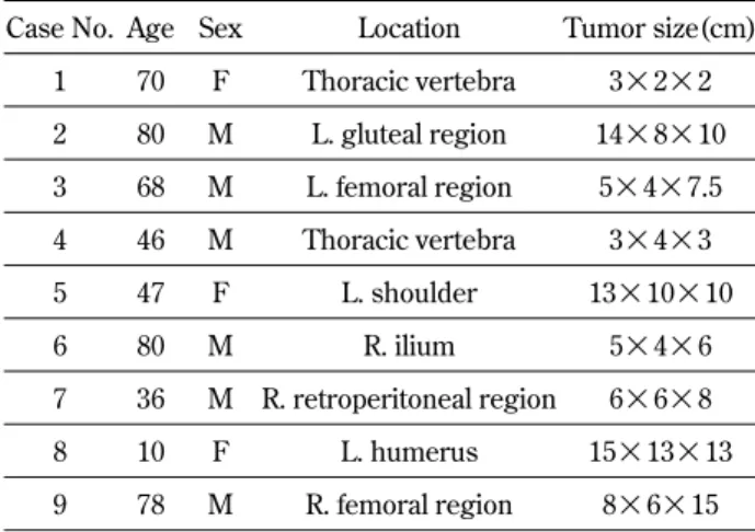
10 cm ; a : enhanced CT, b : MPR coronal view.Figure 2 :Case 5. DSA of right external iliac artery ; a : pre - embolization, b : postembolization. GS particles and microcoils were used. The actual amount of intraoperative bleeding was 350 mL and the average estimated amount of bleeding was 1217 mL.
Figure 3 :Case 9. A 78 - year - old man with a liposarcoma of the right femoral region. Tumor size was 8
!
6!
15 cm ; a : MRI CE Fat Sat T1WI axial & sagittal view, b : enhanced CT.DISCUSSION
Usefulness of embolization
One of the earliest reports of selective transcathe-ter artranscathe-terial embolization for musculoskeletal bone tumors was in 1975 when the technique was em-ployed to reduce perioperative blood loss. Reports of transcatheter arterial embolization prior to exci-sion of musculoskeletal tumors are rare but useful-ness is reported in every case (2-7). Location, size, and vascularity of the tumors vary widely. Operative time and blood loss during surgery also vary widely according to the degree of surgical intervention. In our study, arterial embolization significantly de-creased the amount of intraoperative blood loss com-pared with the estimated amount of bleeding.
The interval from embolization to operation was 5 days on account of the schedule of embolization and excision in the first case, only. Metallic embolic coils are permanent and GS particles are temporary, with the embolic effect lasting at least 1 week with the latter. Generally speaking, the appropriate inter-val would be at least one day because collateral path-ways readily develop, we believe the usefulness is the same if risks, such as infection, pain, and fever, are not considered.
Advantages and indications
The advantages of transcatheter arterial emboli-zation include minimal invasion and that hemostasis is often possible even with difficult surgical ligation. We consider the ideal indications for arterial emboli-zation prior to excision of musculoskeletal tumors are : 1. When large amounts of intraoperative bleed-ing are expected and where bleedbleed-ing would be dif-ficult to stop. 2. Cases with a lower risk of serious
complications following embolization. 3. Cases with hypervascular tumors of the trunk, pelvis and proxi-mal limbs excluding the peripheral spinal region.
Embolization of bone tumors, especially vertebral tumors
Gellad, et al. reported that in patients who under-went adequate embolization, an average of 1,850 mL of estimated blood loss was reported. In those who underwent inadequate or no embolization, greater than 3,500 mL of estimated blood loss oc-curred (8).
We performed embolization in 2 cases with metas-tasis to the thoracic vertebrae. Angio-CT imaging and xylocaine infusion testing was used before em-bolization to identify the Adamkiewicz artery and confirm that it was not involved with the tumor, thus avoiding complications. We also used embolization in the intercostal and lumbar arteries without com-plications. Careful consideration of blood supply en-abled us to perform embolization of vertebral tumors safely and we feel this intervention should be con-sidered in the treatment of primary or secondary bone tumors (9).
Embolic materials
The main purpose of embolization is to achieve thrombus formation and occlusion by administering embolizing materials through a selective catheter placed in an artery or vein (10). Our interventional radiologists determined the appropriate artery and the release site of the metallic coils after discussion with the orthopedic surgeons.
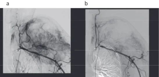
The advantages of using metallic coils include a stronger embolus effect than with the single use of GS particles. Also, the position of the coils can be Figure 4 :Case 9. DSA of the right external iliac artery ; a : pre - embolization, b : postembolization. GS particles and microcoils were used. The actual amount of intraoperative bleeding was 340 mL and the average estimated amount of bleeding was 1433 mL.
easily confirmed by the surgeon using intraopera-tive fluoroscopy and palpation. The disadvantages include the high expense and the possibility of coil migration intraoperatively. Permanently placed coils may also cause CT and MR imaging artifacts.
In conclusion, arterial embolization prior to resec-tion of hypervascular musculoskeletal tumors de-creases the amount of bleeding during the operation and contributes to patient safety.
CONFLICT OF INTEREST
For all authors : No conflicts of interest are pre-sent.
REFERENCES
1. Almgard LE, Fernström I, Haverling M, Ljungqvist A : Treatment of renal adenocarci-noma by embolic occlusion of the renal circu-lation. Br J Urol 45 : 475-479, 1973
2. Fenoy AJ, Greenlee JD, Menezes AH, Donovan KA, Sato Y, Hitchon PW, Chaloupka JC : Pri-mary bone tumors of the spine in children. J Neurosurg 105 Suppl : 252-260, 2006
3. Reuter M, Heller M, Heise U, Beese M : Tran-scatheter embolization of tumors of the muscu-lar and skeletal systems. Rofo 156 : 182-188, 1992
4. Bowers TA, Murray JA, Charnsangavej C, Soo CS, Chuang VP, Wallace S : Bone metastasis from renal carcinoma. The preoperative use of transcatheter arterial occlusion. J Bone Joint Surg 64-A : 749-754, 1982
5. Feldman F, Casarella WJ, Dick HM, Hollander BA : Selective intra-arterial embolization of bone tumors. AJR Am J Roentgenol 123 : 130-139, 1975
6. Dick HM, Bigliani LU, Michelsen WJ, Johnston AD, Stinchfield FE : Adjuvant arterial emboliza-tion in the treatment of benign primary bone tumors in children. Clin Orthop 139 : 133-141, 1979
7. Carpenter PR, Ewing JW, Cook AJ, Kuster AH : Angiographic assessment and control of po-tential operative hemorrhage with pathologic fractures secondary to metastasis. Clin Orthop 123 : 6-8, 1977
8. Gellad FE, Sadato N, Numaguchi Y, Levine AM : Vascular metastatic lesions of the spine : preoperative embolization. Radiology 176 : 683-6, 1990
9. Owen RJ : Embolization of Musculoskeletal Bone Tumors. Semin Intervent Radiol 27 : 111-23, 2010
10. Börüban S, Sancak T, Yildiz Y, Saglik Y : Em-bolization of benign and malignant bone and soft tissue tumors of the extremities. Diagn In-terv Radiol 13 : 164-71, 2007