CASE REPORT
Debridement for Infectious Spondylodiscitis in a 9-year-old Girl
Using Full-Endoscopic Discectomy System :
a Case Report and literature review
Yoshihiro Ishihama, Toshinori Sakai, Hiroaki Manabe, Fumitake Tezuka, Kazuta Yamashita, Yoichiro Takata, Toru Maeda, and Koichi Sairyo
Department of Orthopedics, Institute of Biomedical Sciences, Tokushima University Graduate School, Tokushima, Japan
Abstract : Objective : Infectious spondylodiscitis (IS) is rarely seen in healthy elementary school age children. Conservative treatment with antibiotics is usually preferable but sometimes fails because of the low identifi-cation rate of the pathogen by percutaneous needle biopsy. When surgical treatment is indicated, selecting the appropriate procedure is crucial in terms of invasiveness for such young children. Case report : We present the case of a 9-year-old otherwise healthy girl with IS who successfully underwent debridement and identification of the causative pathogen using full endoscopic discectomy (FED) system. Methicillin-susceptible Staphylococcus aureus was identified on several cultures of samples. Immediately after the surgery, the LBP was significantly decreased and the remittent fever resolved dramatically. At the 1-year follow-up, she had no symptoms and plain radiographs showed bony fusion. Conclusion : This is the first report on IS in elementary school-age children treated with the FED system. Debridement using this system could provide minimally invasive and effective cu-rettage of the infected disc space and can be helpful in identifying the pathogen even for small children. J. Med. Invest. 67 : 351-354, August, 2020
Keywords : Infections spondylodiscitis, Full endoscopic discectomy, Pediatric patient
INTRODUCTION
Infectious spondylodiscitis (IS) is less common in children than in adults. Most commonly found in the lumbar/lumbosacral region (1), IS typically presents as low back pain (LBP) and/or neurological symptoms due to the progressing paravertebral ab-scess as well as fever. It frequently affects the elderly or patients with immunodeficiency and is rarely seen in healthy elementary school age children, which may lead to delayed diagnosis in pe-diatric patients.
Conservative treatment using antibiotics is usually sufficient, and most symptoms resolve without further complications (2). Poor response to conservative treatment, severe persistent LBP with deformity and instability, and progressing neurological deficits are indications for surgical treatment. There are many surgical options available depending on the patient’s condition or specific considerations of the attending surgeon. Although debridement using full-endoscopic discectomy (FED) system for pyogenic discitis has been reported in adults (3), it is rare in children.
In this report, we present the case of a healthy 9-year-old girl with IS who was successfully treated with the FED system.
CASE REPORT
A 9-year-old girl with complaints of persistent LBP for more than 3 months with a slight fever was referred to our hospital. She had consulted several primary care doctors including
or-thopedic surgeons and a pediatrician, but no abnormal findings were seen on plain X-ray of the lumbar spine. The fever was attributed to pharyngitis and the LBP had been treated as a “pharyngitis related-pain” for approximately 3 months prior to presentation. The LBP was refractory to conservative treatment with acetaminophen and antibiotics, and so magnetic resonance imaging (MRI) of the lumbar spine was performed. Subsequent-ly, IS was identified surrounding the L5-S1 intervertebral disc on MRI, and she was referred to our department.
The patient had no medical or surgical history that would affect the immune system. In addition, she had no antecedent infection such as urinary tract infection. At the first medical examination in our outpatient clinic, she presented with a remit-tent fever and LBP that was worsened by motion.
Neurological examination demonstrated weakness of the iliopsoas bilaterally (manual muscle testing : 4 on either side). There was no sensory disturbance and tension test was neg-ative. Laboratory examination revealed that white blood cell count was within the normal range (WBC : 6,200 / μL ; neu-trophils : 68% ; lymphocytes ; 24%), but C-reactive protein (CRP) and erythrocyte sedimentation rate (ESR) were elevated (CRP : 9.30 mg / dL ; ESR : 112 mm / h). Initial blood cultures and the T-SPOT tuberculosis (TB) test were negative. Chest X-ray showed no abnormal findings suggestive of pulmonary disease such as pulmonary TB.
Lateral plain X-ray of the lumbar spine showed disc space nar-rowing and erosive changes on the endplates of L5 and S1. MRI revealed signal changes around the L5-S1 intervertebral disc including the L5 and S1 vertebral bodies, strongly suggestive of IS (Fig. 1,2).
Based on the above-mentioned findings, we made a diagnosis of IS and planned biopsy and debridement using an FED system via a transforaminal approach under general anesthesia. To avoid exiting nerve (L5) root injury, we monitored motor func-tional integrity using transcranial motor evoked potentials and free-running electromyography (EMG).
The Journal of Medical Investigation Vol. 67 2020
351
Received for publication January 6, 2020 ; accepted February 19, 2020.
Address correspondence and reprint requests to Toshinori Sakai, M.D., Ph.D., Associate Professor, Department of Orthopedics, Institute of Biomedical Sciences, Tokushima University Graduate School, 3-18-15 Kuramoto, Tokushima 770-8503, Japan, and Fax : +81-88-633-0178.
352
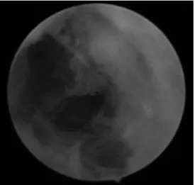
Y. Ishihama, et al. Debridement Using FED System for young childrenThe transforaminal FED procedure was performed at the infected intervertebral disc. Prior to placement of the FED system, a spinal needle was inserted into the annulus fibrosis at a point 4 cm from the midline under fluoroscopic guidance with the patient in the prone position. Yellowish-white pus was aspirated through the needle and endoscope. On the endoscopic views, necrotic tissue and pus were dispersed within the L5-S1 disc space. Debridement of the infected disc structure was done under saline irrigation (Fig. 3).
Immediately after the surgery, the LBP was significantly decreased and she was permitted to ambulate with a corset. In addition, methicillin-susceptible Staphylococcus aureus was identi-fied on several cultures of samples, and antibiotic sensitivity was confirmed. As soon as appropriate antibiotics were prescribed according to those results, the remittent fever resolved dramati-cally and the laboratory data began to improve (Fig. 4). At the 1 month follow-up, MRI demonstrated decreases in inflammatory signal changes and bone destruction (Fig. 5). At the 1-year fol-low-up, she had no symptoms and computed tomography (CT) scans showed bony fusion at L5-S1 (Fig. 6).
Fig 1. Initial image of the lumbar spine
T2-weighted image showing high signal intensity changes in L5, S1 vertebral bodies. Both signal and morphological changes are identified in the L5-S disc space.
Fig 2. Initial image of the lumbar spine
Short tau inversion recovery (STIR) sagittal images of preoperative MRI show high signal intensity in L5, S1 vertebral bodies and intervertebral disc space.
Fig 3. Endoscopic view before aspiration and debridement
Endoscopic findings of the infected disc showing pus accumulation and granulation tissue.
Fig 4. Treatment course after hospitalization (Antibiotics and CRP transition).
The course of C-reactive protein (CRP) during the perioperative period of treatment was shown. Decreasing of CRP showed the effectiveness of surgery and appropriate medication. CEZ: cefazolin; CLDM: clindamycin; RFP: rifampicin; CCL: Cefaclor
353
The Journal of Medical Investigation Vol. 67 August 2020
DISCUSSION
IS in childhood is relatively rare, with a reported incidence of about 1–2 per 32,500 pediatric population per year (4), and the mean age at diagnosis in children is approximately 2 to 8 years old. According to past reports, the lumbar region is most fre-quently involved, followed by the thoracic and then the cervical regions (5). Diagnosis of IS can often be delayed due to its rarity or specific problems in pediatric patients such as nonspecific symptoms / complaints. However, particularly in cases with red flag clinical features such as fever and LBP at rest, a detailed history-taking including the past medical history, laboratory data, and radiological examination.
Treatment for IS is controversial. Basically, conservative treatment, which consists of empirical antibiotics, is preferable for pediatric patients with IS. However, without detection of the causative pathogen, antibiotics may possibly have no effect, and surgical treatment will be required if conservative treatment fails. Therefore, if there is a high index of suspicion for IS, prompt identification of the causative pathogen by various procedures including percutaneous needle biopsy is considered desirable.
Regarding the surgical procedures, there are various op-tions including open debridement via the conventional anterior approach, CT-guided percutaneous catheter drainage, and percutaneous drainage with continuous irrigation (6). Among these, full endoscopic aspiration and drainage as used in our case is considered the most minimally invasive and effective surgical procedure (7). In addition, using this system allows for collection of infected tissue as samples for detecting the causative pathogen. Yang et al. showed that in 90% of patients treated with percutaneous endoscopic technique cultures were positive, in contrast to the causative pathogen being identified in < 50% of patients for whom a CT-guided biopsy was performed (8). Along with these advantages, FED requires no absolute rest as op-posed to other surgical options that require limiting the patients’ daily activities due to invasiveness immediately postoperatively.
FED via the transforaminal approach is usually performed under local anesthesia and conscious sedation (9). However, this is considered difficult for pediatric patients such as the present case, and so FED should be performed under general anesthesia. Nevertheless, performing this surgery under general anesthe-sia poses some risk of exiting nerve injury (10). Furthermore, in pediatric patients, the spinal structure is immature and the foramen is narrow, which further narrows the entry point of the endoscope. To avoid nerve injury while performing the procedure under general anesthesia, we successfully performed surgery using a free-running EMG for intraoperative neurolog-ical monitoring.
As far as we know, this is the first report on IS in elementary school-age children treated with the FED system. Debridement using this system could provide minimally invasive and effective curettage of the infected disc space and can be helpful in identi-fying the pathogen even for small children. However, all steps need to be carefully performed to avoid critical complications.
CONCLUSION
We presented the case of a 9-year-old otherwise healthy girl with IS who successfully underwent debridement and identifica-tion of the causative pathogen using full endoscopic discectomy (FED) system. Debridement with minimally invasive full-endo-scopic technique could provide effective curettage of the infected disc space. In addition, using FED system can also be helpful in identifying the pathogen for children.
Fig 5. Postoperative MRI scan of the lumbar spine
STIR sagittal MRI scan at the 1-month follow-up after surgery showing a decrease in the high signal intensity of the vertebral body, and no increase in the abscess or bone destruction.
Fig 6. Postoperative CT scan of the lumbar spine
CT scan at 12 months postoperatively showing the presence of bridging callus that suggests a tendency to bony fusion.
354
Y. Ishihama, et al. Debridement Using FED System for young childrenCONFLICTS OF INTEREST DISCLOSURE
The authors declare that there is no conflict of interest re-garding the publication of this article. Additionally, no funding was received that could influence the integrity concerning this article.
REFERENCES
1. Fernandez M, Carrol CL, Baker CJ : Discitis and vertebral osteomyelitis in children : an 18-year review. Pediatrics 105 : 1299-1304, 2000
2. Dayer R, Alzahrani MM, Saran N, Ouellet JA, Journeau P, Tabard-Fougere A, Martinez-Álvarez S, Ceroni D : Spinal infections in children : a multicentre retrospective study. Bone Joint J 100-B : 542-548, 2018
3. Ito M, Abumi K, Kotani Y, Kadoya K, Minami A : Clinical outcome of posterolateral endoscopic surgery for pyogenic spondylodiscitis : results of 15 patients with serious comor-bid conditions. Spine (Phila Pa 1976) 32 : 200-206, 2007 4. Cushing AH : Diskitis in children. Clin Infect Dis 17 : 1-6,
1993
5. Chandrasenan J, Klezl Z, Bommireddy R, Calthorpe
D : Spondylodiscitis in children : a retrospective series. J Bone Joint Surg Br 93 : 1122-1125, 2011
6. Hanaoka N, Kawasaki Y, Sakai T, Nakamura T, Nanam-ori K, Nakamura E, Uchida k, Yamada H : Percutaneous drainage and continuous irrigation in patients with severe pyogenic spondylitis, abscess formation, and marked bone destruction. J Neurosurg Spine 4 : 374-379, 2006
7. Yang SC, Chen WJ, Chen HS, Kao YH, Yu SW, Tu YK : Extended indications of percutaneous endoscopic lavage and drainage for the treatment of lumbar infectious spondylitis. Eur Spine J 23 : 846-853, 2014
8. Yang SC, Fu TS, Chen LH, Chen WJ, Tu YK : Identifying pathogens of spondylodiscitis : percutaneous endoscopy or CT-guided biopsy. Clin Orthop Relat Res 466 : 3086-3092, 2008
9. Sairyo K, Chikawa T, Nagamachi A : State-of-the-art trans-foraminal percutaneous endoscopic lumbar surgery under local anesthesia : Discectomy, foraminoplasty, and ventral facetectomy. J Orthop Sci 23 : 229-236, 2018
10. Choi I, Ahn JO, So WS, Lee SJ, Choi IJ, Kim H : Exiting root injury in transforaminal endoscopic discectomy : pre-operative image considerations for safety. Eur Spine J 22 : 2481-2487, 2013