Hattoria 11: 23–29. 2020
On the identity of a little-known Riccia species,
Riccia wichurae
(Ricciaceae) from Japan
Tomoyuki KATAGIRI
Hattori Botanical Laboratory, Obi 6–1–26, Nichinan, Miyazaki 889–2535, Japan Author for correspondence: Tomoyuki KATAGIRI, tomoyuki-katagiri@hattorilab.org
Abstract Detailed morphological descriptions of a little-known Japanese species, Riccia wichurae
Steph. are given based on the type specimen. SEM images of the spores are also provided for the first time. The species is mainly characterized by 0.6–1.0 mm wide thalli with deep median grooves, lacking air chambers, purple or hyaline ventral scales, and large spore size (75–)80–90(–100) µm with irregular thickened wing margins. The author proposes that the species should be treated as a synonym of R. beyrichiana Hampe ex Lehm. which is widely distributed in Japan.
Introduction
Riccia wichurae Steph. was described by Stephani (1898) based on Japanese plants.
Stephani gave a short description of the plants and the rough information on the locality: Hab. Japonia (Wichura) (Fig. 1). It is interesting that below the description Stephani also noted: It is doubtful whether the plant is from Japan. It may be from another part of Asia. At least other liverworts from his collection were described as questionable in this regard. In any case, the plant is new . On the basis of this description Hattori (1943) excluded this species from Japanese flora, and it has long been neglected by Japanese bryologists (Yamada & Iwatsuki 2006), while it was recognized as an independent species in the liverwort world checklist by Söderström et al. (2016). I think that this species should not have been buried but treated as a validly proposed species reported from Japan.
The German botanist, Max Ernst Wichura (1817–1866), visited Japan in 1860 (Shirahata 1994, Stafleu & Cowan 1988) and collected plants from several parts of Japan. Brotherus (1899) showed that he collected bryophytes in such locations as Yedo (currently Tokyo), Yokohama, and Nagasaki. According to Stephani s Icones (Stephani 1985, n. 9005), his description was based on the specimen, Wichura 1492a, which was kept in Stephani s collection in Conservatoire et Jardin Botaniques de la Ville de Genève (G), and the specimen
Wichura 1492a (G-00067889; Fig. 1) was considered the holotype (Turland et al. 2018; Art.
9.1. Note 1). As noted in the specimen label Herb. Berlin , Wichura s collection was in the herbarium of the Botanic Garden and Botanical Museum Berlin-Dahlem (B) but it was
destroyed during World War II.
In this study I examine the holotype of R. wichurae and give morphological information including SEM images of spores for the first time. Based on the morphological information, taxonomic treatment of the species is proposed.
Materials and Methods
The type specimen of Riccia wichurae Steph. was investigated using a Leica M205C stereomicroscope equipped with a Nikon DS-Fi3 digital camera. For SEM images the samples were mounted on an aluminium stage with double-sided carbon tape and sputter-coated with gold at ca. 200 Å using a JFC-1500 Ion Sputtering Device (JEOL, Tokyo). The images were then captured using a JCM-6000 scanning electron microscope (JEOL, Tokyo).
Taxonomy
Riccia wichurae Steph., Bull. Herb. Boissier 6: 330. 1898. Figs. 2–4 Type: Japan (location not specified). Wichura 1492a (G-00067889!, holotype)
Plants yellowish brown in aged material, forming partial rosettes 5–10 mm in diam. or intricate mats, not glistening. Thalli 1–2(–3) times furcate, 3–5(–10) mm long, branches 0.6–1.0 mm wide; median groove deep and narrow at apex, gradually posteriorly becoming shallow and widening to 1/4–1/3 branch-width; cilia absent or scarce. Ventral scales purple or hyaline, not extended beyond margin of thallus. Thallus section 0.4–0.6(–0.8) mm high and 2.0–2.5(–3.0) times as wide, lacking air chambers and consisting of vertical filaments with 1/3–1/2 thallus height; flanks weakly convex; margin acute, with thin-walled cells; epidermal cells collapsing, confirmed only with thin basal walls; ventral tissues parenchymatous, cells
Figure 1. Holotype specimen (A) and close-up of the label (B) of Riccia wichurae Steph.
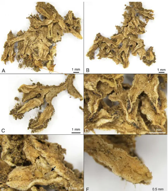
Figure 2. Riccia wichurae Steph. A–B: Habit. C: Single shoot. D–F: Apical parts of thallus. Arrow
shows the projecting part with mature capsule in E. All photographed from Wichura 1492a (G-00067889).
Figure 3. Riccia wichurae Steph. A–B: Cross-sections of thallus. C–D: Spores from distal view with
different focus. E: Spores from proximal view. F: Part of wing from proximal view. All photographed from Wichura 1492a (G-00067889).
(75–)80–90(–100) µm in diam., reddish brown; distal surface with 8–10 incomplete alveolae 7–10(–12) µm wide across surface and tubercles at the angles; proximal surface indistinctly alveolate, with scattered tubercles, with an indistinct trilete scar; wing distinct, 6–8 µm wide, sinuate or subcrenate, obscurely papillose, margin irregularly thickened.
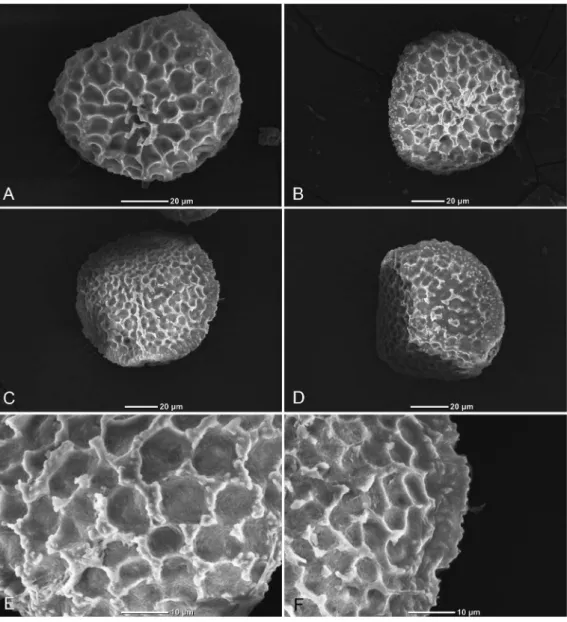
Figure 4. SEM images of spores of Riccia wichurae Steph. A–B: Distal view. C–D: Proximal view. E:
Alveolae on distal surface. F: Close-up of wing from proximal view. All photographed from Wichura 1492a (G-00067889).
Discussion
The detailed morphological study based on the holotype shows that the above-mentioned description basically fits the original description given by Stephani (1898) and of his illustrations of cross-sections of the thallus (Stephani 1985, n. 9005) (Fig. 3 A–B). The morphological characters of the thallus, such as (1) 0.6–1.0 mm wide thallus (Fig. 2A–B), (2) median groove deep and narrow at apex, gradually posteriorly becoming shallow and widening to 1/4–1/3 branch-width (Fig. 2E–F), (3) purple or hyaline ventral scales not extended beyond margin of thallus (Fig. 2D), (4) thallus in cross-section 0.4–0.6(–0.8) mm high, 2.0–2.5(–3.0) times as wide, lacking air chambers (Fig. 3A–B), and (5) slightly projecting dorsal surface of thallus with mature capsule (Fig. 2E) suggest its close relationship to R. bifurca Hoffm. and R. beyrichiana Hampe ex Lehm., both widely distributed in Japan (Tominaga & Furuki 2014). According to Tominaga & Furuki (2014) who gave a taxonomic revision of the genus Riccia sect. Riccia in Japan, R. beyrichiana can be distinguished from R.
bifurca by larger size of spores (75–105 µm in R. beyrichiana vs. 50–80 µm in R. bifurca) and
presence of distinct irregular thickenings of wings. Although Stephani noted the spore size of
R. wichurae as 68 µm, the detailed study shows that the spores of R. wichurae are
characterized by (1) large size of (75–)80–90(–100) µm in diam. (Figs. 3C–E & 4A–D), (2) 8–10 incomplete alveolae on distal surface (Figs. 3C & 4A–B), (3) indistinctly alveolate and an indistinct trilete on proximal surface (Figs. 3E & 4C–D), and (4) distinct wing with irregularly thickened margin (Figs. 3F & 4F), and these data suggest its morphological relationships to R. beyrichiana.
Taking the limited information which was based only on a single old herbarium specimen into consideration and considering morphological characters of spores are more stable than those of the thallus, I propose that R. wichurae should be treated as a synonym of R.
beyrichiana which is one of the most widely ranging holarctic species of the genus with wide
range of morphological variations (Schuster 1992; Paton 1999; Damsholt 2002; Tominaga & Furuki 2014).
Acknowledgements
I am grateful to the curator and staff of Conservatoire et Jardin Botaniques de la Ville de Genève (G) for the specimen loan. I also thank Dr. Yuya Inoue (Hiroshima University) for providing SEM images and Dr. Anders Hagborg (Field Museum) for checking the English text. This work was supported by JSPS KAKENHI Grant Number JP16K18604 and Grant-in-Aid for Scientific Research (Specially Designated Research Promotion) of Japan Society for the Promotion of Science (JSPS).
Hattori S. 1943. Ricciaceae nipponicae. National Science and Museum 14: 138–143. Paton J. A. 1999. The Liverwort Flora of the British Isles. Harley Books, Colchester.
Schuster R. M. 1992. The Hepaticae and Anthocerotae of North America, East of the Hundredth Meridian. Vol. 6. Field Museum of Natural History, Chicago.
Shirahata Y. 1994. Plant Hunters. Kodansha, Tokyo.
Stafleu F. A. & Cowan R. S. 1988. Taxonomic literature: a selective guide to botanical publications and collections with dates, commentaries and types. Ed. 2, Vol. 7: W-Z. Regnum Vegetabile 116: 1–653. Stephani F. 1898. Species hepaticarum 1. Bulletin de l Herbier Boissier 6: 309–343.
Stephani F. 1985. Icones Hepaticarum (microfiche). IDC, Zug.
Söderström L., Hagborg A., von Konrat M., Bartholomew-Began S., Bell D., Briscoe L., Brown E., Cargill D. C., Costa D. P., Crandall-Stotler B. J., Cooper E. D., Dauphin G., Engel J. J., Feldberg K., Glenny D., Gradstein S. R., He X., Heinrichs J., Hentschel J., Ilkiu-Borges A. L., Katagiri T., Konstantinova N. A., Larraín J., Long D. G., Nebel M., Pócs T., Puche F., Reiner-Drehwald E., Renner M. A. M., Sass-Gyarmati A., Schäfer-Verwimp A., Moragues J. G. S., Stotler R. E., Sukkharak P., Thiers B. M., Uribe J., Váňa J., Villarreal J. C., Wigginton M., Zhang L. & Zhu R.-L. 2016. World checklist of hornworts and liverworts. PhytoKeys 59: 1–828.
Tominaga T. & Furuki T. 2014. Taxonomic studies of the genus Riccia sect. Riccia (Ricciaceae, Hepatics) in Japan. Bryological Research 11: 53–62.
Turland N. J., Wiersema J. H., Barrie F. R., Greuter W., Hawksworth D. L., Herendeen P. S., Knapp S., Kusber W.-H., Li D.-Z., Marhold K., May T. W., McNeill J., Monro A. M., Prado J., Price M. J. & Smith G. F. 2018. International Code of Nomenclature for algae, fungi, and plants (Shenzhen Code). Regnum Vegetabile 159: 1–254.
Yamada K. & Iwatsuki Z. 2006. Catalog of the hepatics of Japan. The Journal of the Hattori Botanical Laboratory 99: 1–106.