neopl as i a pr ogr es s i on
著者
Li u Shul i ng, M
i naguc hi Takeo, Lac hkar Bouc hr a,
Zhang Shuang, Xu Chenyang, Tenj i m
bayas hi Yur i ,
Shi kam
a Ayum
i , Tas aka N
obut aka, Aki yam
a Az us a,
Sakur ai M
anabu, N
akao Sar i , O
c hi H
i r oyuki ,
O
nuki M
am
i ko, M
at s um
ot o Koj i , Yos hi kaw
a
H
i r oyuki , Sat oh Toyom
i
j our nal or
publ i c at i on t i t l e
PLO
S O
N
E
vol um
e
13
num
ber
2
page r ange
e0193061
year
2018- 02
権利
( C) 2018 Li u et al . Thi s i s an open ac c es s
ar t i c l e di s t r i but ed under t he t er m
s of t he
Cr eat i ve Com
m
ons At t r i but i on Li c ens e, w
hi c h
per m
i t s unr es t r i c t ed us e, di s t r i but i on, and
r epr oduc t i on i n any m
edi um
, pr ovi ded t he
or i gi nal aut hor and s our c e ar e c r edi t ed.
U
RL
ht t p: / / hdl . handl e. net / 2241/ 00151553
RESEARCH ARTICLE
Separate analysis of human papillomavirus E6
and E7 messenger RNAs to predict cervical
neoplasia progression
Shuling Liu1, Takeo Minaguchi2*, Bouchra Lachkar1, Shuang Zhang1, Chenyang Xu1, Yuri Tenjimbayashi3, Ayumi Shikama2, Nobutaka Tasaka2, Azusa Akiyama2,
Manabu Sakurai2, Sari Nakao2, Hiroyuki Ochi2, Mamiko Onuki3, Koji Matsumoto3, Hiroyuki Yoshikawa2,4, Toyomi Satoh2
1Doctoral Program in Obstetrics and Gynecology, Graduate School of Comprehensive Human Sciences, University of Tsukuba, Tsukuba, Ibaraki, Japan,2Department of Obstetrics and Gynecology, Faculty of Medicine, University of Tsukuba, Tsukuba, Ibaraki, Japan,3Department of Obstetrics and Gynecology, Showa University School of Medicine, Tokyo, Japan,4Ibaraki Prefectural Central Hospital, Kasama, Ibaraki, Japan
Abstract
A few studies previously suggested that human papillomavirus (HPV) E6 messenger RNA (mRNA) may exist uniformly in all grades of cervical intraepithelial neoplasia (CIN), whereas the detection rate of E7 mRNA may increase with disease progression from low-grade CIN to invasive carcinoma. The aim of this study was to clarify the different roles of E6 and E7 mRNAs in cervical carcinogenesis. The presence of each E6 and E7 mRNA was analyzed in 171 patients with pathologically-diagnosed CIN or cervical carcinoma. We utilized a RT-PCR assay based on consensus primers which could detect E6 mRNA (full-length E6/E7 transcript) and E7 mRNAs (spliced E6*/E7 transcripts) separately for various HPV types. E7 mRNAs were detected in 6% of CIN1, 12% of CIN2, 24% of CIN3, and 54% of cervical carcinoma. The presence of E7 mRNAs was significantly associated with progression from low-grade CIN to invasive carcinoma in contrast with E6 mRNA or high-risk HPV (HR-HPV) DNA (p = 0.00011, 0.80 and 0.54). The presence of both E6 and E7 mRNAs was signifi-cantly associated with HPV16/18 DNA but not with HR-HPV DNA (p = 0.0079 and 0.21), while the presence of E6 mRNA was significantly associated with HR-HPV DNA but not with HPV16/18 DNA (p = 0.036 and 0.089). The presence of both E6 and E7 mRNAs showed high specificity and low sensitivity (100% and 19%) for detecting CIN2+ by contrast with the positivity for HR-HPV DNA showing low specificity and high sensitivity (19% and 89%). The positive predictive value for detecting CIN2+ was even higher by the presence of both E6 and E7 mRNAs than by the positivity for HR-HPV DNA (100% vs. 91%). In 31 patients fol-lowed up for CIN1-2, the presence of both E6 and E7 mRNAs showed significant associa-tion with the occurrence of upgraded abnormal cytology in contrast with E6 mRNA, HR-HPV DNA, or HPV16/18 DNA (p = 0.034, 0.73, 0.53, and 0.72). Our findings support previous studies according to which E7 mRNA is more closely involved in cervical carcinogenesis than E6 mRNA. Moreover, the separate analysis of E6 and E7 mRNAs may be more useful
a1111111111 a1111111111 a1111111111 a1111111111 a1111111111 OPEN ACCESS
Citation:Liu S, Minaguchi T, Lachkar B, Zhang S, Xu C, Tenjimbayashi Y, et al. (2018) Separate analysis of human papillomavirus E6 and E7 messenger RNAs to predict cervical neoplasia progression. PLoS ONE 13(2): e0193061.https:// doi.org/10.1371/journal.pone.0193061
Editor:Maria Lina Tornesello, Fondazione IRCCS Istituto Nazionale dei Tumori, ITALY
Received:September 28, 2017
Accepted:February 2, 2018
Published:February 21, 2018
Copyright:©2018 Liu et al. This is an open access article distributed under the terms of theCreative Commons Attribution License, which permits unrestricted use, distribution, and reproduction in any medium, provided the original author and source are credited.
Data Availability Statement:All relevant data are within the paper.
Funding:The Grant-in-Aid for Scientific Research (No. 25462585) from the Ministry of Education, Culture, Sports, Science, and Technology, Tokyo, Japan (KM). https://kaken.nii.ac.jp/grant/KAKENHI-PROJECT-25462585/. The funders had no role in study design, data collection and analysis, decision to publish, or preparation of the manuscript.
than HR-HPV DNA test for detecting CIN2+ precisely and predicting disease progression. Further accumulation of evidence is warranted to validate our findings.
Introduction
Cervical cancer is the fourth most commonly diagnosed cancer and the fourth most common cause of cancer death in women worldwide [1]. Each year, 528,000 women develop cervical cancer, and 266,000 women die of the disease, accounting for 7.5% of all cancer deaths in females [1]. Human papillomavirus (HPV) is classified by the sequence of the L1 gene. Infec-tion with high-risk HPV (HR-HPV), including types 16 and 18, causes development of low-grade cervical intraepithelial neoplasia (CIN), and viral persistence induces cellular transfor-mation resulting in progression to high-grade CIN and invasive cervical cancer [2]. HPV viral genome has 6 early genes, E1, E2, E4, E5, E6, and E7, and 2 late genes, L1 and L2, encoding capsid proteins. Among the early genes, E6 and E7 cause cancer by inactivating the tumor sup-pressor proteins p53 and Rb, respectively [3]. Normal epithelial cells persistently infected with HR-HPV first develop low-grade CIN. When viral DNA is integrated into host chromosome, constant overexpression of E6 and E7 induces abnormal proliferation, transformation and immortalization, and inhibits differentiation, apoptosis and immune response, leading to development of high-grade CIN. Accumulation of genetic and epigenetic alterations further causes progression to invasive cancer [4]. E6 is mainly expressed from full-length E6/E7 mRNA, and E7 is mainly expressed from spliced E6/E7 mRNA [5]. HPV16 expresses two
iso-forms of E7 gene, and the other HPV types including HPV18 express one isoform of E7 gene. To date, only a few studies previously investigated the distinct roles of E6 and E7 mRNAs for cervical carcinogenesis [6,7]. According to Nakagawa et al., E6 transcript is uniformly detected from CIN1 to invasive cancer, but E7 transcripts show a higher detection rate with disease progression from low-grade CIN to invasive cancer [6]. Another previous publication by Sotlar et al. showed that detection rate of E7 transcript increased with disease progression in contrast with E6 mRNA showing only moderate increase [7]. The aim of our study was to investigate the distinct roles of each E6 and E7 mRNAs in the pathogenesis of cervical cancer.
Materials and methods
Patients and samples
according to the Bethesda system [8]. The included patients were treated or followed-up according to the clinical guidelines [9]. Study results of the mRNA analyses did not influenced their management. The median follow-up duration was 194 days (range 0–613). Follow-up data were retrieved until 2017-5-31.
DNA extraction and HPV genotyping
Genomic DNA was extracted using SepaGene kit (Eidia, Tokyo, Japan) according to the man-ufacturer’s instruction. HPV genotyping was performed by L1-PCR and RFLP analyses as described previously [6] or at a clinical testing laboratory (SRL, Tokyo, Japan) by Amplicor lin-ear array HPV genotyping test (Roche Diagnostics, Tokyo, Japan). HR-HPVs are defined as
Fig 1. Study design.HPV: human papillomavirus, CIN: cervical intraepithelial neoplasia, ICC: invasive cervical cancer.
https://doi.org/10.1371/journal.pone.0193061.g001
HPV 16, 18, 31, 33, 35, 39, 45, 51, 52, 56, 58, 59 and 68, which can be detected by Hybrid Cap-ture 2 (HC2).
RNA extraction and Reverse Transcriptase PCR (RT-PCR)
Total RNA extraction and DNase treatment were performed as described previously [6]. RT-PCR was conducted using OneStep RT-PCR kit (QIAGEN, Tokyo, Japan) according to the manufacturer’s instruction. We utilized a RT-PCR assay based on consensus primers designed to maintain around 80–90% homology to the known conserved sequences in E6 and E7 ORFs among multiple oncogenic HPVs [6]. E6 and E7 mRNAs could be separately detected for at least HPV types 16, 18, 31, 33, 35, 51, 52, 56, 58 and 59 [6]. We usedβ2-microglobulin as a control for RT-PCR in order to validate normal RNA extraction and no contamination of DNA which will affect the RT-PCR results, as E6/E7 DNA is the same size as E6 mRNA. Prim-ers used for RT-PCR and PCR are as follows: E6/E7,ACC GAA AAC GGT TGA ACC GAA
AAC GGTandGAG CTG TCG CTT AAT TGC TC;β2-microglobulin,TGT CTT TCA GCA
AGG ACT GGandGAT GCT GCT TAC ATG TCT CG.
Statistical analysis
Differences in proportions were evaluated by the Fisher’s exact test. Diagnostic indices of sen-sitivities, specificities, positive predictive values, and negative predictive values with 95% confi-dence intervals were calculated for detecting CIN2+, CIN3+, and invasive cervical cancer. Disease progression of CIN1-2 was examined as a surrogate by the Kaplan-Meier method cal-culating the intervals from E6/E7 sample collection until patients showed upgraded results of Pap test compared with the cytology at E6/E7 sample collection or they were censored, and the difference was statistically evaluated by the log-rank test.
Results
We first analyzed the E6 and E7 mRNA expression patterns in human cervical cancer cell-lines CaSki and HeLa by RT-PCR (Fig 2), and confirmed that the expression patterns were consistent with data published by Nakagawa et al. [6]. In addition to E6 mRNA, two isoforms of E7 mRNA were detected in HPV 16-positive CaSki cells, and one isoform of E7 mRNA detected in HPV 18-positive HeLa cells. In order to verify that our RT-PCR assay works prop-erly, we further performed sequencing analyses of E6/E7 cDNAs and confirmed that E6 mRNA is actually full-length E6/E7 transcript and that E7 mRNAs are actually spliced E6/E7
transcripts. The E6/E7 DNA is the same size as the full-length RNA, 652 bp for HeLa and 622 bp for CaSki.β2-microglobulin is 148 bp for RNA and 775 bp for DNA (Fig 3).
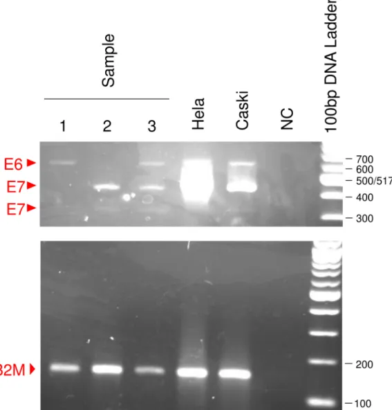
Subsequently we analyzed the E6/E7 mRNA expressions in liquid-based cytology samples from 171 patients.Fig 4shows an example of detection of E6/E7 mRNA from patients. Beta2-microglobulin amplification showed no contamination by genomic DNA in every sample. The detection rate of E7 mRNA significantly increased with disease progression from low-grade CIN to invasive cancer, while those of E6 mRNA and HR-HPV DNA did not change (p = 0.00011, 0.80 and 0.54, respectively;Table 1). We next examined the relationship between E6/E7 mRNA expressions and HPV genotypes. The presence of E6 mRNA showed significant associations with the positivity for HR-HPV DNA but not with the positivity for HPV16/18 DNA (p = 0.0036 and 0.089;Table 2), whereas the presence of both E6 and E7 mRNAs showed significant associations with the positivity for HPV16/18 DNA but not with the positivity for HR-HPV DNA (p = 0.0079 and 0.21;Table 2).
and low sensitivity (100% [95% confidence interval [10], 79–100] and 19% [95% CI, 13–26];
Table 3) in contrast with the positivity for HR-HPV DNA showing high sensitivity and low specificity (19% [95% CI, 4–46] and 89% [95% CI, 83–93];Table 3). Notably, the positive pre-dictive value (PPV) for detecting CIN2+ was even higher by the presence of both E6 and E7 mRNAs than by the positivity for HR-HPV DNA or HPV16/18 DNA (100%, 91% and 91%, respectively;Table 3). Similar trends were also observed about the diagnostic accuracies for detecting CIN3+ and invasive cervical cancer (Tables4and5).
Finally, we examined the impact of the positivity for E6/E7 mRNAs or specific HPV geno-types on disease progression by following up 31 patients with CIN1-2. Since no disease pro-gression was pathologically diagnosed yet in those patients, we compared intervals until the occurrence of upgraded abnormal cytology compared with the cytology at E6/E7 sample col-lection as a surrogate for disease progression. The presence of both E6 and E7 mRNAs showed significant association with the occurrence of upgraded abnormal cytology, the presence of E7 mRNAs showed association without statistical significance, but the presence of E6 mRNA, HR-HPV DNA, or HPV 16/18 DNA showed no such trends (p = 0.034, 0.12, 0.73, 0.53, and 0.72, respectively;Fig 5).
Discussion
Our E6/E7 RT-PCR analyses showed that E7 mRNAs were significantly associated with pro-gression from low-grade CIN to invasive carcinoma in contrast with E6 mRNA showing no
Fig 2. E6/E7 mRNA expression patterns by RT-PCR in human cervical cancer cell lines.H: full-length E6/E7 (E6), M: spliced E6I/E7 (E7), L: spliced E6II/E7 (E7).
https://doi.org/10.1371/journal.pone.0193061.g002
such trend (Table 1). Furthermore, we found that the presence of E6 mRNA was significantly associated with the positivity for HR-HPV DNA but not with the positivity for HPV16/18 DNA, whereas the presence of both E6 and E7 mRNAs was significantly associated with the positivity for HPV16/18 DNA but not with the positivity for HR-HPV DNA (Table 2). These observations suggest that E7 mRNA may be more closely involved in cervical carcinogenesis than E6 mRNA and that the presence of both E6 and E7 mRNAs may be the oncogenic prop-erty specific for HPV16/18, keeping in line with previous publications where the expression of E7 by itself can immortalize human keratinocytes at a low frequency but E6 has no such activ-ity, and the combination of E6 and E7 is highly efficient at immortalizing most types of pri-mary cells [11,12]. Additionally in the transgenic mouse model, E7 alone, but not E6 alone, is reported to be sufficient to induce high-grade CIN and invasive cervical cancers and the addi-tion of E6 results in larger and more extensive cervical cancers [13]. Oncoproteins E6 and E7 are known to cause development of cervical cancer by inactivating the tumor suppressors p53 and Rb, respectively. Accordingly, our findings suggest that Rb may play a more critical role in cervical carcinogenesis than p53, being consistent with the published finding that Rb and Ki67 were the strongest predictive markers for CIN progression among various molecular markers including p53 [14].
Diagnostic indices by our E6/E7 RT-PCR analyses for detecting cervical neoplastic diseases exhibited that the presence of both E6 and E7 mRNAs had high specificity and low sensitivity in contrast with the positivity for HR-HPV DNA having high sensitivity and low specificity (Tables3–5). HC2 is indeed reported to show high sensitivity and relatively low specificity (88.8–95.8% and 38.7–56% for CIN2+) [15–18]. Notably, the PPV for detecting CIN2+ by the presence of both E6 and E7 mRNAs was even higher than by the positivity for HR-HPV DNA (Table 3). Accordingly, the separate analysis of E6 and E7 mRNAs may be more useful than HR-HPV test for detecting CIN2+ precisely. As with the presence of both E6 and E7 mRNAs,
Fig 3. Comparison between DNA and RNA of HPV E6/E7 and humanβ2-microglobulin (B2M) genes.The size of E6/E7 DNA is 652bp for HeLa and 622bp for CaSki, same as E6 mRNA. The size of B2M DNA is 775bp and B2M RNA is 148bp.
Fig 4. Detection of E6/E7 mRNAs from patients.E6 transcript is detected in samples 1 and 3, and 2 kinds of E7 transcript are detected in samples 2 and 3.
https://doi.org/10.1371/journal.pone.0193061.g004
Table 1. E6/E7 mRNA analyses and HPV genotyping in LBC samples from patients with cervical neoplastic diseases.
CIN1 % CIN2 % CIN3 % ICC % P-value
E6 mRNA(+) 7/16 44 16/33 48 35/83 42 20/39 51 0.80
E7 mRNA(+) 1/16 6 4/33 12 20/83 24 21/39 54 0.00011
E6 mRNA(+) and E7 mRNA(+) 0/16 0 4/33 12 12/83 14 14/39 36 0.0047
E6 mRNA(+) and/or E7 mRNA(+) 8/16 50 17/33 52 43/83 52 27/39 69 0.27
HR-HPV DNA(+) 13/16 81 30/33 91 75/83 90 33/39 85 0.54
HPV16/18 DNA(+) 2/16 13 12/33 36 36/83 43 25/39 64 0.030
Abbreviations: mRNA = messenger RNA; HPV = human papillomavirus; LBC = liquid-based cytology; CIN = cervical intraepithelial neoplasia; ICC = invasive cervical cancer; HR-HPV = high-risk HPV.
https://doi.org/10.1371/journal.pone.0193061.t001
Table 2. Relationship between E6/E7 mRNAs and HPV genotypes.
HR-HPV DNA HPV16/18 DNA
(+) (-) P-value (+) (-) P-value
E6 mRNA(+) 75/151 (50%) 3/20 (15%) 0.0036 40/75 (53%) 38/96 (40%) 0.089
E7 mRNA(+) 40/151 (26%) 6/20 (30%) 0.79 26/75 (35%) 20/96 (21%) 0.056
E6 mRNA(+) and E7 mRNA(+) 29/151 (19%) 1/20 (5%) 0.21 20/75 (27%) 10/96 (10%) 0.0079 E6 mRNA(+) and/or E7 mRNA(+) 86/151 (57%) 8/20 (40%) 0.16 46/75 (61%) 48/96 (50%) 0.16
Abbreviations: mRNA = messenger RNA; HPV = human papillomavirus; HR-HPV = high-risk HPV.
https://doi.org/10.1371/journal.pone.0193061.t002
Table 3. Diagnostic indices of E6/E7 mRNA analyses for detecting CIN2+.
Sensitivity Specificity PPV NPV
(% [95% CI]) (% [95% CI]) (% [95% CI]) (% [95% CI])
E6 mRNA(+) 71/155 (46 [38–54]) 9/16 (56 [30–80]) 71/78 (91 [82–96]) 9/93 (10 [5–18]) E7 mRNA(+) 45/155 (29 [22–37]) 15/16 (94 [70–100]) 45/46 (98 [88–100]) 15/125 (12 [7–19]) E6 mRNA(+) and E7 mRNA(+) 30/155 (19 [13–26]) 16/16 (100 [79–100]) 30/30 (100 [88–100]) 16/141 (11 [7–18]) E6 mRNA(+) and/or E7 mRNA(+) 86/155 (55 [47–63]) 8/16 (50 [25–75]) 86/94 (91 [84–96]) 8/77 (10 [5–19]) HR-HPV DNA(+) 138/155 (89 [83–93]) 3/16 (19 [4–46]) 138/151 (91 [86–95]) 3/20 (15 [3–38]) HPV16/18 DNA(+) 73/155 (47 [39–55]) 14/16 (88 [62–98]) 73/75 (97 [91–100]) 14/96 (15 [8–23])
Abbreviations: mRNA = messenger RNA; CIN = cervical intraepithelial neoplasia; PPV = positive predictive value; NPV = negative predictive value; CI = confidence interval; HR-HPV = high-risk HPV.
https://doi.org/10.1371/journal.pone.0193061.t003
Table 4. Diagnostic indices of E6/E7 mRNA analyses for detecting CIN3+.
Sensitivity Specificity PPV NPV
(% [95% CI]) (% [95% CI]) (% [95% CI]) (% [95% CI])
E6 mRNA(+) 55/122 (45 [36–54]) 26/49 (53 [38–67]) 55/78 (71 [59–80]) 26/93 (28 [19–38]) E7 mRNA(+) 41/122 (34 [25–43]) 44/49 (90 [78–97]) 41/46 (89 [76–96]) 44/125 (35 [27–44]) E6 mRNA(+) and E7 mRNA(+) 26/122 (21 [14–30]) 45/49 (92 [80–98]) 26/30 (87 [69–96]) 45/141 (32 [24–40]) E6 mRNA(+) and/or E7 mRNA(+) 70/122 (57 [48–66]) 25/49 (51 [36–66]) 70/94 (74 [64–83]) 25/77 (32 [22–44]) HR-HPV DNA(+) 108/122 (89 [81–94]) 6/49 (12 [5–25]) 108/151 (72 [64–79]) 6/20 (30 [12–54]) HPV16/18 DNA(+) 61/122 (50 [41–59]) 35/49 (71 [57–83]) 61/75 (81 [71–89]) 35/96 (36 [27–47])
Abbreviations: mRNA = messenger RNA; CIN = cervical intraepithelial neoplasia; PPV = positive predictive value; NPV = negative predictive value; CI = confidence interval; HR-HPV = high-risk HPV.
https://doi.org/10.1371/journal.pone.0193061.t004
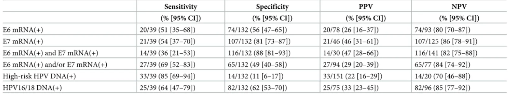
Table 5. Diagnostic indices of E6/E7 mRNA analyses for detecting invasive cervical cancer.
Sensitivity Specificity PPV NPV
(% [95% CI]) (% [95% CI]) (% [95% CI]) (% [95% CI])
E6 mRNA(+) 20/39 (51 [35–68]) 74/132 (56 [47–65]) 20/78 (26 [16–37]) 74/93 (80 [70–87]) E7 mRNA(+) 21/39 (54 [37–70]) 107/132 (81 [73–87]) 21/46 (46 [31–61]) 107/125 (86 [78–91]) E6 mRNA(+) and E7 mRNA(+) 14/39 (36 [21–53]) 116/132 (88 [81–93]) 14/30 (47 [28–66]) 116/141 (82 [75–88]) E6 mRNA(+) and/or E7 mRNA(+) 27/39 (69 [52–83]) 65/132 (49 [40–58]) 27/94 (29 [20–39]) 65/77 (84 [74–92]) High-risk HPV DNA(+) 33/39 (85 [69–94]) 14/132 (11 [6–17]) 33/151 (22 [16–29]) 14/20 (70 [46–88]) HPV16/18 DNA(+) 25/39 (64 [47–79]) 82/132 (62 [53–70]) 25/75 (33 [23–45]) 82/96 (85 [77–92])
liquid-based cytology test is also reported to have high specificity for detecting cervical neo-plastic diseases (84.8–94.1% for CIN2+) [19]. However, while cytology test is considered to reflect the present status of diseases, E6/E7 mRNA analysis may be able to predict future dis-ease progression, as this test examines HPV oncogene expressions with transforming abilities. In this context, we further examined the impact of the presence of E6/E7 mRNAs on disease progression by following up patients with CIN1-2. The presence of both E6 and E7 mRNAs showed significant associations with the occurrence of upgraded abnormal cytology, the pres-ence of E7 mRNA showed association without statistical significance, while positive E6 mRNA, HR-HPV DNA, or HPV 16/18 DNA showed no such trends (Fig 4). Regarding fol-low-up study of HPV mRNA tests, the longitudinal studies have reported that positive mRNA at baseline is an excellent predictor for future development of CIN2+ or CIN3+ in referral or post-treatment populations [20–26]. Moreover, a recent longitudinal screening study has reported that the Aptima HPV test, which collectively detects E6/E7 mRNAs from 14 types of HR-HPV, has a similar sensitivity for detection of CIN2+ or CIN3+ and a significantly higher specificity than the HC2 test [27]. Together with these published findings, our above observa-tions suggest that the separate analysis of E6 and E7 mRNAs may predict disease progression
Fig 5. Kaplan-Meier curves for upgraded Pap-test results in followed-up patients with CIN1-2.A, cases positive for both E6 and E7 mRNAs (n = 3)vs. the remainder (n = 28);B, cases with positive E7 mRNAs (n = 4)vs. negative E7 mRNAs (n = 27);C, cases with positive E6 mRNA (n = 15)vs. negative E6 mRNA (n = 16);D, cases with positive HR-HPV DNA (n = 26)vs. negative HR-HPV DNA (n = 5);E, cases with positive HPV16/18 DNA (n = 8) vs. negative HPV16/18 DNA (n = 23).
https://doi.org/10.1371/journal.pone.0193061.g005
of CIN more precisely than HPV DNA tests. However, further following up patients and path-ologically detecting disease progression are required to clarify the predictive significance of separately analyzing E6 and E7 mRNAs.
The sensitivity of our E6/E7 mRNA test for detecting CIN2+ is lower than those of other reported HPV RNA tests (77.0–96.3% for CIN2+) [10,15–18,23]. However, while almost all other HPV RNA tests examine E6 and E7 mRNAs collectively, our RT-PCR system can detect each E6 and E7 mRNAs separately so that disease progression may be more precisely predicted by individually evaluating E7 mRNA which appears more closely involved in cervical carcino-genesis than E6 mRNA. Moreover, our system using liquid-based cytology specimens will be suitable for clinical application by a “one sample for all” approach.
In conclusion, our separate analyses of E6/E7 mRNAs demonstrated here that the presence of E7 mRNAs was significantly associated with progression from low-grade CIN to invasive carcinoma in contrast with positive E6 mRNA or HR-HPV DNA. Besides, the presence of both E6 and E7 mRNAs was significantly associated with the positivity for HPV16/18 DNA, while the presence of E6 mRNA was significantly associated with the positivity for HR-HPV DNA. The presence of both E6 and E7 mRNAs showed high specificity and low sensitivity for detecting CIN2+ by contrast with the positivity for HR-HPV DNA. Furthermore, the presence of both E6 and E7 mRNAs showed significant association with the occurrence of upgraded abnormal cytology in the patients followed-up for CIN1-2 by contrast with positive E6 mRNA, HR-HPV DNA, or HPV16/18 DNA. Our findings suggest a closer involvement of E7 mRNAs than E6 mRNA in cervical carcinogenesis. Moreover, the separate analysis of E6 and E7 mRNAs may be a more useful tool than HR-HPV DNA test for detecting CIN2+ precisely and predicting disease progression. Further accumulation of evidence is warranted to validate our proposal.
Acknowledgments
This study was partially supported by the Grant-in-Aid for Scientific Research (No. 25462585) from the Ministry of Education, Culture, Sports, Science, and Technology, Tokyo, Japan.
Author Contributions
Conceptualization:Hiroyuki Yoshikawa.
Data curation:Takeo Minaguchi.
Formal analysis:Takeo Minaguchi.
Funding acquisition:Koji Matsumoto.
Investigation:Shuling Liu.
Methodology:Hiroyuki Yoshikawa.
Project administration:Toyomi Satoh.
Resources:Yuri Tenjimbayashi, Ayumi Shikama, Nobutaka Tasaka, Azusa Akiyama, Manabu Sakurai, Sari Nakao, Hiroyuki Ochi, Mamiko Onuki, Koji Matsumoto.
Software:Takeo Minaguchi.
Supervision:Toyomi Satoh.
Validation:Takeo Minaguchi.
Writing – original draft:Shuling Liu.
Writing – review & editing:Takeo Minaguchi, Bouchra Lachkar, Shuang Zhang, Chenyang Xu.
References
1. Ervik M, Lam F, Ferlay J, Mery L, Soerjomataram I, Bray F. Cancer fact sheets: Cervical cancer. Cancer Today. 2016. Available from:http://gco.iarc.fr/today/home.
2. Steenbergen RD, Snijders PJ, Heideman DA, Meijer CJ. Clinical implications of (epi)genetic changes in HPV-induced cervical precancerous lesions. Nat Rev Cancer. 2014; 14(6):395–405. Epub 2014/05/24.
https://doi.org/10.1038/nrc3728PMID:24854082.
3. DeFilippis RA, Goodwin EC, Wu L, DiMaio D. Endogenous Human Papillomavirus E6 and E7 Proteins Differentially Regulate Proliferation, Senescence, and Apoptosis in HeLa Cervical Carcinoma Cells. Journal of Virology. 2003; 77(2):1551–63.https://doi.org/10.1128/JVI.77.2.1551-1563.2003PMID:
12502868
4. Yugawa T, Kiyono T. Molecular mechanisms of cervical carcinogenesis by high-risk human papillomavi-ruses: novel functions of E6 and E7 oncoproteins. Rev Med Virol. 2009; 19(2):97–113. Epub 2009/01/ 22.https://doi.org/10.1002/rmv.605PMID:19156753.
5. Tang S, Tao M, McCoy JP Jr., Zheng ZM. The E7 oncoprotein is translated from spliced E6*I transcripts in high-risk human papillomavirus type 16- or type 18-positive cervical cancer cell lines via translation reinitiation. J Virol. 2006; 80(9):4249–63. Epub 2006/04/14. https://doi.org/10.1128/JVI.80.9.4249-4263.2006PMID:16611884; PubMed Central PMCID: PMCPMC1472016.
6. Nakagawa S, Yoshikawa H, Yasugi T, Kimura M, Kawana K, Matsumoto K, et al. Ubiquitous presence of E6 and E7 transcripts in human papillomavirus-positive cervical carcinomas regardless of its type. J Med Virol. 2000; 62(2):251–8. Epub 2000/09/26. PMID:11002256.
7. Sotlar K, Stubner A, Diemer D, Menton S, Menton M, Dietz K, et al. Detection of high-risk human papillo-mavirus E6 and E7 oncogene transcripts in cervical scrapes by nested RT-polymerase chain reaction. J Med Virol. 2004; 74(1):107–16. Epub 2004/07/20.https://doi.org/10.1002/jmv.20153PMID:15258976. 8. Solomon D, Nayar R. The Bethesda System for Reporting Cervical Cytology. 2nd ed. New York:
Springer; 2004.
9. Takeda T, Wong TF, Adachi T, Ito K, Uehara S, Kanaoka Y, et al. Guidelines for office gynecology in Japan: Japan Society of Obstetrics and Gynecology and Japan Association of Obstetricians and Gyne-cologists 2011 edition. J Obstet Gynaecol Res. 2012; 38(4):615–31. Epub 2012/03/15.https://doi.org/ 10.1111/j.1447-0756.2012.01858.xPMID:22414139.
10. Broccolo F, Fusetti L, Rosini S, Caraceni D, Zappacosta R, Ciccocioppo L, et al. Comparison of onco-genic HPV type-specific viral DNA load and E6/E7 mRNA detection in cervical samples: results from a multicenter study. J Med Virol. 2013; 85(3):472–82. Epub 2013/01/03.https://doi.org/10.1002/jmv. 23487PMID:23280876.
11. Hawley-Nelson P, Vousden KH, Hubbert NL, Lowy DR, Schiller JT. HPV16 E6 and E7 proteins cooper-ate to immortalize human foreskin keratinocytes. EMBO J. 1989; 8(12):3905–10. Epub 1989/12/01. PMID:2555178; PubMed Central PMCID: PMCPMC402081.
12. Munger K, Phelps WC, Bubb V, Howley PM, Schlegel R. The E6 and E7 genes of the human papilloma-virus type 16 together are necessary and sufficient for transformation of primary human keratinocytes. J Virol. 1989; 63(10):4417–21. Epub 1989/10/01. PMID:2476573; PubMed Central PMCID:
PMCPMC251060.
13. Riley RR, Duensing S, Brake T, Munger K, Lambert PF, Arbeit JM. Dissection of human papillomavirus E6 and E7 function in transgenic mouse models of cervical carcinogenesis. Cancer Res. 2003; 63 (16):4862–71. Epub 2003/08/28. PMID:12941807.
14. Kruse AJ, Skaland I, Janssen EA, Buhr-Wildhagen S, Klos J, Arends MJ, et al. Quantitative molecular parameters to identify low-risk and high-risk early CIN lesions: role of markers of proliferative activity and differentiation and Rb availability. Int J Gynecol Pathol. 2004; 23(2):100–9. PMID:15084837. 15. Ratnam S, Coutlee F, Fontaine D, Bentley J, Escott N, Ghatage P, et al. Clinical performance of the
Pre-Tect HPV-Proofer E6/E7 mRNA assay in comparison with that of the Hybrid Capture 2 test for identifica-tion of women at risk of cervical cancer. J Clin Microbiol. 2010; 48(8):2779–85.https://doi.org/10.1128/ JCM.00382-10PMID:20573862; PubMed Central PMCID: PMCPMC2916571.
17. Coquillard G, Palao B, Patterson BK. Quantification of intracellular HPV E6/E7 mRNA expression increases the specificity and positive predictive value of cervical cancer screening compared to HPV DNA. Gynecol Oncol. 2011; 120(1):89–93.https://doi.org/10.1016/j.ygyno.2010.09.013PMID:
20950847.
18. Ratnam S, Coutlee F, Fontaine D, Bentley J, Escott N, Ghatage P, et al. Aptima HPV E6/E7 mRNA test is as sensitive as Hybrid Capture 2 Assay but more specific at detecting cervical precancer and cancer. J Clin Microbiol. 2011; 49(2):557–64.https://doi.org/10.1128/JCM.02147-10PMID:21147950; PubMed Central PMCID: PMCPMC3043526.
19. Whitlock EP, Vesco KK, Eder M, Lin JS, Senger CA, Burda BU. Liquid-based cytology and human papil-lomavirus testing to screen for cervical cancer: a systematic review for the U.S. Preventive Services Task Force. Ann Intern Med. 2011; 155(10):687–97, W214-5. https://doi.org/10.7326/0003-4819-155-10-201111150-00376PMID:22006930.
20. Persson M, Elfstrom KM, Brismar Wendel S, Weiderpass E, Andersson S. Triage of HR-HPV positive women with minor cytological abnormalities: a comparison of mRNA testing, HPV DNA testing, and repeat cytology using a 4-year follow-up of a population-based study. PLoS One. 2014; 9(2):e90023. Epub 2014/03/04.https://doi.org/10.1371/journal.pone.0090023PMID:24587193; PubMed Central PMCID: PMCPMC3936009.
21. Johansson H, Bjelkenkrantz K, Darlin L, Dilllner J, Forslund O. Presence of High-Risk HPV mRNA in Relation to Future High-Grade Lesions among High-Risk HPV DNA Positive Women with Minor Cytological Abnormalities. PLoS One. 2015; 10(4):e0124460. Epub 2015/04/22.https://doi.org/10. 1371/journal.pone.0124460PMID:25893988; PubMed Central PMCID: PMCPMC4404139. 22. Waldstrom M, Christensen RK, Ornskov D. Evaluation of p16(INK4a)/Ki-67 dual stain in comparison
with an mRNA human papillomavirus test on liquid-based cytology samples with low-grade squamous intraepithelial lesion. Cancer Cytopathol. 2013; 121(3):136–45. Epub 2012/09/19.https://doi.org/10. 1002/cncy.21233PMID:22987560.
23. Waldstrom M, Ornskov D. Comparison of the clinical performance of an HPV mRNA test and an HPV DNA test in triage of atypical squamous cells of undetermined significance (ASC-US). Cytopathology. 2012; 23(6):389–95.https://doi.org/10.1111/j.1365-2303.2011.00923.xPMID:21933290.
24. Cubie HA, Canham M, Moore C, Pedraza J, Graham C, Cuschieri K. Evaluation of commercial HPV assays in the context of post-treatment follow-up: Scottish Test of Cure Study (STOCS-H). J Clin Pathol. 2014; 67(6):458–63. Epub 2014/01/18.https://doi.org/10.1136/jclinpath-2013-202014PMID:
24436334.
25. Zappacosta R, Sablone F, Pansa L, Buca D, Buca D, Rosini S. Analytic and Diagnostic Performances of Human Papillomavirus E6/E7 mRNA Test on up-to 11-Year-Old Liquid-Based Cervical Samples. A Biobank-Based Longitudinal Study. Int J Mol Sci. 2017; 18(7). Epub 2017/07/12.https://doi.org/10. 3390/ijms18071480PMID:28696386; PubMed Central PMCID: PMCPMC5535970.
26. Giorgi Rossi P, Benevolo M, Vocaturo A, Caraceni D, Ciccocioppo L, Frega A, et al. Prognostic value of HPV E6/E7 mRNA assay in women with negative colposcopy or CIN1 histology result: a follow-up study. PLoS One. 2013; 8(2):e57600. Epub 2013/03/06.https://doi.org/10.1371/journal.pone.0057600
PMID:23460880; PubMed Central PMCID: PMCPMC3583834.
27. Reid JL, Wright TC Jr., Stoler MH, Cuzick J, Castle PE, Dockter J, et al. Human papillomavirus onco-genic mRNA testing for cervical cancer screening: baseline and longitudinal results from the CLEAR study. Am J Clin Pathol. 2015; 144(3):473–83. Epub 2015/08/16.https://doi.org/10.1309/