ORIGINAL ARTICLE
COMPARISON BETWEEN THE EFFECT OF COLLAGEN TRIPEPTIDE AND SODIUM HYALURONAN TO PREVENT KNEE OSTEOARTHRITIS:
A PRELIMINARY IN VIVO STUDY
Takuya Naraoka
1),Yasuyuki Ishibashi
1),Eiichi Tsuda
1),Yuji Yamamoto
1), Tomomi Kusumi
2)and Satoshi Toh
1)Abstract Objective Collagen peptides have recently been shown to have several biological activities, and have been used as preservatives and immunotherapeutic agents. The purpose of this study was to investigate the ability of weekly intra-articular injections of collagen tripeptide (Ctp) to prevent knee osteoarthritis (OA) compared with sodium hyaluronan (HA).
Methods Thirty rabbits with anterior cruciate ligament (ACL) transection were randomly divided into three groups:
Ctp, HA, and saline (control). All animals were administered the same amount of each reagent once weekly. Articular cartilages of the medial condyle of femur were examined by gross morphological and histopathological examination conducted at 5 (n=6) and 10 (n=4) weeks.
Results The Ctp and HA injection groups tended to have improved gross morphological scores and exhibited preventive effects compared with control groups at 10 weeks after ACL transection. But the scores were not significantly different among the 3 groups. The overall means of histological grading scores were not significantly different among the 3 groups however, the score for the Ctp group had improved from 5 weeks to 10 weeks.
Conclusions Intra-articular injections of Ctp may inhibit progression of knee OA, and further examination is needed to clarify the physical characteristics and regenerative functions of Ctp, and to determine the optimal dose and duration of Ctp injection therapy for the knee OA.
Hirosaki Med.J. 62:107―116,2011
Key words: Osteoarthritis; collagen peptide; hyaluronic acid; cartilage; injection.
原 著
コラーゲン・トリペプチドおよびヒアルロン酸関節内投与における変形性 膝関節症予防効果の比較
奈良岡 琢 哉
1)石 橋 恭 之
1)津 田 英 一
1)山 本 祐 司
1)楠 美 智 巳
2)藤 哲
1)抄録 目的 コラーゲントリペプチド(Ctp)関節内投与による変形性膝関節症(膝OA)予防効果を,現在臨床応用されて いるヒアルロン酸(HA)と比較した.
方法 日本白色家兎30匹を用い右膝関節前十字靱帯切離による OA 発症モデルを作製.それらを生食,Ctp,HA 投与 群に分類し,週 1 回関節内投与を施行した. 6 匹を 5 週投与後, 4 匹を10週投与後屠殺し,大腿骨内側顆を用いて OA 進行度を肉眼的,組織学的に評価した.
結果 肉眼的評価では10週投与時で Ctp 及び HA 投与群で生食投与群に比し膝 OA の進行は軽度であったが,統計学的 有意差は認めなかった.組織学的評価でも 3 群間に有意差は認めなかったが,Ctp 投与群では 5 週から10週にかけてス コアの改善を認めた.
考察 Ctp 関節内投与による OA 予防効果が示唆された.生理活性の詳細な検討と,治療に向けた至適な投与濃度及び 投与期間の更なる検討が必要である.
弘前医学 62:107―116,2011
キーワード:変形性膝関節症;コラーゲンペプチド;ヒアルロン酸;軟骨;注射.
1)Department of Orthopaedic Surgery, Hirosaki University Graduate School of Medicine
2)Department of Pathology, Hirosaki University Graduate School of Medicine
Correspondence: T. Naraoka
Received for publication, November 4, 2010 Accepted for publication, January 5, 2011
1)弘前大学大学院医学研究科整形外科学講座
2)弘前大学大学院医学研究科病理学講座
別刷請求先:奈良岡琢哉 平成22年11月 4 日受付 平成23年 1 月 5 日受理
Introduction
Osteoarthritis (OA) of the knee is a major cause of disability worldwide
1). Knee OA is induced by complex mechanisms such as proteoglycan (PG) degradation and disruption of the collagen network, all of which lead to progressive destruction of joints and functional loss
2). Although several symptomatic therapies have been attempted for Knee OA, no radical treatment methods have been established, with the exception of total knee arthroplasty. Much research has been performed to intervene in the disease process and retard or even prevent progression of joint damage. Intraarticular injection of sodium hyaluronan (HA), which is one of these strategies, has been used for the treatment of pain associated with knee OA, and has been shown to have an anti-inflammatory effect and reduces pain clinically
3, 4).
Collagen peptides, which are enzymatic degradation products of collagen, have recently been shown to have several biological activities and have been used as preservatives and immunotherapeutic agents
5). Collagen tripeptide
(Ctp) prepared from porcine type I collagen, using a bacterial collagenase that degrades the peptide bonds of collagen at the amino- terminal end of Gly, is a highly purified, non- antigenic, and low-allergenic tripeptide fraction containing Gly-Xaa-Yaa sequences. A previous study showed that chondrocyte proliferation and synthetic ability were stimulated by supplementation of culture medium with tripeptide
6).
We hypothesized that Ctp administration within the joint would suppress the loss of cartilage matrix and consequently prevent knee OA progression. The purpose of this study was to examine the preventive effect of knee OA development by intra-articular injection of Ctp compared with HA used for the treatment of knee OA. This study was conducted using an
experimental model in which knee OA develops as a result of anterior cruciate ligament (ACL)
transection
7-9)in Japanese white rabbits.
Materials and Methods
Experimental materials
Ctp was provided by Central Research Institute, Jellice Corp. (Sendai, Japan). The tripeptide content consisting of Gly-Xaa-Yaa sequences was more than 90%. Solutions of 3.0
ȝg/ml Ctp and HA (Supartz, Seikagaku Corp., Tokyo, Japan) were used.
Experimental animals and ACL transection (ACLT) surgery for induction of osteoarthritis
Thirty mature female Japanese white rabbits
(body weight 3.3 ± 0.8 kg) were utilized in the study. Unilateral ACL transection was performed under anesthesia induced by intravenous injection of 30 mg/kg sodium pentobarbital (Dainippon Sumitomo Pharma, Osaka, Japan) . The ACL was exposed through a medial parapatellar incision and transected at the midsubstance with a sharp blade. Complete transection of the ACL was confirmed by a positive anterior drawer sign.
The capsule was sutured to render it watertight, followed by skin closure. All animals were allowed normal cage activity.
All animal experiments in this study followed the Guidelines for Animal Experimentation of Hirosaki University.
Experimental protocol for treatment
After ACL transection, all the rabbits were divided into three groups of 10 rabbits each:
Group 1 was injected with sterile physiological
normal saline as a control, group 2 with Ctp,
group 3 with HA. Using a 27-gauge needle
inserted through the lateral infrapatellar area
toward the intercondylar space of the femur
in each animal in a deep knee-flexed position,
0.3 ml of each reagent was administrated
intra-articularly into the right knee with ACL transection. The first injection was given immediately after ACL transection; six animals of each group were administered once weekly for 5 weeks and four animals of each group for 10 weeks, and all animals were sacrificed one week after the final injection was administered, by an overdose of sodium pentobarbital. The knee joints were then harvested and evaluated.
The dose of 900
ȝg of Ctp was chosen based on the local effective concentration of the previous in vitro preliminary study that fibroblastsʼ gene expressions were stimulated by supplementation of culture medium with Ctp of this concentration.
Gross morphological examination
Gross morphological changes of the medial condyles of femur were assessed and graded as previously described
10)after application of india ink (grade 1, 2, 3, 4a, 4b, 4c = score 0, 1, 2, 3, 4, 5)―grade 1 (intact surface): surface appears normal and does not retain any ink;
grade 2 (minimal fibrillation): site appears normal before staining, but retains the india ink as elongated specks or light gray patches; grade 3 (overt fibrillation): the cartilage is velvety in appearance and retains ink as intense black patches; grade 4 (erosion): loss of cartilage exposing the underlying bone; grade 4a: erosion of 0 to 2 mm; grade 4b: erosion of 2 to 5 mm;
and grade 4c: erosion of >5mm. In a blinded manner, the assessment was conducted by two independent examiners, who were blinded to each otherʼs findings and to the treatment group assignment of the animals. Finally, the two scores from the examiners were averaged to obtain an overall score.
Histopathological examination
Dissected medial condyles of femur were fixed in 10% neutral buffered formalin after gross morphological examination. Specimens
w e r e d e c a l c i f i e d i n 4 % E D T A s o l u t i o n , dehydrated with a gradient ethanol series, and embedded in paraffin blocks. Based on macroscopic observation, 4
ȝm sections including the most severely degenerated area were stained with hematoxylin and eosin (H&E)
and with Alcian blue for light microscopic e x a m i n a t i o n . H i s t o l o g i c a l s e c t i o n s w e r e visualized using an Olympus BX41 microscope
(Olympus, Tokyo, Japan) and Olympus DP2- BSW software (Olympus). Histological sections were assessed in a blinded manner by a pathologist who was unaware of the treatment group assignment of the animals, and quantified using the histological grading method proposed by Mankin and coworkers
11)(Table. 1).
Statistical analysis
All data are expressed as mean ± standard deviation. A Tukeyʼs honestly significant difference test was used to evaluate the statistical significance of difference in the macroscopic and histologic results. P values less than 0.05 were considered statistically significant.
Results
There were no adverse effects due to injections in any rabbit, and no evidence of postoperative infection was noted. At sacrifice, all ACLT knees showed complete transection of the ACL, with only a stump remaining.
Gross morphological assessment
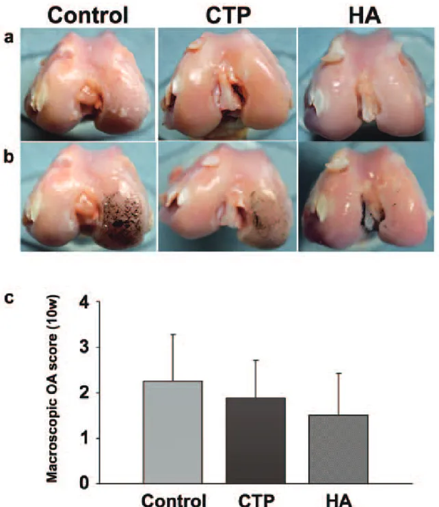
Specimens injected for 5 weeks of all groups showed no morphological change (Fig. 1), however specimens injected for 10 weeks exhibited changes consistent with the develop- ment of knee OA and showed mild to severe degradation of the condyle cartilage (Fig. 2).
Both treatment groups injected for 10 weeks
after ACL transection exhibited slightly
resistance to the changes of knee OA than saline group (saline group 2.3 ±1.0, Ctp group 1.9 ±0.8 and HA group 1.5 ±0.9 and saline group) (p>0.05).
Histopathological assessment
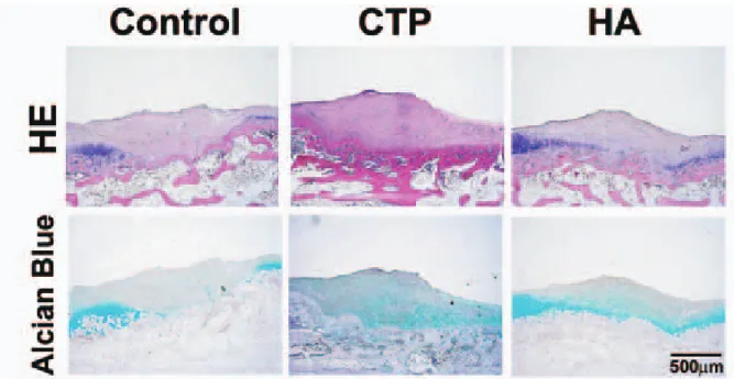
The cartilage on the medial condyle of femur in the ACL transection knees of all groups exhibited pathological changes of some degree of knee OA. In specimens injected for 5
weeks of all groups, fibrous tissue was formed over the degenerated area (Fig. 3), in which fibroblast-like cells were increased (Fig. 4). The degenerative area of saline group was extended to more deeper zone compared with that of Ctp group and HA group. In case of 10 weeks injection, the fibrous tisuue was decreased and the structure and matrix staining of deep layer were improved in Ctp group (Fig. 5).
The overall means of Mankin grading scores
Table 1 Mankin scale 1. Cartilage Anatomy
0: Normal
1: Surface roughness, fissures extending into the radial layer 2: Pannus
3: Superficial layer of the cartilage is lost
4: Mild disorganization, loss of columnar alignment of cells, scarcity of cellular clusters 5: Fissures extending to the calcified cartilage layer
6: Complete loss of cellular organization, clusters of cells, osteoclastic activity 2. Cellular abnormalities
0: Normal
1: Hipercellularity, including small clusters of superficial cells 2: Clusters of cells
3: Hypercellularity 3. Matrix Staining
0: Normal/slightly decreased staining 1: Decreased stained of radial layer
2: Decreased stained of interterritorial matrix 3: Only pericellular matrix is stained
4: Unstained 4. Tidemark integrity 0: Intact
1: Destruction
Figure 1 Representative macroscopic appearances at 5 weeks after anterior cruciate ligament transection. Ctp=collagen tripeptide; HA=sodium hyaluronan.
were not significantly different among groups, but HA group injected for 5 weeks tended to decrease in comparison with other 5 injected groups (saline group 2.8 ±0.8, Ctp group 2.3 ± 1.4 and HA group 1.3±1.0) (Fig. 6). Furthmore,
in case of 10 weeks injection, the score for the Ctp in addition to HA groups were lower than that for saline group (saline group 2.8±1.5, Ctp group 1.8 ±0.5 and HA group 2.0 ±0.8) (Fig.
6). While the overall mean score of HA group
Figure 2 Evaluation of knee OA grades by gross morphological assessment of the articular cartilage from the medial condyle of femur. (a) Representative macroscopic appearances at 10 weeks after anterior cruciate ligament transection. (b) Surface of the cartilage was stained with India ink to identify any fibrillation and erosion. (c)
Quantification of macroscopic analysis (grade 1, 2, 3, 4a, 4b, 4c = score 0, 1, 2, 3, 4, 5). The scores are displayed as means ± standard deviation (10 weeks: n=4). There were no statistically significant differences in any scores among the three groups. Ctp=collagen tripeptide; HA=sodium hyaluronan.
was increased from 5 weeks to 10 weeks, that of Ctp group was decreased. The mean score for criteria related to matrix staining and tidemark integrity of Ctp group injected for 10 weeks were lower than that of saline group and HA group (Table. 2). On the contrary, the mean score for cellular abnormalities of Ctp group was higher than that of saline group and HA group in both 5 and 10 injected groups.
Discussion
The Ctp used in this study was a highly purified tripeptide fraction containing Gly- Xaa-Yaa sequences
12). Articular cartilage has extremely small pores (estimated at 50 Å) in the superficial zone, and so only low-molecular- weight compounds (<20 kDa) in synovial fluid may diffuse into the tissue
13). Our experimental material, Ctp, has a possibility to move freely through the tissue because they are not heavy molecular compounds. Transporter systems for amino acids in cartilage chondrocytes have not yet been identified, but glycine, proline, glutamine and glutamate transporters in chondrocytes have recently been investigated
14, 15). While the Ctp- treated group tended to have a higher score in gross morphological assessment than the HA- treated group, the Mankin score had decreased from 5 weeks to 10 weeks after ACLT, and the score was lower than in the HA-treated group at 10 weeks. Concerning the mean scores for criteria related to cellular abnormalities, Ctp groups had higher cell numbers than the
Figure 3 Representative histologic appearance of cartilage of medial condyle of femur (H-E and Alcian blue staining) of control, Ctp and HA group at 5 weeks. Ctp=collagen tripeptide; HA=sodium hyaluronan.
Figure 4 High magnification of fibrous tissue in Ctp group injected for 5 weeks.
non-Ctp groups. Furthermore, the Ctp group treated for 10 weeks had lower scores for criteria related to matrix staining and tidemark integrity. We speculated that Ctp had a regenerative effect by stimulating proliferation
of cells containing chondrocytes and fibroblast- like cells and repairing cartilage matrix.
Previous study have shown that amino acids are expected not only to control chondrocyte gene expression
16)but also to control synthesis
Figure 5 Representative histologic appearance of cartilage of medial condyle of femur (H-E and Alcian blue staining) of control, Ctp and HA group at 10 weeks. Ctp=collagen tripeptide; HA=sodium hyaluronan.
Figure 6 Quantification of histological analysis using the Mankin grading method at 5 and 10 weeks after anterior cruciate ligament transection. The scores are displayed as overall means ± standard deviation (5 weeks: n=6, 10 weeks: n=4). There were no statistically significant differences in any scores among the three groups. Ctp=collagen tripeptide; HA=sodium hyaluronan.
of collagen by chondrocytes
17). And, amino acids stimulate fibroblasts proliferation
18). Therefore, amino acids in Ctp might be selected to provide substrates for fibril forming collagen and proteoglycan, based on their prevalence in the triple-helical structure of collagen, and depending on their specific biochemical and physiologic characteristics
19, 20).
The total Mankin score of the HA-treated group was lowest among the 3 groups at 5 weeks after ACLT, and maintained the low level until 10 weeks. HA covers cartilage surfaces as an amorphous layer, and partially protects cartilage against cartilaginolytic enzymes and penetration of inflammatory cells
21, 22). Furthermore, a decrease in HA level in the synovial fluid of OA may result in damage to the cartilage surface and increase the permeability of cartilage, allowing proteins and other high molecular weight substances to penetrate into the cartilage matrix
23). Intraarticularly injected HA binds free proteoglycan in the cartilage matrix, and inhibits further proteoglycan release, thus protecting the cartilage matrix and cells
24, 25). Exogenous HA is known to stimulate endogenous HA production by positive feedback
26). From these findings, the lower score of the HA-treated group in this study might also be brought about by its cartilage protective effect.
Further examination is needed to clarify the physical characteristics and regenerative
functions of Ctp, and to determine the optimal dose and duration of Ctp injection therapy for the OA knee, based on in vitro studies and several additional in vivo studies.
Experimental osteoarthritis induced by ACL transection is a useful tool in the investigation of knee OA development
7-9). The biochemical and pathological changes that PG degradation and disruption of the collagen network lead to loss of hyaline cartilage, are identical to those seen in human knee OA and the lesion site, as in humans, is mainly on the medial condyle of femur, and eventually extends over the lateral condyle of femur
10). Therefore, medial condyles of femur were used to examine the knee OA development in this study.
The Mankin grading scores of the HA and saline groups were lower than those of previous study
27-29). The possible reasons for the difference might be due to: (1) duration after ACLT for histology examination: 5 and 10 weeks were used in our study versus more than 20 weeks in other studies; (2) examined site: unilateral knees were used in our study, and as the knee OA progressed, rabbits avoided weight-bearing on the affected legs. However, the improved Mankin scores in the HA-treated ACLT knees in our study are in agreement with previous reports
27-29).
I n c o n c l u s i o n , o u r r e s u l t s s h o w t h e regenerative effects of intra-articular injections of Ctp on degenerative changes in the cartilage,
Table 2 Histological evaluation by the Mankin grading method at 5 and 10 weeks after anterior cruciate ligament transection. There were no statistically significant differences in any scores among the three groups. Data represent the means ± standard deviation for each group: Ctp=collagen tripeptide; HA=sodium hyaluronan.
Group Control CTP HA
weeks(n) 5(n=6) 10(n=4) 5(n=6) 10(n=4) 5(n=6) 10(n=4)
Structure 0.8±0.4 0.8±1.0 0.3±0.5 0.3±0.5 0.2±0.4 0.3±0.5 Cell abnormalities 0.7±0.5 1.0±0.8 1.2±0.4 1.3±0.5 0.8±0.4 0.8±0.5 Matrix staining 0.5±0.5 0.5±0.6 0.5±0.5 0.3±0.5 0.0±0.0 0.5±0.6 Tidemark integrity 0.8±0.4 0.5±0.6 0.3±0.5 0.0±0.0 0.3±0.5 0.5±0.6
Total 2.8±0.8 2.8±1.5 2.3±1.4 1.8±0.5 1.3±1.0 2.0±0.8
in the ACL transection joint with no obvious adverse effects. These results suggest that Ctp injection may be able to modify the progression of OA of the knee and may potentially be applied as an effective treatment.
Acknowledgement
The authors would like to acknowledge Dr.
Ryoko Uesato for technical advice with the experimental knee OA model and to thank Jellice Co. (Sendai, Japan) and Seikagaku Co.
(Tokyo, Japan) for providing experimental materials.
References
1)Kamekura S, Hoshi K, Shimoaka T, Chung U, Chikuda H, Yamada T, et al. Osteoarthritis development in novel experimental mouse models induced by knee joint instability. Osteoarthritis Cartilage 2005 Jul;13(7):632-41.
2)Park YS, Lim SW, Lee IH, Lee TJ, Kim JS, Han JS. Intra-articular injection of a nutritive mixture solution protects articular cartilage from osteoarthritic progression induced by anterior cruciate ligament transection in mature rabbits:
a randomized controlled trial. Arthritis Res Ther 2007;9(1):R8.
3)McCourt PA, Ek B, Forsberg N, Gustafson S.
Intercellular adhesion molecule-1 is a cell surface receptor for hyaluronan. J Biol Chem 1994 Dec 2;269(48):30081-4.
4)Fujii K, Fujii Y, Hubscher S, Tanaka Y. CD44 is the physiological trigger of Fas up-regulation on rheumatoid synovial cells. J Immunol 2001 Aug 1;167(3):1198-203.
5)Gaffney PJ, Edgell TA, Dawson PA, Ford AW, Stocker E. A pig collagen peptide fraction. A unique material for maintaining biological activity during lyophilization and during storage in the liquid state. J Pharm Pharmacol 1996 Sep;48
(9):896-8.
6)Pesakova V, Novotna J, Adam M. Effect of
the tripeptide glycyl-L-histidyl-L-lysine on the proliferation and synthetic activity of chick embryo chondrocytes. Biomaterials 1995 Aug;16
(12):911-5.
7)Setton LA, Elliott DM, Mow VC. Altered mechanics of cartilage with osteoarthritis: human osteoarthritis and an experimental model of joint degeneration. Osteoarthritis Cartilage 1999 Jan;7
(1):2-14.
8)Sah RL, Yang AS, Chen AC, Hant JJ, Halili RB, Yoshioka M, et al. Physical properties of rabbit articular cartilage after transection of the anterior cruciate ligament. J Orthop Res 1997 Mar;15
(2):197-203.
9)McDevitt CA, Muir H. Biochemical changes in the cartilage of the knee in experimental and natural osteoarthritis in the dog. J Bone Joint Surg Br 1976 Feb;58(1):94-101.
10)Yoshioka M, Coutts RD, Amiel D, Hacker SA.
Characterization of a model of osteoarthritis in the rabbit knee. Osteoarthritis Cartilage 1996 Jun;4
(2):87-98.
11)Mankin HJ, Dorfman H, Lippiello L, Zarins A. Bio- chemical and metabolic abnormalities in articular cartilage from osteo-arthritic human hips. II.
Correlation of morphology with biochemical and metabolic data. J Bone Joint Surg Am 1971 Apr;53
(3):523-37.
12)Tsuruoka N, Nakayama T, Ashida M, Hemmi H, Nakao M, Minakata H, et al. Collagenolytic serine-carboxyl proteinase from Alicyclobacillus sendaiensis strain NTAP-1: purification, character- ization, gene cloning, and heterologous expression.
Appl Environ Microbiol 2003 Jan;69(1):162-9.
13)Buckwalter JA, Mankin HJ. Articular cartilage:
tissue design and chondrocyte-matrix interactions.
Instr Course Lect 1998;47:477-86.
14)Barker GA, Wilkins RJ, Golding S, Ellory JC.
Neutral amino acid transport in bovine articular chondrocytes. J Physiol 1999 Feb 1;514 (Pt 3):795- 808.
15)Hinoi E, Wang L, Takemori A, Yoneda Y. Func- tional expression of particular isoforms of excit- atory amino acid transporters by rodent cartilage.
Biochem Pharmacol 2005 Jul 1;70(1):70-81.
16)Fafournoux P, Bruhat A, Jousse C. Amino acid regulation of gene expression. Biochem J 2000 Oct 1;351 (Pt 1):1-12.
17)Haussinger D. The role of cellular hydration in the regulation of cell function. Biochem J 1996 Feb 1;313 (Pt 3):697-710.
18)Ohara H, Ichikawa S, Matsumoto H, Akiyama M, Fujimoto N, Kobayashi T, et al. Collagen-derived dipeptide, proline-hydroxyproline, stimulates cell proliferation and hyaluronic acid synthesis in cultured human dermal fibroblasts. J Dermatol 2010 Apr;37(4):330-8.
19)Bhattacharjee A, Bansal M. Collagen structure:
the Madras triple helix and the current scenario.
IUBMB Life 2005 Mar;57(3):161-72.
20)Persikov AV, Ramshaw JA, Kirkpatrick A, Brodsky B. Amino acid propensities for the collagen triple- helix. Biochemistry 2000 Dec 5;39(48):14960-7.
21)Guidolin DD, Ronchetti IP, Lini E, Guerra D, Frizziero L. Morphological analysis of articular cartilage biopsies from a randomized, clinical study comparing the effects of 500-730 kDa sodium hyaluronate (Hyalgan) and methylprednisolone acetate on primary osteoarthritis of the knee.
Osteoarthritis Cartilage 2001 May;9(4):371-81.
22)Kirwan J. Is there a place for intra-articular hyaluronate in osteoarthritis of the knee? Knee 2001 Jun;8(2):93-101.
23)Yoshimi T, Kikuchi T, Obara T, Yamaguchi T, Sakakibara Y, Itoh H, et al. Effects of high- molecular-weight sodium hyaluronate on experi-
mental osteoarthrosis induced by the resection of rabbit anterior cruciate ligament. Clin Orthop Relat Res 1994 Jan(298):296-304.
24)Balazs EA, Denlinger JL. Viscosupplementation: a new concept in the treatment of osteoarthritis. J Rheumatol Suppl 1993 Aug;39:3-9.
25)Ghosh P, Read R, Numata Y, Smith S, Armstrong S, Wilson D. The effects of intraarticular administration of hyaluronan in a model of early osteoarthritis in sheep. II. Cartilage composition and proteoglycan metabolism. Semin Arthritis Rheum 1993 Jun;22(6 Suppl 1):31-42.
26)Iwata H. Pharmacologic and clinical aspects of intraarticular injection of hyaluronate. Clin Orthop Relat Res 1993 Apr(289):285-91.
27)Amiel D, Toyoguchi T, Kobayashi K, Bowden K, Amiel ME, Healey RM. Long-term effect of sodium hyaluronate (Hyalgan) on osteoarthritis progression in a rabbit model. Osteoarthritis Cartilage 2003 Sep;11(9):636-43.
28)Jean YH, Wen ZH, Chang YC, Lee HS, Hsieh SP, Wu CT, et al. Hyaluronic acid attenuates osteoarthritis development in the anterior cruciate ligament-transected knee: Association with excitatory amino acid release in the joint dialysate. J Orthop Res 2006 May;24(5):1052-61.
29)Sen C, Gunes T, Saygi B, Erdem M, Koseoglu RD, Kilic N. The chondroprotective effect of intra-articular hyaluronic acid at early stages of osteoarthritis: an experimental study in rabbits.
Acta Orthop Traumatol Turc 2004;38(5):348-52.