1
Gene polymorphisms of mannose-binding lectin confer susceptibility to Pneumocystis pneumonia in
HIV-infected patients
マンノース結合レクチン遺伝子多型は
HIV感染者のニューモシスチス肺炎発症に影響する
指導教員 野島美久教授 平成26年3月
群馬大学大学院医学系研究科 平成22年入学
環境病態制御系・生体統御内科学
栁澤 邦雄
2
INTRODUCTION
Pneumocystis pneumonia (PCP), caused by the fungus Pneumocystis jirovecii
is still the most prevalent indicator disease that defines acquired immunodeficiency syndrome (AIDS) in the patients infected with human immunodeficiency virus (HIV) [1, 2]. A cut-off level of <200 cells/μL for CD4 cell counts is well established as a risk factor for PCP [3]; therefore, CD4 cells probably play a pivotal role in the defense against Pneumocystis organisms [4]. CD4 cell count is now routinely considered in the decision to start preventive therapy for PCP. However, CD4 count in HIV patients is broadly distributed at the onset of PCP. Some patients are free from PCP even after their CD4 count has decreased severely, but other patients develop PCP even while their CD4 count remains around 200/μL. This variability raises the possibility that some risk factors other than CD4 count contribute to the development of PCP. However, very few published studies address this issue.
Pneumocystis organisms invading host lung alveoli are recognized by pattern
recognition receptors (PRR) [5]. Mannose-binding lectin (MBL) is one of well-studied PRR [5, 6] that plays a pivotal role in innate immunity by directly opsonizing pathogens or by activating complement system via the mannose-associated serine protease (MASP), called lectin pathway [7, 8]. Most mammals have two genes (MBL1 and MBL2) that encode functional MBL; however, MBL1 is a pseudogene in humans.
3
Genetic mutations in MBL2 influence functional MBL levels in human serum.
Polymorphisms in exon1 of MBL2 that are significantly associated with low serum MBL levels have been identified, one each in codons 52, 54, and 57; these single nucleotide polymorphisms (SNPs) rs5030737, rs1800450, and rs1800451 are referred to as variants D, B, and C, respectively; the wild type is designated A. Additionally, three SNP sites in the promoter to the 5’-untranslated region (5’-UTR) at nt-550, nt-221, and nt+4 (rs11003125, rs7096206, and rs7095891, referred to as H /L, Y/X, and P/Q, respectively) also affect MBL production. From these six alleles, seven typical haplotypes have been determined as follows: HYPA and LYQA result in high MBL levels; LYPA results in intermediate levels; LXPA results in low levels; LYPB, HYPD, and LYQC result in MBL deficiency [9]. With these seven haplotypes, we can categorize MBL production into two classes: 1) high and 2) low to deficient [9, 10].
Associations between clinical prevalence, MBL2 polymorphisms and MBL serum concentrations, or both have been studied for a variety of infectious disease; however, the clinical relevance of MBL productivity at the onset of PCP in HIV infected patients remained unclear.
Here, we retrospectively examined MBL2 genotypes, serum concentrations of MBL and the development of PCP in advanced HIV patients. We also performed in vitro experiments to determine whether MBL enhanced macrophage-mediated phagocytosis of rat-type Pneumocystis organisms.
4
MATERIALS AND METHODS
Selection of patients and blood sampling
This study was approved by the Medical and Human Genome Ethics Committee of Gunma University, Faculty of Medicine (#144, 3 October 2012). We performed a retrospective chart review of a total of 179 HIV-infected patients consecutively admitted to our hospital from November 1991 to July 2012. We selected 111 regular outpatients who had a baseline CD4 count <200/μl (high-risk range for PCP, n=59) at their first admission. Patients with hemophilia (n=5) or a history of exposure to antiretroviral therapy (ART, n=1) were excluded. Then, a total of 53 patients were analyzed in this study. Written informed consent was obtained from participating patients. For each patient, identification of PCP was made based on the following criteria: 1) chest radiograph and/or CT findings consistent with PCP; 2) elevation of serum β (1→6) glucan; 3) detection of Pneumocystis organisms in respiratory specimens by polymerase chain reaction (PCR) or microscope; 4) successful treatment by trimethoprim/sulfamethoxazole (TMP-SMX) and/or pentamidine (Table 1).
Of 53 patients, 27 patients met at least three of four criteria; 15 patients met all four criteria, 12 others met three. Three patients met only two of four criteria but included with PCP, either because their treatment response with anti-PCP drug (TMP-SMX and/or pentamidine) were favorable, or because other pathogen
5
(cytomegalovirus, Mycoplasma, Chlamydia, or Legionella) was not identified. All of their CT findings indicated with PCP, but serum β (1→6) glucan was not elevated (1 patient) or not evaluated (2 patients). Therefore, 30 of 53 patients were identified PCP upon the first admission, and the other 23 patients were identified non-PCP in this study (Figure 1).
Peripheral blood was collected from each patient at each regular visit. To determine serum MBL levels, serum samples were obtained more than 3 months after the onset of PCP to avoid the acute phase reaction. Sepa Gene kits (Sanko Junyaku, Tokyo, Japan) were used to extract genomic DNA from whole blood samples. All serum and DNA samples were stored at -20°C until use.
Genotyping and classification of production group
Genotyping of MBL2 at the six aforementioned SNPs was performed by genomic PCR and direct sequencing. To analyze these six MBL2 SNPs the following primer pair was used: forward: 5’-GGGGAATTCCTGCCAGAAAGT-3’, reverse:
5’-ATCAGTCTCCTCATATCCCC-3’.
Each PCR mixture contained 5 µL of supplied buffer, 0.2 mM of deoxyribonucleotide triphosphate (dNTP), 7.5 mM of magnesium chloride (MgCl2), and 2.5 U of AmpliTaq® Gold PCR en¬zyme (all purchased from Applied Biosystems/Life Technologies, Foster City, CA, USA), DNA template (20 ng), and 0.2
6
µM of primer pair in a final volume of 50 µL. Each PCR was run in a GeneAmp® PCR System 2700 (Applied Biosystems), with the following cycling conditions: 95°C for 12 min, followed by 40 cycles of denaturing at 95°C for 30 sec, annealing at 60°C for 30 sec, and extension at 72°C for 45 sec.
Purified PCR products were processed for direct sequencing reactions with ABI PRISM BigDye Terminator version 1.1 (Applied Biosystems) and 3.2 pmol/l of PCR primers. After the reactions, products were purified using an AutoSeq G-50 Dye Terminator Removal kit (GE Healthcare) and analyzed by the ABI 310 Prism Genetic Analyzer (Applied Biosystems). For SNP rs1800450 in codon 54, PCR products were treated with Ban I (New England Biolabs, Beverly, MA, USA) to confirm the result of direct sequencing [11]; in each case, enzyme-digestion results confirmed the direct-sequencing results.
We classified the results of genotyping into two production groups, 1) high-producing and 2) low to deficient producing, according to each combination of haplotypes [10].
Serum MBL measurement by enzyme-linked immunosorbent assay
MBL-oligomer ELISA kits (BioPorto, Gentofte, Denmark) were used according to the manufacturer’s instructions to measure MBL levels in each serum sample.
7
Preparation of phagocytic cells
THP-1, a human monocyte cell line, was obtained from the RIKEN Cell Bank (Tsukuba, Ibaraki, Japan) and maintained in RPMI-1640 medium (Sigma-Aldrich, St.
Louis, MO, USA) supplemented with 20% of heat-inactivated fetal bovine serum (FBS), 100 U/mL of penicillin, and 100 µg/mL of streptomycin (Sigma-Aldrich). To induce a macrophage-like phenotype, THP-1 cells were cultured with phorbol-12-myristate-13-acetate (PMA, Sigma-Aldrich) at a concentration of 200 nM for 4 days; on the fourth day, the medium was thoroughly discarded and replaced with culture medium without PMA for 4 more days of incubation. On the day 8, cells were collected after short incubation with 0.25% trypsin-EDTA (Life Technologies) at 37°C.
FITC labeling of Pneumocystis organisms
Rat-type Pneumocystis organisms were purchased from ATCC® (Manassas, VA, USA), and these have been used as the target in phagocytic experiments as described previously [12-14]. In brief, the Pneumocystis organisms were thawed quickly at 37°C, centrifuged at 1000g for 5 min, and then suspended in phosphate-buffered saline (PBS) containing fluorescein isothiocyanate (FITC, Sigma-Aldrich) at a concentration of 0.1 mg/mL. After incubation for 30 min at 37°C, the Pneumocystis organisms were extensively and repeatedly washed with cold (4°C) PBS.
8
MBL opsonization and phagocytosis assay
To distinguish the effect of the complement system for opsonization and phagocytosis, we have set two conditions using heat-inactivated serum and using unheated serum. The heat-inactivated serum was fetus bovine serum (FBS) simply heat-inactivated in water bath (56°C for 30 min); the unheated serum was human serum provided from an MBL-deficient volunteer. The concentrations of complements and immunoglobulins in serum from the volunteer were confirmed to be within normal range based on clinical laboratory tests (data not shown).
FITC-labeled organisms were mixed with recombinant human MBL (rhMBL, R&D Systems, Minneapolis, MN, USA) at the indicated concentrations (0-3000 ng/mL) in 25 µL of Hanks balanced salt solution (HBSS) containing 5 mM of calcium chloride (HBSS+) and incubated in a 37°C water bath with continuous shaking for 60 min. Then, 50 μl of RPMI 1640 was added to each mixture, which was then centrifuged at 1000g for 15 min to remove unbound rhMBL. Each sample of pre-treated organisms was added to THP-1-derived macrophages (distributed 5×10⁴ cells/well) on 96-well black plate (BD, Franklin Lakes, NJ, USA), and medium supplemented with 20 % serum (heat-inactivated serum in RPMI1640, or unheated serum in HBSS with 5 mM of calcium chloride and 5 mM of magnesium chloride) was added to each well to a final volume of 100 µL per well. We also prepared conditions that macrophages were
9
pre-incubated with indicated concentrations of rh MBL (0-3000 ng/mL) prior to the addition of FITC-labeled organisms both the serum was heat-inactivated or unheated.
After each opsonization with rhMBL were performed, finally 5×10⁴ of THP-1-derived macrophages and equally distributed FITC-labeled organisms from one preparation were incubated in 100µLvolume per well for 120 min at 37°C, using a 5%
CO2 incubator. Then, 100 μl of cold PBS was added to each well to stop the phagocytic reaction, and each plate was then centrifuged at 400g for 5 min. To quench extracellular fluorescence, 1 mg/mL of trypan blue (Wako Junyaku, Tokyo, Japan) was added to each well, the plates were incubated for 10 min at 37°C [15, 16], and then centrifuged at 400g for 5 min. Assay buffer (Cayman Chemical, Ann Arbor, MI, USA) was used to wash each well; plates were then centrifuged at 400g for 5 min, and each supernatant was removed. Each distinct phagocytic reaction (distinguished by the concentration of rhMBL) was run in triplicate. An ARVO 1420 plate reader system (Wallac/PerkinElmer, Inc., Waltham, MA, USA) was used to measure the fluorescence intensity (FI) in each well; the excitation wavelength was 485 nm, and the emission wavelength was 535 nm.
The average FI was calculated for each group of triplicate wells; mean FI values for different rhMBL concentrations (0-3000 ng/mL) were compared.
A part of phagocytic reactions were performed in clear dishes. After 120 min, the supernatants were discarded, and the samples were fixed in 4% of paraformaldehyde for 10 min. Then, the dishes were inverted and observed the phagocytic reactions by
10
fluorescence microscopy.
Statistical analysis
The Mann-Whitney U test was used to assess between-group differences (PCP versus non-PCP) in clinical characteristics and serum MBL levels. Genotype distribution and production-type distributions were counted directly and analyzed by Fisher’s two-tailed exact test. The χ2 tests were used to assess the significance of deviations from the Hardy-Weinberg equilibrium (HWE). Odds ratios (ORs) and 95%
confidence intervals (CIs) were also determined. Data from phagocytosis assay were compared with control conditions using a two-tailed Student’s t-test. P values <0.05 were considered significant. SPSS software version 19 (SPSS Inc., Chicago, IL, USA) was used to perform most of analyses; however, SAS (SAS Institute, Cary, NC, USA) was used for testing HWE.
RESULTS
Clinical characteristics of patient population
The 53 patients who met the inclusion-exclusion criteria were each assigned to one of two subgroups, those with PCP (PCP) or those without PCP (non-PCP) at the time of admission; clinical characteristics of each group are presented in Table 2. There
11
were no significant differences between the PCP (n = 30) and Non-PCP (n = 23) groups in gender, age, CD4 count, HIV viral load, or leukocyte, lymphocyte or monocyte counts. Two patients (one in each group) had additional risk factors that may have been relevant to immunocompromised states: one patient in the PCP group had been treated with corticosteroids for adrenocorticotropic hormone (ACTH) deficiency, and one patient in the non-PCP group had diabetes mellitus.
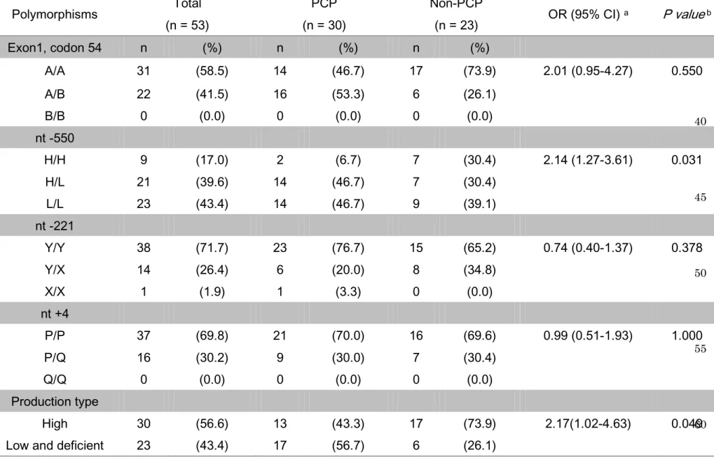
MBL2 gene polymorphisms: genotypes and production types
The distributions of genotypes and of MBL production types were determined based on directly sequenced data for six SNPs in the MBL2 gene (Table 3). Each patient was assigned to one of five typical haplotypes, HYPA, LYQA, LYPA, LXPA, or LYPB; production types were then assigned based on the haplotype assignments. Then, each patient was assigned to one of two production groups, 1) high or 2) low to deficient.
Among 30 patients with PCP, the frequencies of assignments to the high versus to the low to deficient group were 43.3% (13 of 30) and 56.7% (17 of 30), respectively.
Among the 23 patients without PCP, these frequencies were 73.9% (17 of 23, high) and 26.1% (6 of 23, low to deficient). Based on these results of genotyping, the frequencies of the H/H genotype and the high producing type were significantly associated with the non-PCP group (P = 0.031, OR 2.14, 95% CI, 1.27-3.61 (H/H genotype), and P = 0.049,
12
OR 2.17, 95% CI, 1.02-4.63 (high producing type)).
Serum MBL concentrations
The distributions of serum MBL concentrations among patients with high or low to deficient genotypes and in patients with or without PCP are shown in Figure 2.
The median MBL level for patients with high-producing genotypes was 2155.0 ng/mL (range, 1012.5-2394.3 ng/mL), which was significantly higher than that of patients with low-to-deficient genotypes (579.6 ng/mL, range, 1.5-1788.2 ng/mL, P < 0.001).
Similarly, the median MBL level for the non-PCP group was significantly higher than that for the PCP group (2102.6 ng/mL vs 1038.7 ng/mL, respectively, P = 0.039). Based on these results, MBL2 genotypes were confirmed to be strongly associated with serum MBL levels, and patients without PCP had significantly higher levels of MBL than those with PCP.
MBL enhances macrophage-mediated phagocytosis of rat-type Pneumocystis organisms
To examine the effects of MBL concentration on the phagocytic activity via the direct opsonin effect for Pneumocystis organisms, we conducted a phagocytic assay by using a plate reader system. When FITC-labeled Pneumocystis organisms were pre-treated with rhMBL (0 to 3000 ng/mL), the maximum efficacy of phagocytosis was
13
evident with 3000 ng/mL of rhMBL. Significant dose-dependent increases in macrophage-mediated phagocytosis were evident with macrophages cultured in heat-inactivated serum (Figure 3 a, P=0.014). The same trend was observed with macrophages cultured in unheated serum (Figure 3 b, P=0.073) without statistically significant.
To confirm whether rhMBL had a direct effect on macrophages, we treated THP-1-derived macrophages with rhMBL prior to the addition of FITC-labeled Pneumocystis organisms. Enhanced phagocytosis by rhMBL-treated macrophages was
also evident under same range of MBL concentrations (0-3000 ng/ml). The significant increase was observed at the concentration of 1000 ng/mL with either heat-inactivated or unheated serum (P=0.025 and P=0.047, respectively) (Figure 3 c, d).
Whether the MBL pre-treatment was performed with Pneumocystis organisms or with macrophages, MBL-mediated enhancement of phagocytosis was evident with concentrations within the clinically relevant range (0-3000 ng/mL). A representative microscopic picture is shown in Figure 4; FITC-labeled organisms pre-treated with 1000 ng/mL rhMBL were internalized by macrophages.
DISCUSSION
MBL deficiency is associated with susceptibility to fungal infections by
14
Aspergillus, Candida, or Cryptococcus [7]. However, only one clinical study has addressed relationships between MBL levels and the prevalence of PCP among the kidney transplantation recipients [17]. Here, we confirmed that MBL gene polymorphisms determine serum MBL levels among advanced HIV-infected patients, as in previous studies observed at other populations [8]. And we also showed a significant association between MBL gene polymorphisms and susceptibility to Pneumocystis organisms in HIV-infected patients. The frequencies and distribution determined for each MBL2 genotype in this study were similar to those previously determined for Japanese populations [18].
Regardless of the presence or absence of HIV infection, Pneumocystis organisms invading host lung alveoli are recognized by PRRs in soluble forms or expressed on the surface of alveolar macrophages [4]. However, to our knowledge, no studies have focused on the associations between PRR variants and the prevalence of PCP in HIV-infected patients. Findings from a mouse model of invasive lung aspergillosis indicate that exogenous replenishment of MBL enhances phagocytosis of invading organisms, TNFα and IL-8 production, and the survival rate of mice [19].
These effects of MBL can be regarded as protective roles of MBL in cases of fungal infection, and results of our in vitro experiments confirmed a part of these protective roles of MBL.
Here, we showed that MBL resulted in enhanced phagocytosis with clinically
15
relevant concentrations of MBL. Additionally, we used two opsonizing conditions involving heat-inactivated or unheated serum to evaluate the contributions of complement systems on macrophage-mediated phagocytosis of invading organisms;
notably, the trend and rate of phagocytosis enhancement were similar for all conditions with heat-inactivated or unheated serum (Figure 3 a, c compared with Figure 3 b, d).
According to these results, the protective role of MBL seems to predominantly depend on direct binding between invading organisms and macrophages, and less on the activation of complement system at the onset of PCP. Of course these results are insufficient to explain the all risks associated with low MBL concentrations at the onset of PCP. However, our results indicated that a lack of MBL attenuated the direct opsonin effect and thereby disturbed the eradication of Pneumocystis organisms. The lower clinical prevalence of PCP among patients with high producing MBL genotypes probably depends on increased MBL-mediated opsonization of invading organisms.
Therefore, we believe that our findings warrant consideration of the stratification of treatment of and prophylaxis for PCP based on MBL genotype and serum concentrations.
Subsequently, we can propose some clinical practices using our findings. For example, the situation that CD4 recovery was insufficient after the initiation of ART in HIV-infected patient is conceivable. We often encounter such a situation when the baseline CD4 count of a patients is below 50/µL or in elderly patients [20, 21].
16
Furthermore, regardless as infected with HIV or not, systemic chemotherapy and a hematopoietic stem cell transplant for hematological malignancies are also conceivable situations. We have to assess the priority of PCP prophylaxis in these situations, while considering the patient’s general condition and the many other medications in use.
TMP-SMX often causes damage to liver, kidney, bone marrow, or some combination thereof [22]; moreover, inhalation of pentamidine requires special room and often induces a cough [23]. Consequently, long-term administration of these anti-PCP drugs can heavily burden a patient. In order to implement a sophisticated anti-PCP prevention strategy, treatment stratification based on MBL genotype, serum MBL concentration, or both may be useful in designing an appropriate PCP prophylaxis.
Here, we elucidated at least one component of the anti-PCP mechanisms of MBL, and some of the clinical roles of MBL at the onset of PCP in advanced HIV-infection. MBL genotype and serum MBL may be used as novel predictive factor for PCP, in addition with CD4 cell levels.
Summary
Genotypes associated with low or deficient production of MBL were significantly more common in the PCP group than in the non-PCP group. Serum MBL levels were significantly higher in the non-PCP group. Findings from in vitro
17
experiments indicated that MBL enhanced macrophage-mediated phagocytosis of Pneumocystis organisms. Genetic MBL deficiency influence susceptibility to PCP in
patients with advanced HIV infection, and can be regarded as a risk factor for PCP other than CD4 cells counts.
Acknowledgments
This work was performed with the generous advice of Dr. Takashi Takahashi (Graduate School of Infection Control Sciences, Kitasato University, Tokyo, Japan) regarding the experimental design. Dr. Kunihiko Hayashi (Gunma University School of Health Science, Gunma, Japan) and Dr. Yasunori Sato (School of Medicine, Chiba University, Chiba, Japan) provided the helpful advice on the statistical analyses. We also thank the study patients and all laboratory staff for their contributions.
References
1. Schwarcz L, Chen MJ, Vittinghoff E, Hsu L, Schwarcz S. Declining incidence of AIDS-defining opportunistic illnesses: results from 16 years of population-based AIDS surveillance. AIDS 2013; 27:597-605.
2. Huang L, Cattamanchi A, Davis JL, et al. HIV-associated Pneumocystis pneumonia.
Proc Am Thorac Soc 2011; 8:294-300.
18
3. Masur H, Ognibene FP, Yarchoan R, et al. CD4 counts as predictors of opportunistic pneumonias in human immunodeficiency virus (HIV) infection. Ann Intern Med 1989;
111:223-31.
4. Thomas CF, Limper AH. Current insights into the biology and pathogenesis of Pneumocystis pneumonia. Nat Rev Microbiol 2007; 5:298-308.
5. Willment JA, Brown GD. C-type lectin receptors in antifungal immunity. Trends Microbiol 2008; 16:27-32.
6. Kerrigan AM, Brown GD. C-type lectins and phagocytosis. Immunobiology 2009;
214:562-75.
7. Eisen DP, Minchinton RM. Impact of mannose-binding lectin on susceptibility to infectious diseases. Clin Infect Dis 2003; 37:1496-505.
8. Garred P, Larsen F, Seyfarth J, Fujita R, Madsen HO. Mannose-binding lectin and its genetic variants. Genes Immun 2006; 7:85-94.
9. Verdu P, Barreiro LB, Patin E, et al. Evolutionary insights into the high worldwide prevalence of MBL2 deficiency alleles. Hum Mol Genet 2006; 15:2650-8.
10. Ou XT, Wu JQ, Zhu LP, et al. Genotypes coding for mannose-binding lectin deficiency correlated with cryptococcal meningitis in HIV-uninfected Chinese patients.
J Infect Dis 2011; 203:1686-91.
11. Gomi K, Tokue Y, Kobayashi T, et al. Mannose-binding lectin gene polymorphism is a modulating factor in repeated respiratory infections. Chest 2004; 126:95-9.
19
12. Koziel H, Eichbaum Q, Kruskal BA, et al. Reduced binding and phagocytosis of Pneumocystis carinii by alveolar macrophages from persons infected with HIV-1
correlates with mannose receptor downregulation. J Clin Invest 1998; 102:1332-44.
13. Koziel H, Phelps DS, Fishman JA, Armstrong MY, Richards FF, Rose RM.
Surfactant protein-A reduces binding and phagocytosis of pneumocystis carinii by human alveolar macrophages in vitro. Am J Respir Cell Mol Biol 1998; 18:834-43.
14. Koziel H, Li X, Armstrong MY, Richards FF, Rose RM. Alveolar macrophages from human immunodeficiency virus-infected persons demonstrate impaired oxidative burst response to Pneumocystis carinii in vitro. Am J Respir Cell Mol Biol 2000; 23:452-9.
15. Rajagopalan R, Nyaundi T, Salvi VP, Rawal N. Recombinant form of human wild type mannan-binding lectin (MBL/A) but not its structural variant (MBL/C) promotes phagocytosis of zymosan by activating complement. Mol Immunol 2010; 47:2505-14.
16. Walenkamp AM, Scharringa J, Schramel FM, Coenjaerts FE, Hoepelman IM.
Quantitative analysis of phagocytosis of Cryptococcus neoformans by adherent phagocytic cells by fluorescence multi-well plate reader. J Microbiol Methods 2000;
40:39-45.
17. Schürmann M, Schürmann D, Schindler R, et al. Impaired thymic function and CD4+ T lymphopenia, but not mannose-binding lectin deficiency, are risk factors for Pneumocystis jirovecii pneumonia in kidney transplant recipients. Transpl Immunol 2013; 28:159-63.
20
18. Thorisson GA, Smith AV, Krishnan L, Stein LD. The International HapMap Project Web site. Genome Res 2005; 15:1592-3.
19. Kaur S, Gupta VK, Thiel S, Sarma PU, Madan T. Protective role of mannan-binding lectin in a murine model of invasive pulmonary aspergillosis. Clin Exp Immunol 2007;
148:382-9.
20. Lawn SD, Myer L, Bekker LG, Wood R. CD4 cell count recovery among HIV-infected patients with very advanced immunodeficiency commencing antiretroviral treatment in sub-Saharan Africa. BMC Infect Dis 2006; 6:59.
21. Grabar S, Weiss L, Costagliola D. HIV infection in older patients in the HAART era.
J Antimicrob Chemother 2006; 57:4-7.
22. Hardy WD, Feinberg J, Finkelstein DM, et al. A controlled trial of trimethoprim-sulfamethoxazole or aerosolized pentamidine for secondary prophylaxis of Pneumocystis carinii pneumonia in patients with the acquired immunodeficiency syndrome. AIDS Clinical Trials Group Protocol 021. N Engl J Med 1992; 327:1842-8.
23. Monk JP, Benfield P. Inhaled pentamidine. An overview of its pharmacological properties and a review of its therapeutic use in Pneumocystis carinii pneumonia. Drugs 1990; 39:741-56
Figure Legends
21
Figure 1
Selection, inclusion, and exclusion of study patients
Figure 2
Serum MBL concentrations of study patients
Low to deficient and high-producing genotype groups were compared with regard to median MBL levels; the PCP and non-PCP groups were also compared with regard to median MBL levels. All serum samples were collected at least 3 months after the onset of PCP to avoid acute phase reactions.
Figure 3
MBL opsonization enhances the phagocytosis of Pneumocystis organisms
FITC-labeled Pneumocystis organisms were pre-treated with rhMBL in medium with heat-inactivated serum a) or unheated serum b). In both, the maximum enhancement of phagocytosis was observed at a concentration of 3000 ng/mL rhMBL. Alternatively, THP-1 derived macrophages were pre-treated with rhMBL in medium with heat-inactivated serum c) or unheated serum d) prior to the addition of FITC-labeled Pneumocystis organisms; for both serum types, maximum enhancement of phagocytosis was observed at a concentration of 1000 ng/mL rhMBL. The significance of mean FIs compared to control conditions (MBL=0 ng/mL) were as follows: *P=0.014, **P=0.025,
22
***P= 0.047. ★P=0.073.
Figure 4
Microscopic detection of phagocytosis
FITC-labeled Pneumocystis organisms pre-treated with 1000 ng/mL rhMBL were internalized by THP-1-derived macrophages. This image represents two images merged with Adobe Photoshop CS6 (Adobe Systems, San Jose, CA); one image was taken in visible light mode and the other in FITC-excitation mode via fluorescence microscopy.
23
Figures
Figure.1
Figure.2
24
Figure.3
Figure.4
25
Tables:
Table 1. Diagnostic criteria of PCP
N
Xp/CT findings consistent with PCP 30
ground-glass opacity 26
interstitial infiltration 1
Consolidation 2
multiple cavity 1
Detection of Pc in respiratory specimen 17
Microscopic detection 5
PCR positive 9
Both 3
Elevation of serum β-D glucan (>11pg/mL) 26
11-100 17
>100 9
Successful treatment by TMP-SMX or pentamidine 30
Meet the criteria above Total 30
4 of 4 15
3 of 4 12
2 of 4 3
Abbreviations: Xp, chest radiograph; CT, computed tomography; Pc, Pneumocystis jirovecii organisms: PCP, Pneumocystis pneumonia;
TMP-SMX, trimethoprim/sulfamrthoxazole
26
Table 2. Clinical Profiles of all Patients and Categorized According to the Development of PCP
5
10
15
20
Abbreviations: PCP, Pneumocystis pneumonia; WBC, white blood cells. Data represent the number or median (range) of clinical information in the PCP group and non-PCP group.
a) One patient had received cortisol for idiopathic adrenocorticotropic hormone (ACTH) deficiency.
b) One patient had previously been diagnosed with diabetes mellitus.
25
c) Each of seven listed characteristics was compared between the two groups, and the Mann-Whitney U-test was used to assess the significance of each differences. P values are two-tailed, and the significance level was set at <0.05.
Total (n = 53)
PCP (n = 30)
Non-PCP
(n = 23) P value c
Male / Female 44 / 9 27 / 3 17 / 6 0.122
Age (range) 42(19-69) 42 (19-69) 46 (22-64) 0.578
Numbers of patients having additional risk
factor of Immunocompromised state 3 1 a 1 b -
CD4 count (/μl, range) 31(1-192) 20 (1-185) 42 (1-192) 0.132
HIV-RNA load (log10 copies/mL, range) 4.82 (3.31-6.39) 5.04 (3.58-6.39) 4.70 (3.31-6.23) 0.062 WBC (/μl, range) 4600 (1600-11700) 5050 (1600-11700) 4300 (1800-5500) 0.062 Lymphocytes (/μl, range) 776 (208-2364) 660 (208-1760) 893 (310-2364) 0.287
Monocytes (/μl, range) 261 (32-763) 305 (32-763) 254 (137-434) 0.260
27
Table 3. Genotype and Production Group in HIV Patients
30
35
40
45
50
55
60
Polymorphisms Total
(n = 53)
PCP (n = 30)
Non-PCP
(n = 23) OR (95% CI) a P value b
Exon1, codon 54 n (%) n (%) n (%)
A/A 31 (58.5) 14 (46.7) 17 (73.9) 2.01 (0.95-4.27) 0.550
A/B 22 (41.5) 16 (53.3) 6 (26.1)
B/B 0 (0.0) 0 (0.0) 0 (0.0)
nt -550
H/H 9 (17.0) 2 (6.7) 7 (30.4) 2.14 (1.27-3.61) 0.031
H/L 21 (39.6) 14 (46.7) 7 (30.4)
L/L 23 (43.4) 14 (46.7) 9 (39.1)
nt -221
Y/Y 38 (71.7) 23 (76.7) 15 (65.2) 0.74 (0.40-1.37) 0.378
Y/X 14 (26.4) 6 (20.0) 8 (34.8)
X/X 1 (1.9) 1 (3.3) 0 (0.0)
nt +4
P/P 37 (69.8) 21 (70.0) 16 (69.6) 0.99 (0.51-1.93) 1.000
P/Q 16 (30.2) 9 (30.0) 7 (30.4)
Q/Q 0 (0.0) 0 (0.0) 0 (0.0)
Production type
High 30 (56.6) 13 (43.3) 17 (73.9) 2.17(1.02-4.63) 0.049
Low and deficient 23 (43.4) 17 (56.7) 6 (26.1)
28
a) The values are for comparison between wild-type genotypes to heterozygous and homozygous variant-type genotypes.
b) P values were analyzed with Fisher’s two-tailed exact tests. Odds ratio (OR) and 95% confidence intervals were determined
29 65