1
STXBP4 drives tumor growth and is associated with poor prognosis through PDGF Receptor signaling in lung squamous cell carcinoma
Yukihiro Otaka1,2*, Susumu Rokudai1,3*, Kyoichi Kaira4, Michiru Fujieda1, Ikuko Horikoshi1, Reika Iwakawa-Kawabata1,5, Shinji Yoshiyama5, Takehiko Yokobori1,5, Yoichi Ohtaki6, Kimihiro Shimizu6, Tetsunari Oyama7, Jun’ichi Tamura2, Carol Prives3 and Masahiko Nishiyama1,5
1Department of Molecular Pharmacology and Oncology, Gunma University, Gunma, Japan. 2Department of General Medicine, Gunma University, Gunma, Japan. 3Department of Biological Sciences, Columbia University, NY, USA.
4Department of Oncology Clinical Development, Gunma University, Gunma, Japan. 5Division of Integrated Oncology Research, Gunma University Initiative for Advanced Research (GIAR), Gunma, Japan. 6Department of Thoracic Visceral Organ Surgery, Gunma University, Gunma, Japan. 7Department of Pathology, Gunma University, Gunma, Japan. *These authors are equally contribution.
Running title: STXBP4 as a key regulator of lung squamous cell carcinoma pathogenesis
Key words: TP63, STXBP4, PDGFRα, Lung Squamous Cell Carcinoma (Lung SCC), p40
Grant support: This work was supported by New Paradigms - Establishing Center for Fostering Medical Researchers of the Future Programs by Ministry of Education, Culture, Sports, Science and Technology of Japan, Promotion Plan for the Platform of Human Resource Development for Cancer, and Technology of Japan, and Gunma University Initiative for Advanced Research (GIAR), and Yasuda Memorial Medical Foundation. This work was also supported by NIH grant CA87497.
Correspondence: Susumu Rokudai, Department of Molecular Pharmacology and Oncology, Graduate School of Medicine, Gunma University, 3-39-22 Showa, Maebashi, Gunma 371-8511, Japan. E-mail: [email protected]; Phone:
2 +81-27-220-7962, Fax: +81-27-220-7963
Disclosure of Potential Conflicts of Interest: M. Nishiyama receives commercial research support from Yakult Honsha Co. Ltd. No potential conflicts of interest were disclosed by the other authors.
Word count (excluding references): 4781
Total of figures and tables: 6
3 Abstract
Purpose: Expression of the ∆N isoform of p63 (ΔNp63) is a diagnostic marker highly specific for lung squamous cell carcinoma (SCC). We previously found that Syntaxin Binding Protein 4 (STXBP4) regulates ΔNp63 ubiquitination, suggesting that STXBP4 may also be a SCC biomarker. To address this issue, we investigated the role of STXBP4 expression in SCC biology and the impact of STXBP4 expression on SCC prognosis.
Experimental design: We carried out a clinicopathological analysis of STXBP4 expression in 87 lung SCC patients. Whole transcriptome analysis using RNA-seq was performed in STXBP4-positive and STXBP4-negative tumors of lung SCC. Soft agar assay and xenograft assay were performed using overexpressing or knockdown SCC cells.
Results: Significantly higher levels of STXBP4 expression were correlated with accumulations of ΔNp63 in clinical lung SCC specimens (Spearman’s rank correlation ρ=0.219). Notably, STXBP4-positive tumors correlated with three important clinical
parameters: T factor (P<0.001), disease stage (P=0.030) and pleural involvement (P=0.028). Whole transcriptome sequencing followed by pathway analysis indicated that STXBP4 is involved in functional gene networks that regulate cell growth, proliferation, cell death and survival in cancer. Platelet-Derived Growth Factor Receptor alpha (PDGFRα) was a key downstream mediator of STXBP4 function. In line with this, shRNA mediated STXBP4 and PDGFRA knockdown suppressed tumor growth in soft agar and xenograft assays.
4
Conclusions: STXBP4 plays a crucial role in driving SCC growth and is an independent prognostic factor for predicting worse outcome in lung SCC. These data suggest that STXBP4 is a relevant therapeutic target for patients with lung SCC.
Translational Relevance
ΔNp63 is a diagnostic marker highly specific for lung squamous cell carcinoma (SCC), but the regulation of p63 protein stability and the pathologic relevance of p63 in tumorigenesis remains unclear. We report here for the first time that Syntaxin Binding Protein 4 (STXBP4) expression increases the oncogenic potential of ΔNp63, and is STXBP4 an independent negative prognostic marker for predicting poor outcome in lung SCC. Transcriptional analysis (RNA-seq) using Next Generation Sequencing in STXBP4-positive and STXBP4-negative lung SCC indicated that Platelet-Derived Growth Factor Receptor α (PDGFRα) is a key downstream mediator of STXBP4 function. The data suggest that STXBP4 is a new diagnostic marker in lung SCC, and STXBP4 might be a relevant therapeutic target for the treatment of patients with this disease.
5 Introduction
Non-small cell lung cancer (NSCLC) accounts for approximately 85% of all
cases of lung cancer, and is mainly sub-classified into adenocarcinoma (AC) and
squamous cell carcinoma (SCC) (1). Current treatment strategies for NSCLC include
chemotherapy, depending on the histological tumor type, and targeted agents for
patients whose tumors carry a specific targetable genomic alteration. Although there
have been significant advances in the treatment of lung SCC, further improvements in
prognosis are dependent upon the identification of SCC specific molecules or genomic
alterations that can be used as therapeutic biomarkers and/or targets (2).
Several immunohistochemical markers have been investigated for their utility
in distinguishing lung SCC from lung AC, including TTF-1, napsin A and CK5/6 and
the ∆N isoform of p63 (∆Np63) (3-5). The latter is a highly specific marker for lung
SCC and genomic regions containing the TP63 gene are frequently amplified in a
variety of SCCs, including lung, head and neck, bladder and cervical cancers (4, 6-8).
Although these findings suggest that ΔNp63 is a lung SCC oncogene, the pathologic
relevance of p63 in tumorigenesis remains unclear (9, 10).
Alternative splicing of the TP63 gene generates transcripts encoding two
opposing classes of proteins: one containing the transactivation domain (TAp63) and the
6
other lacking the domain (ΔNp63) (11-13). Early studies showed that ΔNp63 acts as a
dominant-negative transcriptional repressor to inhibit p53- or TAp63-mediated
transcription in vitro and in vivo, consistent with a potential oncogenic role for the
ΔNp63 isoform (12, 14). However, the ∆Np63 isoform also has transcriptional activity
that is independent of the second transactivation domain (15).
∆Np63 is regulated in a coordinated manner by two scaffold proteins, Syntaxin
Binding Protein 4 (STXBP4) and Receptor of activated kinase C1 (RACK1; encoded by
the GNB2L1), which bind to ΔNp63 (16, 17). STXBP4, originally identified as a
glucose transporter, is localized on human chromosome 17q22 and plays a role in the
translocation of transport vesicles from the cytoplasm to the plasma membrane (18, 19).
While ΔNp63 plays a role in maintaining the viability and proliferative capacity of basal
epithelial cells, STXBP4 is a positive regulator of ΔNp63 stability and is also crucial for
keratinocyte proliferation (16, 20).
In this report, we focused on STXBP4 and its oncogenic function in lung SCC,
with a particular emphasis on the interactions between STXBP4 and p63. We also
addressed the relevance of STXBP4 expression to patient prognosis. Initially, we
assessed the expression of STXBP4 and ΔNp63 in SCC tumors by
immunohistochemistry, and found that positive STXBP4 expression signified worse
7
Overall Survival (OS) and Progression-Free Survival (PFS). We further performed a
genome-wide transcriptome analysis (RNA-seq) using Next Generation Sequencing
(NGS) and found that Platelet-Derived Growth Factor Receptor α (PDGFRα) was
positively correlated with the expression of STXBP4. In line with this, shRNA mediated
depletion of PDGFRα suppressed the growth of a lung SCC cell line in soft agar and
xenograft tumor assays, similar to the findings obtained when the expression of STXBP4
or ∆Np63 were knocked down. Taken together, our data address the physiological role
and diagnostic potential of STXBP4 in lung SCC, and suggest that PDGFRα may be a
key mediator of STXBP4-mediated oncogenic activity.
8 Materials and Methods
Cell Culture
The human lung SCC cell lines, RERF-LC-Sq1 and EBC-1 were obtained from
the Japanese Collection of Research Bioresources (JCRB). The cell lines were last
authenticated by short tandem repeat (STR) analysis on December 22, 2015
(RERF-LC-Sq1), or on June 10, 2016 (EBC-1). RERF-LC-Sq1 cells were cultured in
RPMI1640 with 10% fetal bovine serum (FBS), and EBC-1 cells were cultured in
Eagle’s minimal essential medium (EMEM) with 10% FBS at 37°C in a 5% CO2
incubator.
Patients
Human tissue specimens were surgically resected from 87 lung SCC patients at
Gunma University Hospital and its affiliated hospitals between August 2003 and
December 2010 (21). The main eligibility criteria were as follows: age 20 to 85 years;
performance status based on Eastern Cooperative Oncology Group ≤ 2; estimated life
expectancy ≥ 3 months; adequate hepatic, cardiac, renal, and bone marrow functions.
The study was conducted in compliance with the Declaration of Helsinki, and was
approved by the Institutional Review Board of the participating hospitals and
9
institutions. All patients provided written informed consent before registration. Tumor
samples were stored at −80°C until use.
Immunohistochemistry
Immunohistochemical analysis was performed on formalin-fixed and
paraffin-embedded SCC sections. The sections were de-paraffinized, blocked in PBS
containing 5% FBS for 1 hr, and incubated overnight with diluted primary antibodies at
4°C in a humidified chamber. Staining reactions were developed using Vectastain
universal ABC Kit (Vector Laboratories, Burlingame, CA) and then DAB Kit (Vector
Laboratories) for immunohistochemistry. Meyer’s hematoxylin (IHC world, Woodstock,
MD) was used as a nuclear counterstain. STXBP4, p63, ∆Np63 levels were assessed by
immunohistochemical staining and scored using a semi-quantitative method: 1 ≤ 10%, 2
= 10-25%, 3 = 25-50%, 4 = 51-75% and 5 ≥ 75% of positive cells. The tumors in which
the stained cancer cells were scored as 3, 4, or 5 were defined as STXBP4-positive; 1
and 2 were defined as STXBP4-negative.
We used antibodies specific for p63 (4A4) (Santa Cruz Biotechnology, Dallas,
TX) and STXBP4 (Abcam, Cambridge, MA). Rabbit polyclonal ΔNp63 antibody was
previously described (16). CD147 (Santa Cruz Biotechnology) and mTOR (Cell
10
Signaling Technology, Danvers, MA) immunohistochemical staining was performed
according to the procedures described in a previous report (22). The following diluted
antibodies were used: p63 (1:100 dilution); ∆Np63 (1:100 dilution); STXBP4 (1:100
dilution); CD147 (1:100 dilution); mTOR (1:80 dilution). Highly cellular areas of the
sections were evaluated for Ki-67 expression. All epithelial cells with nuclear staining
of any intensity were defined as high expression. Approximately 1,000 nuclei were
counted on each slide. Proliferative activity was assessed as the percentage of
Ki-67-stained nuclei (Ki-67 labeling index) in the sample. The median value of the
Ki-67 labeling index was evaluated, and tumor cells with greater than the median value
were defined as high expressors. The sections were assessed using light microscopy in a
blind fashion by at least two of the authors.
Plasmids and antisense oligonucleotides
Human cDNAs encoding FLAG-tagged or HA-tagged ∆Np63α, STXBP4 and
PDGFRα were cloned into the LPCX retroviral expression vector (Takara Bio, Shiga,
Japan). The sequences of the above constructs were verified using DNA sequencing. For
siRNA experiments, 19 nucleotide siRNA duplexes with 3’dTdT overhangs were
synthesized by Dharmacon (GE Dharmacon, Lafayette, CO). The siRNA
11
oligonucleotide sequences for Luciferase control (LUC), ∆Np63, STXBP4 and
PDGFRα, are described in the Supplementary Information. For siRNA transfection,
RERF-LC-Sq1 or EBC-1 cells were transfected with 50 nM siRNA using DharmaFECT
1 siRNA transfection reagent (GE Dharmacon) according to the manufacturer’s
instruction. For shRNA experiments, the shRNAs for Luciferase (LUC), ∆Np63,
STXBP4 and PDGFRα oligonucleotides were cloned into the pLKO.1 puro lentivirus
expression vector between Age I site and Eco RI site. The sequences of the above
constructs were verified using DNA sequencing. The target sequences of the shRNA
oligonucleotides are described in Supplementary Information.
Immunoblotting analysis
Immunoblotting analysis was performed as previously described (23). In short,
cells were solubilized with lysis buffer (20 mM sodium phosphate [pH 7.0], 125 mM
NaCl, 30 mM sodium pyrophosphate, 0.1% NP-40, 5 mM EDTA, 10 mM sodium
fluoride, 0.1 mM Na3VO4 and 1 mM phenylmethylsulfonyl fluoride) supplemented with
Complete protease inhibitor cocktail (Roche, Penzberg, Germany), and homogenized by
passage through a 20G needle. The eluates were then concentrated and separated by
SDS-PAGE. Transfer to nitrocellulose membranes and screening using rabbit polyclonal
12
antibodies for ∆Np63 and STXBP4 were carried out as previously described (24). We
used antibodies specific for p63 (4A4), ∆Np63α, STXBP4, Phospho-PDGFRα (Tyr849)
(Cell Signaling Technology), PDGFRα (Abcam), phospho-p38MAPK (Thr180/Tyr182)
(Cell Signaling Technology), p38MAPK (Cell Signaling Technology) and β-Actin
(Sigma-Aldrich, St. Louis, MO).
Genome-wide transcriptome analysis (RNA-seq) and real-time RT-PCR
Total RNA was prepared from surgically resected samples using a RNeasy
Mini kit (Qiagen, Hilden, Germany) after homogenizing with Mixer Mill MM400
(Qiagen). RNA quality was assessed using an Agilent Bioanalyzer (Agilent
Technologies, Santa Clara, CA). High quality RNA (RNA integrity numbers > 7.0) from
six STXBP4-positive and six STXBP4-negative samples were used for genome-wide
transcriptome analysis (RNA-seq experiments). mRNAs were captured using a
Dynabeads mRNA DIRECT Micro Purification Kit (Thermo Fisher Scientific, Waltham,
MA). The mRNA was then used to generate sequencing libraries of barcoded fragments
using the Ion Total RNA-Seq Kit v2 (Thermo Fisher Scientific) following the
manufacturer’s instructions. Libraries were sequenced on an Ion Proton System using
four libraries per Ion PI Chip v2, Ion PI Template OT2 200 kit v3 and Ion PI
13
Sequencing 200 kit v3 (Thermo Fisher Scientific). BAM files generated by the Ion
Proton System were converted to FASTQ files using bam2fastq software (v1.1.0,
https://gsl.hudsonalpha.org/information/software/bam2fastq), and reads shorter than 21
nucleotides were removed. Quantitation of each gene was undertaken as previously
described (25). Briefly, the reads were aligned to the UCSC reference human genome
19 (hg19) using a combination of Tophat2 (v2.0.11,
http://ccb.jhu.edu/software/tophat/index.shtml), and the Bowtie2 (2.2.2.0,
http://bowtie-bio.sourceforge.net/index.shtml) pipelines. The read counts were obtained
using Partek Genomics Suite software (http://www.partek.com/). Differentially
expressed genes were detected using edgeR software (26) and genes with a FDR < 0.50
(p < 0.01) were analyzed by Ingenuity Pathway Analysis (Qiagen).
For real-time RT-PCR, relative RNA quantities were measured by Universal
Probe Library set (Roche) with KAPA Master mix (KAPA Biosystems, Wilmington,
MA) on a StepOne real-time PCR system (Thermo Fisher Scientific). The Universal
Probe Library Human ACTB Gene Assay (Roche) was used for an endogenous
normalization control. Sequence detection software was utilized for data analysis, and
relative fold induction was determined by the comparative threshold cycle method using
standard curves, which were generated by plotting the observed Ct values against the
14
standard dilutions of a positive control sample. In all experiments, the average of three
independent reactions is shown with error bars indicating standard deviation. Gene
expression data were downloaded from the Gene Expression Omnibus database
(GSE84339).
Subcutaneous xenografts
A total of 5 x 10⁶ lentivirally transduced or retrovirally expressed cells were injected
subcutaneously into nude mice (BALB/c-nu/nu, CLEA Japan, Tokyo, Japan) and tumor
size was measured after 20 days (RERF-LC-Sq1) or 14 days (EBC-1). All animal
procedures were performed with the approval of the Animal Ethics Committee of
Gunma University.
Anchorage-independent growth
RERF-LC-Sq1 cells were transduced with lentiviruses carrying shRNAs for Luciferase
(LUC), ∆Np63, STXBP4 or PDGFRα. For soft agar assays, the cells were grown in
triplicate for 12 days, after which anchorage-independent growth was quantified with a
CytoSelect-96 kit (Cell Biolabs, San Diego, CA).
15 Statistical analysis
Probability values (P-value) < 0.05 indicated a statistically significant difference.
Fisher’s exact test was used to examine the association of two categorical variables. The
correlation between different variables was analyzed using the nonparametric
Spearman’s rank test. Follow-up for the 87 patients was conducted using the patient
medical records. The Kaplan-Meier method was used to estimate survival as a function
of time, and survival differences were analyzed by the log-rank test. The day of surgery
was defined as the starting day for measuring postoperative survival. OS was
determined as the time from tumor resection to death from any cause. PFS was defined
as the time between tumor resection and first disease progression or death. Multivariate
analyses were performed using a stepwise Cox proportional hazards model to identify
independent prognostic factors. Statistical analysis was performed using JMP 8 (SAS)
software.
16 Results
Survival outcomes according to STXBP4 and p63 expression
The clinicopathological features of the 87 patients included in this study are
shown in Table 1. The median age of the patients was 72 (range 56 to 84), the majority
of patients were male (92.0 %), and former or current smokers (98.9 %). All patients
received radical surgery with evidence of pathological stage IA/B in 54.0 %, stage
IIA/B in 26.4 %, and stage IIIA in 18.4 % of patients. Pleural involvement, lymphatic
permeation and venous invasion were observed in 41 patients (47.1%), 47 patients
(54.0%), and 40 patients (46.0%), respectively.
Frequently, lung SCCs exhibit simultaneous up-regulation of both TAp63 and
∆Np63, and ∆Np63 in particular, is a putative diagnostic marker for pulmonary SCC (10). To address the clinical significance of STXBP4 expression, we investigated
whether high expression of this gene correlates with ∆Np63 status. We found that
59.8 % (52/87) of all patients were STXBP4-positive, and STXBP4 expression was
detected in those tumors that showed an accumulation of p63 (Fig. 1A).
Statistical correlation analysis between STXBP4 expression and
clinicopathological features revealed that pathological local tumor factor stage (Disease
stage), pathological tumor-node-metastasis (TNM) stage, and pleural involvement as a
17
local invasion factor, were correlated with STXBP4-positivity (Table 1). On the other
hand, age, gender, and pathological differentiation were not correlated with STXBP4
expression. The expression of ΔNp63 was significantly correlated with age and
differentiation status, but not with clinical stage of disease. Remarkably, patient survival
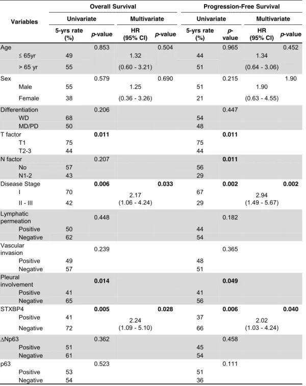
was significantly associated with T factor, disease stage, pleural involvement, and
STXBP4 expression, as assessed by univariate analysis. Multivariate analysis confirmed
that STXBP4 expression and disease stage were independent prognostic factors in lung
SCC patients with poor OS and PFS (Table 2).
Kaplan-Meier analysis of OS and PFS according to STXBP4 expression
revealed a statistically significant difference in OS and PFS between the patients who
were STXBP4-positive compared with those who were STXBP4-negative (Fig. 1B and
1C). The five-year survival rate and median survival time for all patients were 50.2 %
and 38.3 months (0.75 to 111.5 months), respectively. The median PFS and OS (21.2
months versus 52.2 months; P < 0.05) were shorter in STXBP4-positive patients
compared with STXBP4-negative patients (Fig. 1B and 1C). STXBP4-positive patients
showed poor OS (log-rank P < 0.01) and PFS (log-rank P < 0.01) compared with those
with STXBP4-negative patients. Interestingly, STXBP4 levels significantly predicted
outcome in patients with tumors expressing high ΔNp63 levels (Supplementary Fig. 1A
18
and 1B), while the complementary analysis showed that ΔNp63 levels did not
significantly predict OS (P = 0.35) and PFS (P = 0.54) in the STXBP4 high-expressing
group (Supplementary Fig. 1C and 1D). Thus, these results indicate that STXBP4 could
be an independent prognostic marker for predicting poor outcome in lung SCC.
We observed significantly higher levels of STXBP4 expression in those tumors
that showed an accumulation of ∆Np63 (Spearman’s ρ = 0.219; P < 0.05), while among
all p63 isoforms, no significant correlations were observed (P > 0.5) (Fig. 1D).
Interestingly, high mammalian target of rapamycin (mTOR), a major controller of
growth and is often deregulated in cancer (27), was significantly correlated with
STXBP4-positivity (Spearman’s ρ = 0.220; P < 0.05), while other tumor markers,
including CD147, a member of the immunoglobulin superfamily involved in
angiogenesis (P < 0.5), and Ki-67, a general marker for cell division (P < 0.1), were not
significantly correlated (Fig. 1D).
Transcriptional profiling and functional screening to identify possible downstream
mediators of STXBP4
Hierarchical cluster analysis after alignment of a total of 15,346 genes to the
reference sequence, showed that STXBP4-positive and STXBP4-negative tumors had
19
distinctly different gene expression profiles (Fig. 2A). Among the differentially
expressed genes (P-value < 0.05, False Discovery Rate (FDR) < 0.5), we identified a
total of 172 genes that were either significantly up-regulated (79 genes) or
down-regulated (93 genes) in the STXBP4-positive tumors. These candidate genes
potentially represent a network involved in STXBP4-mediated biology (Fig. 2B). To
address this possibility in more detail, we carried out Ingenuity Pathway Analysis (IPA),
which revealed that more than 30% of the affected genes were classified in the
functional class of “Cell Death and Survival”. This finding supported our experimental
observations that STXBP4 could be linked to the poor prognosis of lung SCC (Fig. 2C).
Additionally, other significant functional classes identified by IPA, including
“Cellular Movement” and “Cell to Cell Signaling and Interaction”, may be relevant to
the correlation of STXBP4-positivity with local tumor progression related to local
tumor size (T factor) and disease stage (Fig. 2C). The canonical pathway analysis
characterized two signaling pathways as the functional relationship of
STXBP4-positivity. “Cellular Movement” and “Cell Morphology”, have been predicted
as the most significantly activated canonical pathways (Supplementary Fig. 2).
IPA revealed that STXBP4-positivity was also correlated with the expression of
growth factor receptors and components of downstream pathways. Among these genes
20
listed in descending order of normalized expression, PDGFRA was a significant
up-regulated gene (FDR < 0.1) (Fig. 2D and 2E), and a most relevant candidate for
addressing the growth of STXBP4-positive lung SCC cells (Supplementary Fig. 3).
PDGFRα is a receptor tyrosine kinase that is a critical regulator of growth and
proliferation of certain cell types during embryonal development (28, 29). In subsequent
experiments described later in this study, PDGFRα proved to be a key mediator of
STXBP4 oncogenic activity.
STXBP4 regulates PDGF-PDGFR signaling in lung SCC
PDGF and PDGFR isoforms have important functions in the regulation of
growth and survival of certain cell types (28, 29), and upregulation of PDGF-PDGFR
signaling drives tumor cell growth. Indeed, the oncogenic properties of mutated or
amplified PDGFRα have been studied in several tumor types, and PDGFRβ has been
linked to not only tumor angiogenesis via paracrine effects, but also cancer metastasis
(30, 31).
We measured mRNA expression levels by real-time RT-PCR in a total of 52
available samples from lung SCC patients for which high quality RNA was available
(RIN > 2.0). The mRNA levels of STXBP4 were also correlated with ΔNp63 mRNA
21
levels in those 52 samples (Supplementary Fig. 4A). Interestingly, we observed that
PDGFRA expression was significantly up-regulated in STXBP4-positive lung SCC
samples compared with STXBP4-negative samples (P < 0.05) (Fig. 3A and 3B). On the
other hand, PDGFRB, VEGFR1 (FLT1), VEGFR2 (KDR) and VEGFR3 (FLT4) were
consistently, but not significantly, up-regulated in STXBP4-positive lung SCC samples.
STXBP4-positive samples defined by immunohistochemistry also had high STXBP4
mRNA expression levels compared with STXBP4-negative samples, and interestingly,
PDGFRA mRNA levels were also significantly correlated with STXBP4 mRNA levels
(Fig. 3C). Additionally, analyses of The Cancer Genome Atlas (TCGA) datasets of lung
SCC (n = 488), supported in part, our finding that PDGFRA mRNA levels are
significantly correlated with STXBP4 mRNA levels (P = 0.015) (Supplementary Fig.
4B).
To confirm the enhanced expression levels of PDGFRA in STXBP4 high
expressing tumors, the lung SCC cell line, EBC-1, was transduced with STXBP4 and
∆Np63α retroviruses. As shown in Fig. 3D and 3E, the STXBP4 transduced stable
clones showed high induction levels of both PDGFRA mRNA level (Fig. 3D) and
PDGFRα protein levels (Fig. 3E), consistent with our findings in 52 resected patient
lung SCC samples (Fig. 3B and 3C). Correspondingly, high expressing STXBP4 cells
22
had elevated ∆Np63 protein levels, but not mRNA levels. Additionally, the relative
increase in PDGFRA mRNA expression is more marked in STXBP4 low-expressing
EBC-1 cells compared with STXBP4 high-expressing RERF-LC-Sq1 cells
(Supplementary Fig. 5A and 5B). The results indicate that STXBP4 regulates PDGFRα
expression in lung SCC, most likely in a ∆Np63-dependent manner, and also suggest
that STXBP4 may serve as a new SCC biomarker.
STXBP4-depletion represses lung SCC tumor growth in vivo
We next examined the oncogenic role of STXBP4 in regulating the expression
of PDGFRα in a lung SCC cell line using a loss-of-function approach. Two independent
siRNAs against STXBP4 (STXBP4#1 and STXBP4#2) were transfected into the lung
SCC cell line, RERF-LC-Sq1, and a siRNA targeting luciferase (siLUC) was used as a
control. The STXBP4 knockdown cells showed low expression of STXBP4, which
correlated with down-regulation of both PDGFRA mRNA (Fig. 4A) and PDGFRα
protein levels (Fig. 4B), consistent with our findings in resected patient lung SCC
samples.
In order to evaluate the functional relevance of STXBP4 and PDGFRα
expression during tumor formation, we monitored the colony formation of
23
RERF-LC-Sq1 cells lentivirally transduced with STXBP4, ∆Np63 or PDGFRα shRNAs.
As shown in Fig. 4C, PDGFRα, STXBP4 or ∆Np63 knockdown in RERF-LC-Sq1 cells,
led to decreased anchorage-independent colony formation in soft agar. Subcutaneous
transplantation of PDGFRA or STXBP4 knockdown clones into immunodeficient mice
resulted in suppressed tumor formation compared with control luciferase shRNA
xenografts (Fig. 4D). Additionally, the knockdown effect of STXBP4 and suppression of
tumorigenesis are more marked in high STXBP4 expressing RERF-LC-Sq1 cells
compared with low STXBP4 expressing EBC-1 cells (Fig. 4 and Supplementary Fig. 6).
These data suggest that downregulation of STXBP4 decreases PDGFRα expression and
suppresses tumor formation.
Overall, our results indicate that STXBP4 has oncogenic activity both in vitro
and in vivo, and further suggest that STXBP4 could be a critical driver of tumor
propagation through regulating the PDGFRα pathway.
24 Discussion
We demonstrated that STXBP4 expression in clinical specimens was closely
associated with T factor (P < 0.001), disease stage (P = 0.030), and pleural involvement
(P = 0.028). Furthermore, univariate and multivariate analysis indicated that STXBP4
expression was an independent prognostic factor for OS and PFS. While p63 is essential
for normal epidermal stratification and the proliferative potential of epithelial stem cells,
∆Np63 is thought to maintain the proliferative potential of basal regenerative cells,
including stem cells, in skin, thymus, breast, prostate, and urothelial stratified
epithelium (20, 32-36). STXBP4 can physically interact with ∆Np63 and is
indispensable for stabilizing ∆Np63, which is consistent with a putative diagnostic role
for STXBP4 in lung SCC.
Polymorphisms of STXBP4/COX11 (rs6504950; AA/AG-genotype) were
associated with a significantly decreased risk of carcinogenesis in a meta-analysis of
breast cancer patients (37). Although functional assessments of these polymorphisms
were not undertaken, and the extent of loss of STXBP4 function in tumors was not
studied, the data suggested that STXBP4 could play a role in carcinogenesis and tumor
progression in breast cancer patients. Our study provides diagnostic role of STXBP4
alongside ∆Np63 in lung SCC.
25
The pathological function of STXBP4 in human cancers remains unclear.
However, STXBP4 can physically interact with p63 and is indispensable for stabilizing
ΔNp63 even in normal conditions (16). Consistent with STXBP4 localization in both
the nucleus and cytoplasm, it has been suggested that nuclear STXBP4 has
p63-mediated functions, and that cytoplasmic STXBP4 could facilitate other functions
in a p63-independent manner (16). In fact, our data indicated that STXBP4 induction
partially increased tumor growth even in the absence of elevated ΔNp63
(Supplementary Fig. 7A), and that PDGFRA induction also partially increased tumor
growth even in STXBP4 knockdown cells (Supplementary Fig. 7B). Thus, STXBP4 may
contribute to the susceptibility and severity of cancer in a p63-dependent and
independent manner. Indeed, amplification and overexpression of p63 has frequently
been observed in a variety of SCCs, including lung cancers and head and neck cancers
(8, 38). However, p63 expression is decreased during progression to invasion and
metastasis of lung, breast and bladder cancer, and loss of p63 expression is associated
with worse prognosis in some cases (35, 39, 40). It could be the balance between the TA
isotype (tumor suppressive) and ∆N isotype (oncogenic), as well as the tissue context,
which is critical for proliferation and differentiation in both epithelial stem cells and
cancer stem cells.
26
Global transcriptome profiling using next-generation sequencing technologies
has become more common for comprehensive gene expression analysis to explore novel
regulators and target genes in different types of cancers. In this report, genome-wide
transcriptome analysis identified mediators of STXBP4 activity, including PDGFRα,
which contributes to cell growth and metastasis in a p63-dependent manner. PDGF
family proteins consist of several disulfide-bonded, dimeric isoforms (PDGF AA,
PDGF AB, PDGF BB, PDGF CC and PDGF DD) that bind in a specific pattern to two
related receptor tyrosine kinases, PDGFRα and PDGFRβ (41). PDGFRα homodimers
bind to all PDGF isoforms except those containing PDGF D (42). A number of different
signaling pathways, including mTOR (Fig. 1D) and MAPK (Fig. 3E), are initiated by
activated PDGF receptors, and stimulate cell growth, actin reorganization, migration,
and differentiation (43, 44).
PDGF receptors are expressed at low levels in normal lung epithelial cells,
however, increased PDGFRα expression has been reported in lung cancer. PDGFRβ
expression is observed mainly in stromal cells, but also in the sarcomatoid type of
NSCLC (45). Based on recent evidence, inhibition of the p53/NF-Y complex by mutant
gain of function p53 enhances PDGFRβ expression and promotes metastasis in a subset
of pancreatic cancers (31). In addition, the interaction of mutant p53 with p63 regulates
27
the expression of p63 target genes to enhance invasion and metastasis (46). Hence, the
oncogenic activity of mutant p53 is a consequence of the physical association between
mutant p53 and the p53 family members, p63 and p73.
Treatment strategies for lung cancer are based on the assumption that an
individual patient's cancer is purely of one subtype. Since many cancers are
heterogeneous and relatively resistant to chemotherapy or radiation, there is strong
interest in molecular-targeted therapies based on tumor biology. In particular, targeted
agents that inhibit the epidermal growth factor receptor (EGFR) or anaplastic lymphoma
kinase (ALK) are approved for the treatment of NSCLC harboring genetic alterations in
the genes encoding these proteins (47). EGFR inhibitors, such as erlotinib and gefitinib,
are only effective against NSCLCs with EGFR mutations, which occur almost
exclusively in lung AC. Similarly, the recently identified EML4-ALK rearrangement,
which predicts susceptibility to the targeted agent crizotinib, also occurs only in lung
AC. Unfortunately, therapeutic advances in the treatment of lung SCC have lagged
behind those for AC (48). Therefore, the capacity to distinguish between lung AC and
SCC is particularly important for the effective use of novel targeted therapies to treat
patients with these NSCLC subtypes.
Inhibition of the PDGFRα signaling pathway by treatment with a neutralizing
28
PDGFRα antibody, MEDI-575, had minimal effect on tumor cell proliferation in
preclinical models of NSCLC (49). Lung SCC histology also identified patients at a
higher risk of bleeding during treatment with bevacizumab, a monoclonal anti-VEGF
antibody (50). Thus, more studies are required to determine whether specific inhibition
of PDGF receptors, without inhibition of VEGF receptors, is of any benefit for lung
cancer patients. These issues highlight the growing importance of accurate identification
of NSCLC subtypes for assigning patients to appropriate histology-based therapies and
the triage of tissue for appropriate molecular studies.
Acknowledgements
We thank Dr. Arito Yamane for instrument settings of Ion Proton system, and
Gombodorj, N., Umezawa, S., Suto, Y., Nakamura, M., Ito, M., Ichihara, A. and
Horigome, E. for expert technical assistances. We also thank Drs. Vincenzo Castronovo, Andrei Turtoi and Akeila Bellahcene in University of Liege for valuable discussions. This work was supported, in part, by the Promotion Plan for the Platform of Human Resource Development for Cancer, New Paradigms - Establishing
Center for Fostering Medical Researchers of the Future Programs by Ministry of
Education, Culture, Sports, Science and Technology of Japan, and Gunma University
29
Initiative for Advanced Research (GIAR). This work was also supported, in part, by The
Yasuda Medical Foundation and by NIH grant CA87497.
30 References
1. Molina JR, Yang P, Cassivi SD, Schild SE, Adjei AA. Non-small cell lung cancer:
epidemiology, risk factors, treatment, and survivorship. Mayo Clinic proceedings 2008;83(5):584-94.
2. Recondo G, Jr., Recondo G, Sr., Galanternik F, Greco M, de la Vega M, Canton ED, et al. Immunotherapy for Non-Small Cell Lung Cancer - Finally a Hint of Hope.
Rev Recent Clin Trials 2016.
3. Mukhopadhyay S, Katzenstein AL. Subclassification of non-small cell lung carcinomas lacking morphologic differentiation on biopsy specimens: Utility of an immunohistochemical panel containing TTF-1, napsin A, p63, and CK5/6. Am J Surg Pathol 2011;35(1):15-25.
4. Massion PP, Taflan PM, Jamshedur Rahman SM, Yildiz P, Shyr Y, Edgerton ME, et al. Significance of p63 amplification and overexpression in lung cancer development and prognosis. Cancer Res 2003;63(21):7113-21.
5. Sniezek JC, Matheny KE, Westfall MD, Pietenpol JA. Dominant negative p63 isoform expression in head and neck squamous cell carcinoma. Laryngoscope 2004;114(12):2063-72.
6. Candi E, Dinsdale D, Rufini A, Salomoni P, Knight RA, Mueller M, et al. TAp63 and DeltaNp63 in cancer and epidermal development. Cell Cycle 2007;6(3):274-85.
7. Mills AA. p63: oncogene or tumor suppressor? Curr Opin Genet Dev 2006;16(1):38-44.
8. Di Como CJ, Urist MJ, Babayan I, Drobnjak M, Hedvat CV, Teruya-Feldstein J, et al. p63 expression profiles in human normal and tumor tissues. Clin Cancer Res 2002;8(2):494-501.
9. Nonaka D. A study of DeltaNp63 expression in lung non-small cell carcinomas.
Am J Surg Pathol 2012;36(6):895-9.
31
10. Bishop JA, Teruya-Feldstein J, Westra WH, Pelosi G, Travis WD, Rekhtman N. p40 (DeltaNp63) is superior to p63 for the diagnosis of pulmonary squamous cell carcinoma. Mod Pathol 2012;25(3):405-15.
11. Melino G, Lu X, Gasco M, Crook T, Knight RA. Functional regulation of p73 and p63: development and cancer. Trends Biochem Sci 2003;28(12):663-70.
12. Yang A, Kaghad M, Wang Y, Gillett E, Fleming MD, Dotsch V, et al. p63, a p53 homolog at 3q27-29, encodes multiple products with transactivating, death-inducing, and dominant-negative activities. Mol Cell 1998;2(3):305-16.
13. Schmale H, Bamberger C. A novel protein with strong homology to the tumor suppressor p53. Oncogene 1997;15(11):1363-7.
14. McKeon F. p63 and the epithelial stem cell: more than status quo? Genes Dev 2004;18(5):465-9.
15. Duijf PH, Vanmolkot KR, Propping P, Friedl W, Krieger E, McKeon F, et al.
Gain-of-function mutation in ADULT syndrome reveals the presence of a second transactivation domain in p63. Hum Mol Genet 2002;11(7):799-804.
16. Li Y, Peart MJ, Prives C. Stxbp4 regulates DeltaNp63 stability by suppression of RACK1-dependent degradation. Mol Cell Biol 2009;29(14):3953-63.
17. Fomenkov A, Zangen R, Huang YP, Osada M, Guo Z, Fomenkov T, et al. RACK1 and stratifin target DeltaNp63alpha for a proteasome degradation in head and neck squamous cell carcinoma cells upon DNA damage. Cell Cycle 2004;3(10):1285-95.
18. Min J, Okada S, Kanzaki M, Elmendorf JS, Coker KJ, Ceresa BP, et al. Synip: a novel insulin-regulated syntaxin 4-binding protein mediating GLUT4 translocation in adipocytes. Mol Cell 1999;3(6):751-60.
32
19. Saito T, Okada S, Yamada E, Ohshima K, Shimizu H, Shimomura K, et al. Syntaxin 4 and Synip (syntaxin 4 interacting protein) regulate insulin secretion in the pancreatic beta HC-9 cell. J Biol Chem 2003;278(38):36718-25.
20. Westfall MD, Mays DJ, Sniezek JC, Pietenpol JA. The Delta Np63 alpha phosphoprotein binds the p21 and 14-3-3 sigma promoters in vivo and has transcriptional repressor activity that is reduced by Hay-Wells syndrome-derived mutations. Mol Cell Biol 2003;23(7):2264-76.
21. Shimizu K, Kaira K, Tomizawa Y, Sunaga N, Kawashima O, Oriuchi N, et al. ASC amino-acid transporter 2 (ASCT2) as a novel prognostic marker in non-small cell lung cancer. Br J Cancer 2014;110(8):2030-9.
22. Kaira K, Sunose Y, Arakawa K, Sunaga N, Shimizu K, Tominaga H, et al.
Clinicopathological significance of ASC amino acid transporter-2 expression in pancreatic ductal carcinoma. Histopathology 2015;66(2):234-43.
23. Rokudai S, Aikawa Y, Tagata Y, Tsuchida N, Taya Y, Kitabayashi I. Monocytic leukemia zinc finger (MOZ) interacts with p53 to induce p21 expression and cell-cycle arrest. J Biol Chem 2009;284(1):237-44.
24. Rokudai S, Laptenko O, Arnal SM, Taya Y, Kitabayashi I, Prives C. MOZ increases p53 acetylation and premature senescence through its complex formation with PML. Proc Natl Acad Sci U S A 2013;110(10):3895-900.
25. Trapnell C, Roberts A, Goff L, Pertea G, Kim D, Kelley DR, et al. Differential gene and transcript expression analysis of RNA-seq experiments with TopHat and Cufflinks. Nat Protoc 2012;7(3):562-78.
26. Anders S, McCarthy DJ, Chen Y, Okoniewski M, Smyth GK, Huber W, et al.
Count-based differential expression analysis of RNA sequencing data using R and Bioconductor. Nat Protoc 2013;8(9):1765-86.
27. Zoncu R, Efeyan A, Sabatini DM. mTOR: from growth signal integration to cancer, diabetes and ageing. Nat Rev Mol Cell Biol 2011;12(1):21-35.
33
28. Heldin CH. Targeting the PDGF signaling pathway in tumor treatment. Cell communication and signaling : CCS 2013;11:97.
29. Dai Y. Platelet-derived growth factor receptor tyrosine kinase inhibitors: a review of the recent patent literature. Expert opinion on therapeutic patents 2010;20(7):885-97.
30. Cao R, Bjorndahl MA, Religa P, Clasper S, Garvin S, Galter D, et al. PDGF-BB induces intratumoral lymphangiogenesis and promotes lymphatic metastasis.
Cancer Cell 2004;6(4):333-45.
31. Weissmueller S, Manchado E, Saborowski M, Morris JPt, Wagenblast E, Davis CA, et al. Mutant p53 drives pancreatic cancer metastasis through cell-autonomous PDGF receptor beta signaling. Cell 2014;157(2):382-94.
32. Mills AA, Zheng B, Wang XJ, Vogel H, Roop DR, Bradley A. p63 is a p53 homologue required for limb and epidermal morphogenesis. Nature 1999;398(6729):708-13.
33. Yang A, Schweitzer R, Sun D, Kaghad M, Walker N, Bronson RT, et al. p63 is essential for regenerative proliferation in limb, craniofacial and epithelial development. Nature 1999;398(6729):714-8.
34. Tonon G, Wong KK, Maulik G, Brennan C, Feng B, Zhang Y, et al. High-resolution genomic profiles of human lung cancer. Proc Natl Acad Sci U S A 2005;102(27):9625-30.
35. Barbieri CE, Tang LJ, Brown KA, Pietenpol JA. Loss of p63 leads to increased cell migration and up-regulation of genes involved in invasion and metastasis.
Cancer Res 2006;66(15):7589-97.
36. Higashikawa K, Yoneda S, Tobiume K, Taki M, Shigeishi H, Kamata N.
Snail-induced down-regulation of DeltaNp63alpha acquires invasive phenotype of human squamous cell carcinoma. Cancer Res 2007;67(19):9207-13.
34
37. Tang L, Xu J, Wei F, Wang L, Nie WW, Chen LB, et al. Association of STXBP4/COX11 rs6504950 (G>A) polymorphism with breast cancer risk:
evidence from 17,960 cases and 22,713 controls. Arch Med Res 2012;43(5):383-8.
38. Yamaguchi K, Wu L, Caballero OL, Hibi K, Trink B, Resto V, et al. Frequent gain of the p40/p51/p63 gene locus in primary head and neck squamous cell carcinoma. Int J Cancer 2000;86(5):684-9.
39. Karni-Schmidt O, Castillo-Martin M, Shen TH, Gladoun N, Domingo-Domenech J, Sanchez-Carbayo M, et al. Distinct expression profiles of p63 variants during urothelial development and bladder cancer progression. Am J Pathol 2011;178(3):1350-60.
40. Deyoung MP, Ellisen LW. p63 and p73 in human cancer: defining the network.
Oncogene 2007;26(36):5169-83.
41. Deuel TF, Silverman NJ, Kawahara RS. Platelet-derived growth factor: a multifunctional regulator of normal and abnormal cell growth. BioFactors 1988;1(3):213-7.
42. Bergsten E, Uutela M, Li X, Pietras K, Ostman A, Heldin CH, et al. PDGF-D is a specific, protease-activated ligand for the PDGF beta-receptor. Nat Cell Biol 2001;3(5):512-6.
43. Ostman A, Heldin CH. Involvement of platelet-derived growth factor in disease:
development of specific antagonists. Advances in cancer research 2001;80:1-38.
44. Andrae J, Gallini R, Betsholtz C. Role of platelet-derived growth factors in physiology and medicine. Genes Dev 2008;22(10):1276-312.
45. Donnem T, Al-Saad S, Al-Shibli K, Andersen S, Busund LT, Bremnes RM.
Prognostic impact of platelet-derived growth factors in non-small cell lung cancer tumor and stromal cells. Journal of thoracic oncology : official publication of the International Association for the Study of Lung Cancer 2008;3(9):963-70.
35
46. Adorno M, Cordenonsi M, Montagner M, Dupont S, Wong C, Hann B, et al. A Mutant-p53/Smad complex opposes p63 to empower TGFbeta-induced metastasis. Cell 2009;137(1):87-98.
47. Solomon BJ, Mok T, Kim DW, Wu YL, Nakagawa K, Mekhail T, et al. First-line crizotinib versus chemotherapy in ALK-positive lung cancer. The New England journal of medicine 2014;371(23):2167-77.
48. Socinski MA, Stinchcombe TE, Moore DT, Gettinger SN, Decker RH, Petty WJ, et al. Incorporating bevacizumab and erlotinib in the combined-modality treatment of stage III non-small-cell lung cancer: results of a phase I/II trial.
Journal of clinical oncology : official journal of the American Society of Clinical Oncology 2012;30(32):3953-9.
49. Laing N, McDermott B, Wen S, Yang D, Lawson D, Collins M, et al. Inhibition of platelet-derived growth factor receptor alpha by MEDI-575 reduces tumor growth and stromal fibroblast content in a model of non-small cell lung cancer.
Molecular pharmacology 2013;83(6):1247-56.
50. Johnson DH, Fehrenbacher L, Novotny WF, Herbst RS, Nemunaitis JJ, Jablons DM, et al. Randomized phase II trial comparing bevacizumab plus carboplatin and paclitaxel with carboplatin and paclitaxel alone in previously untreated locally advanced or metastatic non-small-cell lung cancer. Journal of clinical oncology : official journal of the American Society of Clinical Oncology 2004;22(11):2184-91.
36 Figures Legends
Figure 1. STXBP4 expression is correlated with p63 expression and poor
prognosis in lung SCC. (A) Representative immunohistochemical staining of a
lung SCC. STXBP4 immunostaining demonstrates a nuclear and cytoplasmic
pattern with a score of 5. Scale bars are 200 µm. (B, C) Kaplan-Meier analysis of
Overall Survival (OS) and Progression Free Survival (PFS) defined according to
STXBP4 expression. A statistically significant difference in OS and PFS was
observed between the STXBP4-positive patients and those with low STXBP4
expression [OS, p = 0.0056(A); PFS, p = 0.0068 (B)]. P-values were obtained by
log-rank test. (D) Spearman’s rank correlation was performed based on the
expression levels of STXBP4 and ∆Np63.
Figure 2. Gene expression profiling of clinical samples from lung SCC patients.
(A) A cluster diagram of RNA-seq data from six pairs of STXBP4-positive and
STXBP4-negative samples. The color bars represent relative expression levels:
Red indicates higher than average expression and blue indicates lower than
average expression. (B) 79 significantly differentially expressed upregulated
genes and 93 downregulated genes (P < 0.05 and FDR < 0.5) were identified.
(C) Functional analysis of differentially expressed genes was performed by
37
Ingenuity Pathway Analysis. (D) The genes are listed in descending order of
normalized expression. *FDR < 0.1. (E) A cluster diagram of PDGFR and
VEGFR expression from RNA-seq analysis. The color bars represent relative
expression levels: red indicates higher than average expression and blue
indicates lower than average expression.
Figure 3. PDGFRA expression is significantly upregulated in STXBP4-positive
samples from lung SCC Patients. (A) A cluster diagram of relative expression of
PDGFRs and VEGFRs by real-time RT-PCR analysis. Tumor specimens were
collected from lung SCC patients with surgical resection. A total of 52 high RNA
integrity number (RIN > 2.0) STXBP4-positive samples (n=34) and
STXBP4-negative samples (n=18), were used for transcriptome profiling by
real-time RT-PCR. (B) PDGFRA mRNA was significantly upregulated in
STXBP4-positive lung SCC samples. Relative mRNA levels of PDGFRs and
VEGFRs of the STXBP4-positive samples (n=35) and STXBP4-negative
samples (n=19); *P < 0.05. (C) Scatter plot of relative mRNA expression levels of
PDGFRA and STXBP4. (D) STXBP4 or ∆Np63α induces PDGFRA expression in a lung SCC cell line, EBC-1. The cells were retrovirally transduced with empty
vector control (Mock), ∆Np63α or STXBP4. The mRNA levels of PDGFRA,
38
STXBP4 or ∆Np63 were determined by real-time RT-PCR. (E) EBC-1 cells
transduced as in (D), were subjected to immunoblotting using antibodies for
phospho-PDGFRα (p-PDGFRα), PDGFRα, phospho-p38MAPK (p-p38MAPK),
p38MAPK, STXBP4, ∆Np63 or β-Actin.
Figure 4. STXBP4-depletion inhibits lung SCC tumorigenesis and modulates
PDGF signaling in vivo. (A) The lung SCC cells, RERF-LC-Sq1, were treated
with siRNAs for Luciferase (siLUC) as a control, STXBP4#1, STXBP4#2, ∆Np63
or PDGFRα. Total RNAs were quantified by real-time RT-PCR analysis and the
induction levels of PDGFRA were determined by the relative Ct method. (B)
RERF-LC-Sq1 cells depleted as in (A), were subjected to immunoblotting using
anti-PDGFRα, STXBP4, ∆Np63 or β-Actin antibodies. (C) The growth of
RERF-LC-Sq1 cells after shRNA mediated PDGFRα, STXBP4 or ∆Np63
knockdown was monitored by soft agar colony formation assays. Standard
deviations (SD) are plotted. *P < 0.05. (D) Representative images of xenografts
from subcutaneously transplanted with lentivirally shRNA transduced Luciferase
as a control (shLUC), Stxbp4, ∆Np63 or PDGFRα knockdown RERF-LC-Sq1
cells (n = 6 for each knockdown). The results of six independent injections of
39
knockdown cells are shown. Twenty days after implantation, the length (L) and
width (W) of the tumor mass were measured, and the tumor volume (TV) was
calculated using the equation: TV = (L x W2)/2. *P < 0.05.
A
D
B C
0 20 40 60 80 100 Months 0
0.2 0.4 0.6 0.8 1.0
p = 0.0056
Overall Survival
Positive STXBP4 (n=52) Negative STXBP4 (n=35)
Months 0 20 40 60 80 100 0
0.2 0.4 0.6 0.8 1.0
p = 0.0068
Progression-Free Survival
Positive STXBP4 (n=52) Negative STXBP4 (n=35)
STXBP4 p63 (4A4) DNp63
vs. STXBP4 DNp63 p63 mTOR CD147
Spearman’s ρ 0.219 0.088 0.220 0.144 P-value 0.042 0.417 0.041 0.183
B
C
E
High Low Positive STXBP4 Negative STXBP4
L101 L100 L106 L082 L078 L092 L075 L060 L080 L095 L050 L087
PDGFRA PDGFRB
VEGFR1 (FLT1)
VEGFR2 (KDR)
VEGFR3 (FLT4)
A
2.92 -2.92
Positive STXBP4 L100 L101 L106 L078 L082 L092 L050 L060 L075 L080 L087 L095 Negative STXBP4
Z-score
Lung Squamous Cell Carcinoma
RNA-sequencing Positive STXBP4
(n = 6)
Negative STXBP4 (n = 6)
93 genes ↓ 79 genes ↑
P < 0.05 FDR < 0.5
D
* FDR < 0.1
Molecular and cellular function
0 10 20 30 40
Cellular Function and Maintenance Cellular Movement
Cell Death and Survival
Cell to Cell Signaling and Interaction Cell Morphology
(%) (P = 1.04E-7−6.88E-3) (P = 6.41E-7−7.49E-3) (P = 7.94E-7−7.62E-3) (P = 2.33E-6−7.06E-3) (P = 7.52E-6−6.82E-3)
B C A
STXBP4
Positive STXBP4 (n = 34) Negative STXBP4 (n = 18)
L004 L049 L047 L024 L051 L002 L090 L015 L076 L019 L091 L069 L059 L102 L061 L031 L081 L084 L077 L074 L057 L083 L097 L066 L098 L103 L100 L101 L106 L088 L082 L092 L099 L078 L033 L067 L062 L060 L050 L017 L075 L094 L048 L104 L087 L085 L016 L079 L080 L095 L107 L096
(n = 52)
0 1 2 3 4 5
PDGFRA PDGFRB
VEGFR1 VEGFR2
VEGFR3 Low High
Relative mRNA levels
VEGFR1 (FLT1) VEGFR2 (KDR) VEGFR3 (FLT4) PDGFRA PDGFRB
*P < 0.05
Nega. Posi. Nega. Posi.
Nega. Posi. Nega. Posi. Nega. Posi.
D E
0 2 4 6
Mock STXBP4
DNp63a
DNp63
0 1
Mock STXBP4
DNp63a
2 3 4 5
PDGFRA
Relative mRNA levels
STXBP4 DNp63a
Mock STXBP4
DNp63a
p-PDGFRa
b-Actin PDGFRa p-p38MAPK p38MAPK
R = 0.610 (95%CI 0.397-0.761)
*P < 0.01
STXBP4
PDGFRA
(n=52)
STXBP4
0 2 4 6 8
Mock STXBP4
DNp63a
B A
C D
PDGFRa
DNp63 b-Actin STXBP4
siLUC STXBP4 #1 DNp63
STXBP4 #2 PDGFRa
siRNA:
0 0.5 1.0
shLUC STXBP4 DNp63 PDGFRa
shRNA:
* * *
Colony Formation
shLUC STXBP4 DNp63 PDGFRa shRNA
Relative mRNA expression
0 0.5 1.0
siRNA:
1.5
PDGFRA
siLUC STXBP4#1 DNp63
STXBP4#2 PDGFRa
* * * *
Tumor Volume (mm3 )
0 200 400 600 800 1000
shLUC STXBP4 DNp63 PDGFRa
shRNA:
*
* * shRNA
shLUC STXBP4 DNp63 PDGFRa
1 Table 1. Patient's demographics according to STXBP4 expression
Variables
STXBP4 DNp63 p63
High No. (%)
Low
No. (%) P-value Positive No. (%) Negative No. (%) P-value Positive No. (%) Negative No. (%) P-value
Age 0.42 0.051 0.22
≤ 65yr 9 (10.3) 9 (10.3) 10 (11.5) 8 ( 9.2) 16 (18.4) 2 ( 2.3) > 65 yr 43 (50.5) 26 (29.9) 20 (23.0) 49 (56.3) 51 (58.6) 18 (20.7)
Sex 0.70 0.69 < 0.99
Male 47 (54.0) 33 (37.9) 27 (31.0) 53 (60.9) 61 (70.1) 19 (21.8) Female 5 ( 5.7) 2 ( 2.3) 3 ( 3.4) 4 ( 4.6) 6 ( 4.6) 1 ( 3.4)
Differentiation 0.080 0.034 0.24
Well or Moderately 43 (49.4) 23 (26.4) 27 (31.0) 39 (44.8) 53 (60.9) 13 (15.0) Poorly 9 (10.3) 12 (13.8) 3 ( 3.4) 18 (20.7) 14 (16.1) 7 ( 8.0)
T factor < 0.001 0.33 < 0.001
T1 8 (9.2) 19 (21.8) 7 ( 8.0) 20 (23.0) 27 (31.0) 0 ( 0.0)
T2-3 44 (50.6) 16 (18.4) 23 (26.4) 37 (42.5) 40 (46.0) 20 (23.0)
N factor > 1 > 1 0.28
N0 36 (41.4) 25 (28.7) 21 (24.1) 40 (46.0) 49 (56.3) 12 (13.8) N1-2 16 (18.4) 10 (11.5) 9 (10.3) 17 (19.5) 18 (20.7) 8 ( 9.2)
Disease stage 0.030 0.37 0.20
I 23 (26.4) 24 (27.6) 14 (16.1) 33 (37.9) 39 (44.8) 8 ( 9.2) II-III 29 (33.3) 11 (12.6) 16 (18.4) 24 (27.6) 28 (32.2) 12 (13.8)
Pleural Involvement 0.028 0.66 0.29
Positive 30 (34.5) 11 (12.6) 13 (14.9) 28 (32.2) 30 (34.5) 11 (13.8) Negative 22 (25.3) 24 (27.6) 17 (19.5) 29 (33.3) 37 (42.5) 9 (10.3)
Lymphatic permeation 0.83 > 1 0.61
Positive 29 (33.3) 18 (20.7) 16 (18.4) 31 (35.6) 35 (40.2) 12 (13.8) Negative 23 (26.4) 17 (19.5) 14 (16.1) 26 (29.9) 32 (36.8) 8 ( 9.2)
Vascular invasion 0.39 > 1 > 1
Positive 26 (29.9) 14 (16.1) 14 (16.1) 26 (29.9) 31 (35.6) 9 (10.3) Negative 26 (29.9) 21 (24.1) 16 (18.4) 31 (35.6) 36 (41.4) 11 (12.6)
*P-values were obtained by Fisher's exact test.
†Clinical stage at the time of initial diagnosis was determined according to the seventh edition of General Rule for Clinical and Pathological Record of Lung Cancer (2010), the Japan Lung Cancer Society.