Introduction
Charcot-Marie-Tooth disease (CMT) is a hereditary peripheral neuropathy presenting distal wasting and weakness, usually with some distal sensory impairment. In most cases, the clinical course is benign and the disease is not life threatening; however, in some cases, severe phenotypes can include respiratory dis-tress, which, in relation to adults, is not widely recognized in the literature1). We describe a unique case characterized by progres-sion of serious symptoms; ultimately, these included facial and
respiratory muscle impairment, and a novel mutation was found in the glycyl-tRNA synthetase gene (the gene is abbreviated as GARS and the protein as GlyRS), which is a causative gene for both CMT type 2D (CMT2D) and distal spinal muscular atrophy type V (dSMA-V).
Case report
A 45-year-old woman initially presented with distal dominant muscle atrophy, which progressed, and facial muscle atrophy and
Brief Clinical Note
A novel mutation in glycyl-tRNA synthetase caused Charcot-Marie-Tooth
disease type 2D with facial and respiratory muscle involvement
Nobuko Kawakami, M.D.
1)2), Kenichi Komatsu, M.D.
2)3)*, Hirofumi Yamashita, M.D., Ph.D.
2),
Kengo Uemura, M.D., Ph.D.
2)4), Nobuyuki Oka, M.D., Ph.D.
2)5),
Hiroshi Takashima, M.D., Ph.D.
6)and Ryosuke Takahashi, M.D., Ph.D.
2)Abstract: BACKGROUND: Charcot-Marie-Tooth disease (CMT) is a hereditary peripheral neuropathy;
symptoms include distal wasting and weakness, usually with some sensory impairment. The clinical course
is typically benign and the disease is not life threatening; however, in some cases, severe phenotypes
include serious respiratory distress. CASE REPORT: Here we describe a 45-year-old woman with a long
course of motor-dominant neuropathy. Distal weakness appeared in childhood and became worse with age.
After a diagnosis of CMT type 2, the symptoms progressed, and in her fourth decade, facial and respiratory
muscle weakness appeared, ultimately requiring non-invasive mechanical ventilation. There was no family
history of CMT. Comprehensive analysis of known CMT-related genes revealed a novel heterozygous
c.815T>A, p.L218Q mutation in glycyl-tRNA synthetase (GARS), a causative gene for both CMT type 2D
(CMT2D) and distal spinal muscular atrophy type V (dSMA-V). This mutation was considered pathogenic
based on molecular evidence; notably, it was unique in that all other reported GARS mutations associated
with severe phenotypes are located in an anticodon-binding domain, while in this case in an apparently
non-functional region of the GARS gene. Not a simple loss-of-function mechanism, but rather gain-of-function
mechanisms have also been reported in GARS mutations. This case provided useful information for
under-standing the mechanism of CMT2D/dSMA-V.
(Clin Neurol: 2014;54:911-915)
Key words: Charcot-Marie-Tooth disease, hereditary sensory and motor neuropathy, glycine-tRNA ligase, spinal muscular atrophy, respiratory distress
*Corresponding author: Department of Neurology, Kitano Hospital, The Tazuke Kofukai Medical Research Institute〔2-4-20 Ohgimachi, Kita-ku, Osaka-shi, Osaka 530-8480〕
1)Department of Neurology, Shizuoka General Hospital
2)Department of Neurology, Kyoto University Graduate School of Medicine
3)Department of Neurology, Kitano Hospital, The Tazuke Kofukai Medical Research Institute 4)Ishiki Hospital
5)Department of Neurology, National Hospital Organization Minami Kyoto Hospital
6)Department of Neurology and Geriatrics, Kagoshima University Graduate School of Medical and Dental Sciences (Received: 14 January 2014)
preschool years. Bilateral foot drop developed around age 7, and distal muscle atrophy developed in all limbs by age 10. She was wheelchair-bound in her third decade. At age 29, she was admitted to our hospital for three months; muscle and nerve biopsies were performed, and she subsequently received a diagnosis of CMT type 2 (CMT2) with evidence of axonal sensorimotor neuro-pathy. At age 36, respiratory muscle dysfunction developed and non-invasive mechanical ventilation was started. At age 45, she was admitted for re-evaluation. Her parents were unrelated to one another (Fig. 1A). Her mother had died at age 46 due to unknown causes. Her father and brother were alive and healthy at the writing of this report. Physical examination showed severe atrophy in all skeletal muscles, including limb, truncal, facial and tongue muscles (Fig. 1B). Dysphagia and nasal voice were evident. Her muscle strength scores, which were based on the Medical Research Council scale, were 2 out of 5 for proximal muscles and 1 out of 5 for distal muscles. Deep tendon reflexes were absent. Sensory disturbance was mild and only evident with distal lower limbs.
In nerve conduction studies, compound muscle action poten-tials (CMAPs) were not evoked from routinely examined muscles, including the abductor pollicis brevis, abductor digiti minimi and flexor hallucis brevis. CMAPs from the flexor carpi radialis had extremely low amplitudes, but the distal latency was normal, and conduction velocities were only slightly decreased (42 m/s) relative to normal values. Sensory nerve action potentials (SNAPs) and sensory conduction velocities (SCVs) from the median nerve were normal. SNAPs from the sural nerve had been recorded when the patient was 29 years old; these SNAPs had very low amplitudes (2.1 mV), but the SCVs were normal (55 m/s). Needle electromyography showed chronic neurogenic patterns.
A muscle biopsy from triceps brachii was performed at age 29. The majority of the muscle fibers ranged from 70 to 100 mV in diameter. Pyknotic clamp was present in the rim of a fascicle. Necrotic or regenerating fibers were not observed. Islands of groups of extremely atrophic fibers and spindles were present in epimysium. Internal nuclei were moderately increased. Muscle fibers occasionally showed fiber splitting. Fatty connective tissue was increased in perimysium and more markedly in epimysium. Trichrome staining added no information. Intermyofibrillar net-work was preserved in NADH dehydrogenase-stained sections. Every fascicle of fibers showed fiber-type grouping as assessed by ATPase staining. These findings were consistent with chronic denervation.
A sural nerve biopsy also taken at age 29 revealed moderate loss of myelinated fibers; however, axonal degeneration and active demyelination were not evident. Perivascular mononuclear
(Fig. 1C). Electron microscopy revealed no obvious mitochondrial abnormalities.
Lung CT scan revealed no abnormalities. Electrocardiogram showed normal sinus rhythm with a tall P wave and right axis deviation. Echocardiogram appeared normal.
A comprehensive sequence analysis of CMT-related genes2)3) revealed a novel heterozygous c.815T>A, p.L218Q mutation in the GARS gene (Fig. 1D). The patientʼs unaffected father and brother did not carry this mutation. HomoloGene (http://www. ncbi.nlm.nih.gov/homologene) was used to conduct a sequence homology search; we found that leucine 218 in GlyRS was highly conserved among species (Fig. 1E). The computational protein function-predicting algorithm MUPro score was −1; this value indicated that the mutant protein was less stable than the wild-type protein (http://www.igb.uci.edu/~baldig/mutation.html). Moreover, the Polyphen-2 score was 1.0; this score indicated that the mutant GlyRS protein was pathogenic (http://genetics. bwh.harvard.edu/pph2/).
Discussion
We present a unique case of CMT that involved a new muta-tion in GARS; the patient initially developed moderate CMT2 symptoms and subsequently developed facial and respiratory muscle impairment.
GARS is one of 37 aminoacyl-tRNA synthetases (ARSs). ARSs are divided into two groups, based upon their cytoplasmic or mitochondrial localization. Among them, GARS and lysyl-tRNA synthetase (KARS) are localized to both the cytoplasm and mitochondria. GlyRS, the product protein of GARS gene, is ubiquitously expressed, including the brain and spinal cord4). It has two isoforms, with and without an N-terminal mitochondrial targeting sequence (MTS), localizing in the mitochondria and cytoplasm, respectively. GlyRS catalyzes attachment of glycine to its cognate tRNA for protein synthesis and non-translational functions of GlyRS include tumor suppression when secreted5)6). Remarkably, all known disease-associated mutations in cyto-plasmic ARSs are associated with CMT and related neuropathies, and the causative genes include GARS, KARS, tyrosyl-tRNA synthetase (YARS), and alanyl-tRNA synthetase (AARS)6). GARS is also one of the genes that, when mutant, can cause CMT2 or distal spinal muscular atrophy (dSMA)3); conditions originating from GARS mutations are called CMT2D or dSMA-V, depending on whether sensory nerves are affected. The majority of pre-viously reported CMT2D/dSMA-V cases involved adolescent onset with upper limb-dominant weakness, and the progression of symptoms was slow4)7)~12). Other organs including brain and
muscle were not involved. Even though mitochondrial isoform of GlyRS localizes in mitochondria, mitochondrial disorders like myopathy and MELAS are not reported in GlyRS mutations,
unlike mutations of other mitochondrial ARSs6). Neither muscle or nerve biopsy in the presented case showed mitochondrial abnormalities.
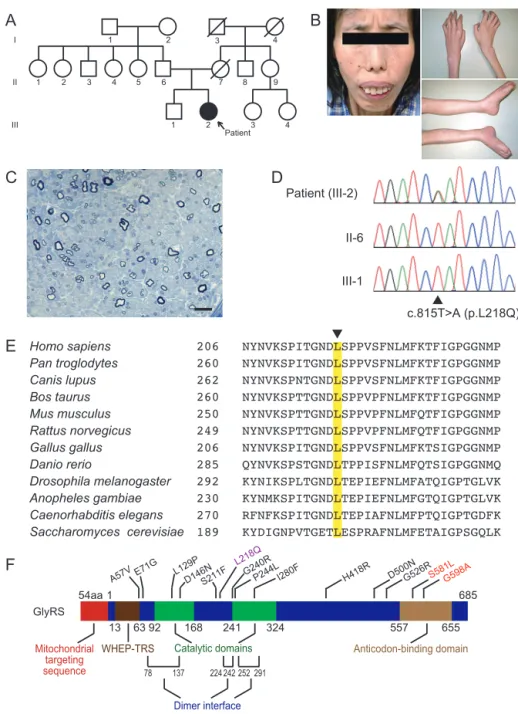
Fig. 1 Clinical, pathological and molecular features of the patient.
(A) Pedigree. (B) Facial involvement with weakness of the orbicularis oris and atrophy of the temporalis and masseter muscles. The patient was instructed to close her mouth. Limbs showed severe muscle atrophy. (C) The sural nerve biopsy at age 29 showed moderate loss of myelinated fibers. Axonal degeneration and active demyelination were not evident. Bar = 20 mm. (D) Chromatogram of the heterozygous c.815T>A (p.L218Q) mutation in exon 7 of GARS; the patient and two unaffected relatives. (E) Comparison of GlyRS from different species. Arrowhead on top of the align-ment indicates amino acid position 218 (Note: numbering differences from related species are because the human annotation does not consider the N-terminal mitochondrial targeting sequence appended through alternative start codon usage). (F) The GlyRS protein contains four functional domains and three dimer interface regions. Mutations identified in GlyRS are distributed across the entire protein; modified from Motley, et al13). L218Q, the mutation found in our patient is shown in purple. It is located in an apparently non-functional region. In contrast, both of two other known mutations that cause early onset and severe clinical phenotypes, shown in red, are located in an anticodon-binding domain.
The GlyRS protein comprises four functional domains and three dimer interface regions13) (Fig. 1F). (Note: numbering of residues starts from the alternative start codon after MTS in human protein). Among 13 reported GARS mutations7)~10)14), two mutations caused early-onset clinical phenotypes in four patients. One patient developed facial and respiratory muscle involvement7), and another developed vocal cord dysfunction14). Both mutations are located in an anticodon-binding domain. In contrast, the mutation described in the current study was located in neither of the functional domains. Even so, we still consider this L218Q mutation a pathogenic mutation based on the following reasons: 1) its close location to the dimer interface region; 2) the high conservation of the affected amino acid; and 3) the fact that neither the unaffected parent nor the unaffected brother carried this mutation. In silico prediction using MUPro and Polyphen-2 suggests pathogenicity of the mutation, but the results from other reported mutations using these algorithms do not necessarily correlate with clinical severity (Table 1) and this approach may not be suitable as far as this gene is concerned.
Mechanisms underlying CMT2D/dSMA-V caused by GARS mutations have been examined from various aspects, including enzyme activity, protein stability and dimerization, but those properties considerably depend on individual mutations and none of these approaches reached consistent results. Moreover, heterozygous mice with a single loss-of-function GARS allele
exhibited reduced synthetase activity but none of the symptoms of CMT15) and overexpression of wild-type GlyRS could not rescue the neuropathy phenotype in mouse models16). These experi-mental results, together with the observations of scattered loca-tions of the mutaloca-tions throughout the gene and the dominant inheritance pattern lead to a consequence that not a simple loss-of-function, but rather a gain-of-function mechanism significantly contributes to the pathogenesis of the disease5)6)13). Recent study analyzing the tertiary structure of GlyRS using hydrogen- deuterium exchange revealed that all five mutations tested promote the same localized conformational opening17). All other mutations untested are also within the opened-up areas, except for some mutations which are not covered in that analysis. They argued that those opened-up areas provide unique surfaces for potential novel interactions that lead to pathological conse-quences. The mutation of our case is also within the “opened-up areas” and that may account for the pathogenicity.
Although both loss-of-function and gain-of-function mechanisms were likely to synergistically give rise to severe phenotypes in the previous cases with mutations in an anticodon-binding domain, gain-of-function predominantly appears to have led to severe phenotypes in our case. Data from this unique case pro-vided new information for understanding the mechanism of CMT2D/dSMA-V and for drug discovery as well.
Method 1 Method 2 Rohkamm, et al. (2007)8)
WHEP-TRS A57V -0.13 -0.76 0.439
Antonellis, et al. (2003)4) E71G -0.76 -0.98 0.788
Antonellis, et al. (2003)4) Catalytic-1 L129P -1.00 -1.00 1.000 Lee, et al. (2012)9) D146N -0.79 -0.86 1.000 Lee, et al. (2012)9) S211F 0.19 0.55 1.000 Presented case L218Q* -1.00 -0.96 1.000 Antonellis, et al. (2003)4) G240R 0.30 0.68 1.000 Abe, et al. (2009)10) Catalytic-2 P244L 0.25 0.67 1.000
James, et al. (2006)7) I280F -1.00 -1.00 1.000
Sivakumar, et al. (2005)11) H418R 0.55 0.73 0.998
Del Bo, et al. (2006)12) D500N -0.53 -0.79 0.048
Antonellis, et al. (2003)4) G526R 0.01 -0.51 1.000
James, et al. (2006)7)
Anticodon-binding S581L* 0.15 0.80 0.420
James, et al. (2006)7); Eskuri, et al. (2012)14) G598A* 0.86 0.82 0.013 Asterisks indicate mutations associated with severe phenotypes. MUPro scores range between -1 and 1. A score less than 0 means that the mutation decreases the protein stability, and vice versa. A larger absolute value indicates more confident prediction. Polyphen-2 scores range between 0 and 1. A larger score indicates that the mutation is more pathogenic. Underlines indicate high scores, predicting unstability and pathogenicity of the mutated proteins.
The patient and family members included in this study gave written informed consent, and the study was approved by the Kyoto University and the Institutional Review Board of Kagoshima University.
Abstract of this work was presented at the 98th Kinki Regional Meeting of the Japanese Society of Neurology and recommended by the conference chairperson for the publication to Rinsho Shinkeigaku.
Acknowledgment: The authors wish to thank the patient and the family described in this report for their cooperation, Dr. Y. Takahashi for sample preparation and study coordination, Dr. S Nakano for the pathological analysis of the muscle biopsy and Dr. Y. Higuchi and Dr. A. Hashiguchi for the molecular analysis.
※ The authors declare there is no conflict of interest relevant to this article.
References
1) Wilmshurst JM, Ouvrier R. Hereditary peripheral neuropathies of childhood: an overview for clinicians. Neuromuscul Disord 2011;21:763-775.
2) Takashima H. Genetic diagnosis and molecular pathology of inherited neuropathy. Rinsho Shinkeigaku 2012;52:399-404. 3) Zhao Z, Hashiguchi A, Hu J, et al. Alanyl-tRNA synthetase
mutation in a family with dominant distal hereditary motor neuropathy. Neurology 2012;78:1644-1649.
4) Antonellis A, Ellsworth RE, Sambuughin N, et al. Glycyl tRNA synthetase mutations in Charcot-Marie-Tooth disease type 2D and distal spinal muscular atrophy type V. Am J Hum Genet 2003;72:1293-1299.
5) Guo M, Schimmel P. Essential nontranslational functions of tRNA synthetases. Nat Chem Biol 2013;9:145-153.
6) Yao P, Fox PL. Aminoacyl-tRNA synthetases in medicine and disease. EMBO Mol Med 2013;5:332-343.
7) James PA, Cader MZ, Muntoni F, et al. Severe childhood SMA and axonal CMT due to anticodon binding domain mutations
in the GARS gene. Neurology 2006;67:1710-1712.
8) Rohkamm B, Reilly MM, Lochmüller H, et al. Further evidence for genetic heterogeneity of distal HMN type V, CMT2 with predominant hand involvement and Silver syndrome. J Neurol Sci 2007;263:100-106.
9) Lee HJ, Park J, Nakhro K, et al. Two novel mutations of GARS in Korean families with distal hereditary motor neuropathy type V. J Peripher Nerv Syst 2012;17:418-421.
10) Abe A, Numakura C, Saito K, et al. Neurofilament light chain polypeptide gene mutations in Charcot-Marie-Tooth disease: nonsense mutation probably causes a recessive phenotype. J Hum Genet 2009;54:94-97.
11) Sivakumar K, Kyriakides T, Puls I, et al. Phenotypic spectrum of disorders associated with glycyl-tRNA synthetase mutations. Brain 2005;128:2304-2314.
12) Del Bo R, Locatelli F, Corti S, et al. Coexistence of CMT-2D and distal SMA-V phenotypes in an Italian family with a GARS gene mutation. Neurology 2006;66:752-754.
13) Motley WW, Talbot K, Fischbeck KH. GARS axonopathy: not every neuron’s cup of tRNA. Trends Neurosci 2010;33:59-66. 14) Eskuri JM, Stanley CM, Moore SA, et al. Infantile onset
CMT2D/dSMA V in monozygotic twins due to a mutation in the anticodon-binding domain of GARS. J Peripher Nerv Syst 2012; 17:132-134.
15) Seburn KL, Nangle LA, Cox GA, et al. An active dominant mutation of glycyl-tRNA synthetase causes neuropathy in a Charcot-Marie-Tooth 2D mouse model. Neuron 2006;51:715-726.
16) Motley WW, Seburn KL, Nawaz MH, et al. Charcot-Marie-Tooth-linked mutant GARS is toxic to peripheral neurons independent of wild-type GARS levels. PLoS Genet 2011;7: e1002399.
17) He W, Zhang HM, Chong YE, et al. Dispersed disease-causing neomorphic mutations on a single protein promote the same localized conformational opening. Proc Nat Acad Sci USA 2011;108:12307-12312.