Abbreviations: DMEM, Dulbecco’s modified Eagle’s medium; HNSCC, head and neck squamous cell carcinoma; LOI, loss of imprinting; PEG1/MEST, paternally expressed gene 1/mesoderm-specific transcript; RFLP, restriction fragment length polymor-phism; RT, reverse transcriptase; SNP, single nucleotide polymorphism
Allele-Specific Expression Analysis of PEG1/MEST
in Head and Neck Squamous Cell Carcinomas
Hideyuki Kataoka, Seiji Nakano*, Yasuomi Kunimoto, Naoko Sugie*, Mitsuhiko Osaki*, Narikazu Uzawa†, Mitsuaki A. Yoshida‡, Mitsuo Oshimura* and Hiroya Kitano Division of Otolaryngology, Head and Neck Surgery, Department of Medicine of Sensory and Mo-tor Organs, School of Medicine, TotMo-tori University Faculty of Medicine, Yonago 683-8503, *Division of Molecular Genetics and Biofunction, Department of Biomedical Science, Institute of Regenerative Medicine and Biofunction, Graduate School of Medical Science, Tottori University, Yonago 683-8503, †Maxillofacial Surgery, Maxillofacial Reconstruction and Function, Division of Maxillofacial and Neck Reconstruction, Graduate School, Tokyo Medical and Dental University, Bunkyo-ku 113-8510 and ‡Biodosimetry Section, Department of Radiation Dosimetry, Research Center for Radiation Emer-gency Medicine, National Institute of Radiological Sciences, Chiba 263-8555 Japan
Genomic imprinting is an epigenetic feature that plays a significant role in carcinogen-esis. In this study, we examined the expression status of an imprinted gene, paternally ex-pressed gene 1/mesoderm-specific transcript (PEG1/MEST), in 38 cases of head and neck squamous cell carcinomas (HNSCCs) and in 17 oral squamous cancer cell lines. Loss of imprinting (LOI) of PEG1/MEST was found in 8 of 10 (80%) in tumor specimens, and 6 of 10 (60%) informative cases even in the extracted normal tissue specimens. As for the oral squamous cancer cell lines, LOI was detected in 5 of 8 (62.5%) informative cases in PEG1/ MEST. Thus, these data showed that abnormal expression of PEG1/MEST was found at a high frequency in the tumor, the extracted normal tissue specimens and the oral squamous cancer cell lines. PEG1/MEST LOI in extracted normal tissue specimens may have a po-tential individual cancer risk for HNSCC.
Key words: head and neck squamous cell carcinoma; loss of imprinting; PEG1/MEST
Head and neck cancers are almost always squamous cell types arising in the oral cavity, tongue, pharynx and larynx. Understanding the molecular mecha-nisms involved in tumor development and progres-sion has enabled the design of new biological ap-proaches. This study focuses on molecular studies on the roles of genomic imprinting. Almost all im-printed genes identified to date can be classified as regulators of embryonic growth, placental growth or adult metabolism (Morison et al., 2005). Mono-allelic expression of either the maternal or paternal
copy occurs while the other copy is silenced in nor-mal somatic tissue specimens. Imprinted genes are often clustered in chromosomal domains and are thought to be coordinately regulated by imprinting control centers. More than 60 types of imprinted genes have been identified in humans (Delaval and Feil, 2004).
Mutations that affect the epigenetic states of imprinted domains underlie a number of human diseases (Walter and Paulsen, 2003). Loss of im-printing (LOI) is one of the most frequent alterations
found in cancers and involves abnormal activation of a normally silent allele. Jaenisch and colleagues have demonstrated that global LOI leads to tumor formation in chimeric mice. All the tumors in the mice were derived from non-imprinted cells. Most importantly, tumors were not seen in the offspring of the chimaeras, as imprinting is reset during gameto-genesis (Goymer, 2005).
Imprinting is therefore an epigenetic tumor-suppressing phenomenon. When imprinting is lost, cells are immortalized through the inappropriate regulation of both tumor suppressors and oncogenes (Holm et al., 2005). The genes exhibiting LOI in human cancer leading to biallelic expression has been reported in a variety of tumors including head and neck squamous cell carcinomas (HNSCCs) (Schofield et al., 2001; Feinberg and Tycko, 2004). Previous studies regarding imprinted genes in HNSCC suggested that LOI of IGF2, H19 and
p57KIP2 loci play an important role in oncogenesis
(el-Naggar et al., 1999; Lai et al., 2000; Rainho et al., 2001). However, the involvement of other imprinted genes is still poorly understood in HNSCC.
Paternally expressed gene 1/mesoderm-specific transcript (PEG1/MEST) is a paternally expressed gene located on human chromosome 7q32 (Kobayashi et al., 1997). PEG1/MEST LOI has been reported in lung cancers (Suda et al., 2003; Nakanishi et al., 2004), breast cancers (Pedersen et al., 1999, 2002) and colorectal cancers (Nishihara et al., 2000). In these previous studies, it was postulated that the disruption of PEG1/MEST might be impli-cated in the etiology of these cancers. In this study, we examined the imprinting status of PEG1/MEST in HNSCC specimens and oral squamous cancer cell lines with special attention to the role of PEG1/MEST LOI on oncogenesis in HNSCC tissue specimens and several oral squamous cancer cell lines.
Materials and Methods
Tissue samples
HNSCCs, diagnosed by histopathologic
examina-tion and matched with extracted normal tissue specimens from 38 patients, were analyzed in this study. They consisted of 16 oral cancers, 4 oropha-ryngeal cancers, 5 hypophaoropha-ryngeal cancers and 13 laryngeal cancers. All tissue specimens were obtained during surgery at the Clinical Depart-ment of Otolaryngology, Head and Neck Surgery, Tottori University Hospital. Each resected tumor was carefully trimmed to remove normal tissue. Then extracted normal tissue was selected as far from the cancerous areas as possible. We usually picked extracted normal tissue from the muscle, which was removed when neck dissections were performed. Informed consent to participate in this study was obtained from each patient. The institu-tional review board of Tottori University approved this study (approval number 746). All tissue speci-mens were stored at –80°C until analysis.
Cell lines
Seventeen oral squamous cancer cell lines were analyzed. HSC-2, HSC-3 and HSC-4 were es-tablished and provided by Uzawa and associates (Uzawa et al., 1995). HO-1-N-1, HO-1-u-1, KON, KOSC-2 cl3-43, OSC-19, OSC-20, SCC-4 and SKN-3 were obtained from the Japanese Collection of Research Bioresources (Osaka, Japan). SAS were obtained from the Cell Resource Center for Biomedical Research, Tohoku University (Sendai, Japan). Eight oral squamous cancer cell lines (2, 3, 4, 5, 6, HSC-7, KON and SCCKN) were grown in Dulbecco’s modified Eagle’s medium (DMEM) with 10% fe-tal bovine serum. Two (HO-1-N-1 and HO-1-u-1) were grown in DMEM-F12 medium (1:1 mix) with 10% fetal bovine serum. Two (19 and OSC-20) were grown in DMEM-F12 medium (1:1 mix) without serum. SCC-4 was grown in DMEM-F12 medium (1:1 mix) with 10% fetal bovine serum and 0.4 µg/mL hydrocortisone. Four (KOSC-2 cl3-43, SAT, SAS and SKN-3) were grown in RPMI 1640 medium with 10% fetal bovine serum. These cells were cultured and growing cells with 70% conflu-ence were harvested for analyses.
DNA and RNA sample preparation
Genomic DNA was isolated using a Puregene DNA Isolation Kit (Gentra Systems, Minneapolis, MN). Total RNA was isolated using an RNeasy Kit (QIAGEN, Valencia, CA) according to the manufacturer’s instructions. Total RNA was treated with RNase-free DNase I (Takara Bio, Shiga, Japan) to remove any DNA contamination. First-strand cDNA synthesis was carried out with or without SuperScript III Reverse Transcriptase (RT+ or –) (Invitrogen, Carlsbad, CA) by using an oligo (dT)15 primer (Roche, Basel, Switzerland).
The sequences of the primers were 5-CACTGAT-GCAGAAAGACGTTC-3 and 5-CAGCACCAT TTGCTCATAGG-3.
Identification of polymorphisms
Imprinting analysis for PEG1/MEST was car-ried out by single nucleotide polymorphism (SNP) analysis for informative heterozygotes at the G/A (exon 12; rs10863) polymorphism. An informative DNA polymorphism at an AflIII site was identified as the AflIII-digestible (ACACGT) or indigestible (ACACAT) sequence. PCR was performed for 30 cycles including denaturation at 95˚C for 30 s, an-nealing at 62˚C for 30 s and extension at 72˚C for 30 s.
Allele-specific expression analysis
Allele-specific expression analysis with RT-PCR and digestion by restriction enzymes was per-formed under the same conditions as those used to detect genomic polymorphisms. To confirm the results of restriction fragment length polymorphism (RFLP), we also performed a method for allele-specific expression analysis of imprinted genes by real-time PCR with Cycling Probe Technology (Takara Bio). In this method, one end of the probe is labeled with a fluorescent substance and the other end is labeled with a quencher, which quenches the fluorescence emitted from the substance. RNaseH specificially cuts the RNA region of this probe, resulting in emission of strong fluorescence, but it does not cut the RNA probe region including any mismatches.
Results
We analyzed 38 primary HNSCCs for the LOI in
PEG1/MEST. First of all we identified genomic polymorphisms using RFLP analysis. Informative cases were then further analyzed to determine their allele-specific expression. As shown in Figs. 1 and 2, we distinguished the 2 alleles and determined whether a case is informative or not. This poly-morphism (G/A; rs10863) results in the presence
Fig. 1.
A: A schematic of the AflIII polymorphism of human PEG1/MEST is shown. The arrow indicates the site of the polymor-phism. PCR products contain this polymorphic site. A-a represents the undigested product (792 bp) and A-b represents product digested with AflIII (383 and 409 bp). PEG1/MEST, paternally expressed gene 1/mesoderm-specific transcript. B: Heterozygosity called informative is necessary for allele-specific analysis.
Extracted specimen
Genome Normal Tumor
+ – + –
Case 1 Case 4
Imprinted LOI LOI LOI
792 409 383 792 409 383 Extracted specimen
Genome Normal Tumor
+ – + –
(bp) (bp)
or absence of an AflIII site. The 3 different bands (792, 409 and 383 bp) after AflIII digestion of the genomic PCR fragment indicated an informative heterozygous case. Next, to determine the expres-sion status of PEG1/MEST, the RT-PCR products are digested with AflIII to determine whether one or both copies are expressed. The presence of 3 bands indicates biallelic expression, that is, PEG1/
MEST LOI. On the other hand, a single band of 792 bp or 2 bands of 409 bp and 383 bp showed monoallelic expression, indicating the normal im-printing status of PEG1/MEST. Among the cases screened, we observed 10 informative cases of
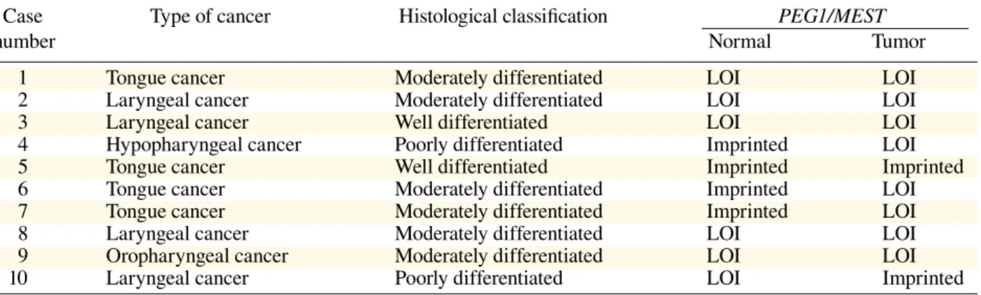
PEG1/MEST. PEG1/MEST LOI was found in 8 informative cases (80%), and in 6 cases (60%) it was detected even in the normal tissue specimens (Table 1), regardless of tumor location or histology.
These data suggest that PEG1/MEST LOI occurs in HNSCC specimens, suggesting its fundamental role in HNSCC oncogenesis.
Next, we analyzed the imprinting status of
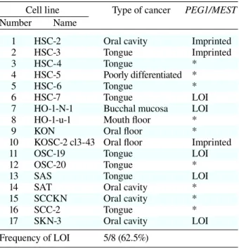
PEG1/MEST in 17 oral squamous cancer cell lines to substantiate the results from the tissue specimens. The results for the histopathologic types of these oral squamous cancer cell lines are summarized in Table 2. PEG1/MEST LOI was observed in 5 of 8 informative cases (62.5%). The results from the cell lines are summarized in Table 2, which were con-sistent with the high frequency of LOI at the PEG1/
MEST loci detected in the HNSCC tissue speci-mens. Thus, abnormal expression of PEG1/MEST was found at a high frequency in the cancer tissue specimens, the extracted normal tissue specimens and the oral squamous cancer cell lines.
Fig. 2. Allele-specific expression analysis of PEG1/MEST in head and neck squamous cell carcinoma tissue specimens. The allele-specific expressions of PEG1/MEST were assessed by restriction fragment length polymorphism analysis. Representa-tive results of PEG1/MEST are shown. +, presence of reverse transcriptase (RT); –, absence of RT; LOI, loss of imprinting;
PEG1/MEST, paternally expressed gene 1/mesoderm-specific transcript.
Table 1. Summary of PEG1/MEST gene expression profiles in informative cases of tissue speci-mens of head and neck squamous cell carcinoma
Case Type of cancer Histological classification PEG1/MEST
number Normal Tumor
1 Tongue cancer Moderately differentiated LOI LOI
2 Laryngeal cancer Moderately differentiated LOI LOI
3 Laryngeal cancer Well differentiated LOI LOI
4 Hypopharyngeal cancer Poorly differentiated Imprinted LOI
5 Tongue cancer Well differentiated Imprinted Imprinted
6 Tongue cancer Moderately differentiated Imprinted LOI
7 Tongue cancer Moderately differentiated Imprinted LOI
8 Laryngeal cancer Moderately differentiated LOI LOI
9 Oropharyngeal cancer Moderately differentiated LOI LOI
Discussion
Several lines of evidence suggest that the disrup-tion of imprinting mechanisms play a critical role in oncogenesis including HNSCC, but none have examined PEG1/MEST in HNSCC. The data from mice studies of Mest have suggested a possible role of Mest as a regulator of embryonic growth.
Mest paternal knockouts have been shown to result in an imprinted phenotype characterized by fetal and placental growth retardation (Lefebvre et al., 1998). In our report, we provide the first evidence regarding the presence of biallelic expression of
PEG1/MEST in HNSCC. PEG1/MEST LOI was identified 8 out of 10 cases in HNSCC tissue speci-mens and 5 out of 8 cases in oral cancer cell lines. Interestingly, PEG1/MEST LOI was observed in the extracted normal tissue surrounding the tumor specimens.
IGF2 and PEG1/MEST are known as im-printed genes important in fetal growth. The cor-relation between IGF2 LOI in normal cells and colorectal cancer has been well documented (Cui et al., 2003; Nakano et al., 2006). IGF2 LOI has been found in 10% of normal individuals (Sakatani et al., 2001) and it appears to be 5 times more common in patients with a family history of colon carcinoma, and 21 times more common in patients with a personal history of colorectal neoplasia (Cui et al., 2003). Moreover, a mouse model with Igf2 LOI suggests that mice with abnormal imprinting acquired twice as many intestinal adenomas as those whose imprinting was normal (Sakatani et al., 2005). These studies indicated that IGF2 LOI might be a valuable predictive marker of an indi-vidual’s risk for colorectal cancer.
PEG1/MEST LOI in both extracted normal and tumor tissue specimens suggests that the detec-tion of PEG1/MEST LOI in extracted normal tissue specimens may also pose a potential cancer risk in individuals for HNSCC. IGF2 LOI has been found in 10% of normal individuals. However, there are no reports indicating the incidence of PEG1/MEST LOI in normal individuals. It will be necessary to
clarify the incidence of PEG1/MEST LOI in nor-mal individuals in order to elucidate its predictive value for an individual’s cancer risk for HNSCC.
References
1 Cui H, Cruz-Correa M, Giardiello FM, Hutcheon DF, Kafonek DR, Brandenburg S, et al. Loss of IGF2 im-printing: a potential marker of colorectal cancer risk. Science 2003; 299:1753–1755.
2 Delaval K, Feil R. Epigenetic regulation of mammalian genomic imprinting. Curr Opin Genet Dev 2004;14: 188–195.
3 el-Naggar AK, Lai S, Tucker SA, Clayman GL, Goepfert H, Hong WK, et al. Frequent loss of im-printing at the IGF2 and H19 genes in head and neck squamous carcinoma. Oncogene 1999;18:7063–7069. 4 Feinberg AP, Tycko B. The history of cancer
epigenet-ics. Nat Rev Cancer 2004;4:143–153.
5 Goymer P. Cancer epigenetics: dangerous unmarked genes. Nature Reviews Genetics 2005; 6:878-878. 6 Holm TM, Jackson-Grusby L, Brambrink T, Yamada Y,
Rideout WM 3rd, Jaenisch R. Global loss of imprinting leads to widespread tumorigenesis in adult mice. Can-cer Cell 2005; 8:275-285.
Table 2. Summary of allele-specific expression of PEG1/MEST in 17 oral squamous cancer cell lines
Cell line Type of cancer PEG1/MEST
Number Name
1 HSC-2 Oral cavity Imprinted
2 HSC-3 Tongue Imprinted
3 HSC-4 Tongue *
4 HSC-5 Poorly differentiated *
5 HSC-6 Tongue *
6 HSC-7 Tongue LOI
7 HO-1-N-1 Bucchal mucosa LOI 8 HO-1-u-1 Mouth floor *
9 KON Oral floor *
10 KOSC-2 cl3-43 Oral floor Imprinted
11 OSC-19 Tongue LOI
12 OSC-20 Tongue *
13 SAS Tongue LOI
14 SAT Oral cavity *
15 SCCKN Oral cavity *
16 SCC-2 Tongue *
17 SKN-3 Oral cavity LOI
Frequency of LOI 5/8 (62.5%) * Non-informative case.
LOI, loss of imprinting; PEG1/MEST, paternally expressed gene 1/mesoderm-specific transcript.
7 Kobayashi S, Kohda T, Miyoshi N, Kuroiwa Y, Aisaka K, Tsutsumi O, et al. Human PEG1/MEST, an imprinted gene on chromosome 7. Hum Mol Genet 1997;6:781– 786.
8 Lai S, Goepfert H, Gillenwater AM, Luna MA, El-Naggar AK. Loss of imprinting and genetic altera-tions of the cyclin-dependent kinase inhibitor p57KIP2 gene in head and neck squamous cell carcinoma. Clin Cancer Res 2000;6:3172–3176.
9 Lefebvre L, Viville S, Barton SC, Ishino F, Keverne EB, Surani MA. Abnormal maternal behaviour and growth retardation associated with loss of the imprinted gene
Mest. Nat Genet 1998;20:163–169.
10 Morison IM, Ramsay JP, Spencer HG. A census of mammalian imprinting. Trends Genet 2005; 21:457– 465.
11 Nakanishi H, Suda T, Katoh M, Watanabe A, Igishi T, Kodani M, et al. Loss of imprinting of PEG1/MEST in lung cancer cell lines. Oncol Rep 2004;12:1273–1278. 12 Nakano S, Murakami K, Meguro M, Soejima H,
Higashimoto K, Urano T, et al. Expression profile of LIT1/KCNQ1OT1 and epigenetic status at the KvDMR1 in colorectal cancers. Cancer Sci 2006;97:1147–1154. 13 Nishihara S, Hayashida T, Mitsuya K, Schulz TC,
Ikeguchi M, Kaibara N, et al. Multipoint imprinting analysis in sporadic colorectal cancers with and without microsatellite instability. Int J Oncol 2000;17:317–322. 14 Pedersen IS, Dervan PA, Broderick D, Harrison M,
Miller N, Delany E, et al. Frequent loss of imprinting of PEG1/MEST in invasive breast cancer. Cancer Res 1999;59:5449–5451.
15 Pedersen IS, Dervan P, McGoldrick A, Harrison M, Ponchel F, Speirs V, et al. Promoter switch: a novel mechanism causing biallelic PEG1/MEST expression in
invasive breast cancer. Hum Mol Genet 2002;11:1449– 1453.
16 Rainho CA, Kowalski LP, Rogatto SR. Loss of imprint-ing and loss of heterozygosity on 11p15.5 in head and neck squamous cell carcinomas. Head Neck 2001;23: 851–859.
17 Sakatani T, Kaneda A, Iacobuzio-Donahue CA, Carter MG, de Boom Witzel S, Okano H, et al. Loss of im-printing of Igf2 alters intestinal maturation and tumori-genesis in mice. Science 2005;307:1976–1978.
18 Sakatani T, Wei M, Katoh M, Okita C, Wada D, Mitsuya K, et al. Epigenetic heterogeneity at imprinted loci in normal populations. Biochem Biophys Res Commun 2001;283:1124–1130.
19 Schofield PN, Joyce JA, Lam WK, Grandjean V, Ferguson-Smith A, Reik W, et al. Genomic imprinting and cancer: new paradigms in the genetics of neoplasia. Toxicol Lett 2001;120:151–160.
20 Suda T, Katoh M, Hiratsuka M, Fujiwara M, Irizawa Y, Oshimura M. Use of real-time RT-PCR for the detec-tion of allelic expression of an imprinted gene. Int J Mol Med 2003;12:243–246.
21 Uzawa N, Yoshida MA, Oshimura M, Ikeuchi T. Sup-pression of tumorigenicity in three different cell lines of human oral squamous cell carcinoma by introduction of chromosome 3p via microcell-mediated chromosome transfer. Oncogene 1995;11:1997–2004.
22 Walter J, Paulsen M. Imprinting and disease. Semin Cell Dev Biol 2003;14:101–110.
Received March 25, 2009; accepted May 20, 2009 Corresponding author: Hideyuki Kataoka, MD