Original Paper
Treatment of M alignant Glioma in the Basal Ganglia
with Hyperthermia and Radiotherapy
HIDEAKI TAKAHASHI , SEIICHI YOSHIDA , TAKEO UZUKA ,
YUKIHIKO FUJII
Niigata Cancer Center, Section of Neurosurgery, 2-15-3 Kawagishi-cho, Niigata 951-8566, Japan
Niigata University, Brain Research Institute, Department of Neurosurgery, 1-757 Asahimachi-dori, Niigata 951-8585, Japan
Abstract : We staged the Malignant gliomas in the thalamus and basal ganglia seen at one institution over a 20-year period were staged, and the clinical features of hyperthermic treatment in these site were examined.
The subjects in this study were inpatients who received treatments at Niigata University. There were 43 patients with grade 3 or 4 glioma in the basal ganglia (24 patients in the thalamus and 19 patients in the putamen). For staging,tumors in the thalamus and putamen were classified by their sizes: less than 2 cm, 2-4 cm, and greater than 4 cm were classified as stages 1 to 3. Tumors which progressed outside of the basal ganglia were classified as stage 4,and those which progressed to the ventricle were classified as stage 5.
The percentage of patients in stage 4 whose tumor invaded to the internal capsule or midbrain was 30%, and this percentage reached 40% when combined with patients in stage 5. Resection was performed in 14 patients. Tumors of the 29 remaining patients were biopsied. In 14 of the biopsied cases,needle electrodes were implanted in the tumors,and interstitial hyperthermia was performed. The electrodes (RF antennas) were stereotactically implanted in the tumor under local anesthesia simultaneously with biopsy. The heating strategy used was to control the RF output in order to heat the rim of the tumor up to a temperature of 43℃ for a 40-60 minute heating period. The heating procedure was repeated twice a week for a total of 3 to 4 times,and combined with conventional radiation therapy with a dose of 50-60 Gy, and MCNU or ACNU chemotherapy.
The Median Survival Time(MST) for overall survival was 11 months(thalamic cases: 17 months; putamen cases: 11 months). The MST for cases receiving hyperthermia was 22 months, and for cases which did not receive hyperthermia, the MST was 9 months.
Few malignant gliomas in the basal ganglia were removed with surgery because of their localization. In more than a few cases, local treatments were difficult and the prognosis was poor. However, there were responsive cases among the patients who underwent hyperthermia.
Key Words : glioma, basal ganglia, hyperthermia, stereobiopsy
Received 12 March 2007, Accepted 23 April 2007. Corresponding author; Tel, +81-25-266-5111; Fax, +81-25-266-9385; e-mail, takahash-nii@umin.ac.jp
Introduction
Basal ganglia tumors are,narrowly defined,those located in the putamen or caudate nucleus. Such basal ganglia tumors and tumors in the thalamus are deep seated and near the internal capsule. In many instances, such tumors in these functionally important areas are collectively called basal ganglia tumors . Many patients with glioma in these sites receive treatments which focus on radiotherapy after biopsies . In addition,since the frequency of basal ganglia tumors is not high in a given institution, accurate clinical features have not been defined . In the retrospective study presented below,features of malignant glioma in the basal ganglia were analyzed and staged, and the effects of hyperthermia treatment, (including in the thalamus) were examined over a 20-year period at one institution.
Methods
The subjects examined in this study were 43 patients with grades 3 or 4 glioma in the basal ganglia, and who were inpatients who underwent surgery at Niigata University Hospital from 1983 to 2004. Of these 43 patients,24 were diagnosed with glioma in the thalamus,and 19 with glioma in the putamen or caudate nucleus (these two areas will be referred to here as the putamen). There were 27 males and 16 females. The subjects were between 18 and 81 years old with a mean age of 53.6 years. The tumor was located on the left side in 25 patients and on the right side in 18 patients. Histologically, 14 patients were grade 3, and 29 patients were grade 4.
The patients were staged from 1 to 5 based on the staging system used in the Nomura Group : Establishment of Standard Treatments for the Prognostic Improvement of Malignant Glioma published by the Ministry of Health, Labor and Welfare. A tumor with a size of less than 2 cm was classified as stage 1,2-4 cm as stage 2,and over 4 cm as stage 3. A tumor was stage 4 if it had progressed outside of the thalamus,putamen,or caudate nucleus,and had reached the internal capsule or midbrain. A tumor was classified as stage 5 if it extended into the ventricle or it was disseminated (Fig. 1).
This departments treatment policies for malignant glioma are to promptly schedule a resection and chemotherapy using ACNU or MCNU via intravenous infusion, in addition to a postoperative conventional external-beam radiation exposure of 60 Gy. However, a stereotactic biopsy is performed in cases in which resection is difficult. After obtaining patient consent,radiotherapy and hyperthermia were used with some patients. These procedures were performed subsequent to obtaining biopsies and
Fig.1. Staging of malignant glioma in the basal ganglia. Stage 1: tumor of less than 2 cm in the basal ganglia; Stage 2: tumor of 2-4 cm in the basal ganglia; Stage 3: tumor of more than 4 cm in the basal ganglia; Stage 4: tumor extending to the internal capsule or midbrain ; Stage 5: dissemination.
placing intracranial electrodes into the tumor for interstitial hyperthermia . Electrodes (RF antennas) were inserted into the malignant glioma (a contrast-enhanced lesion on the CT scan) using a CT-guide stereotactic technique under local anesthesia. Thermosensor catheters were also inserted stereotactically in two dimensions. The temperature of the brain and the tumor along the catheters was continuously monitored with the thermocouple sensors in the catheter during heating. The heating strategy used with this system was to control the RF output in order to heat the rim of the tumor up to 43℃ for a 40-60 minute heating period. In clinical practice, a single RF antenna can effectively heat small tumor with a diameter of less than 3 cm. Heating was repeated twice a week for a total of 3 to 4 times,and combined with conventional radiation therapy with a dose of 50-60 Gy(except in 2 cases)and MCNU or ACNU chemotherapy(except in aged patients). As a general rule, chemotherapy was not used for patients who were 70 years old or older.
To determine survival rates,the median survival times(MSTs)were calculated using Kaplan-Meyer curves, and the log rank test was performed for comparisons.
Results
Clinical features
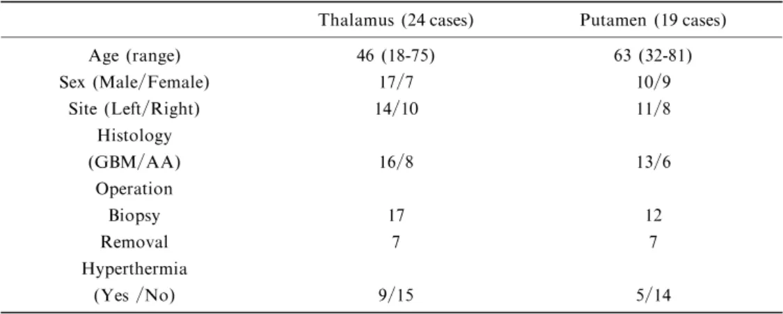
In Table I,patients are divided into 24 thalamic cases and 19 putamen cases,and the number of cases are shown in different categories: age, gender, left or right tumor location, histological diagnosis, surgical method, and whether or not hyperthermia was performed. The distinctive feature is the lower mean age, 46 years old, of the patients with thalamic tumors, indicating a younger age group. The putamen cases were elderly with a mean age of 63 years. In both,the thalamic and putamen groups,the surgical methods used were primarily biopsy,and resections and were performed in only 7 patients from each group. Hyperthermia was indicated and used in 9 patients in the thalamic group and in 5 patients in the putamen group.
Table I. Summary of cases with malignant glioma in the basal ganglia: comparison between the thalamus and putamen.
Thalamus (24 cases) Putamen (19 cases) Age (range) 46 (18-75) 63 (32-81) Sex (Male/Female) 17/7 10/9 Site (Left/Right) 14/10 11/8 Histology (GBM/AA) 16/8 13/6 Operation Biopsy 17 12 Removal 7 7 Hyperthermia (Yes /No) 9/15 5/14 GBM : glioblastoma AA : anaplastic astrocytoma
Staging
Among the 24 thalamic cases,there were 2,6,and 5 patients in stages 1,2,and 3,respectively. There were 7 patients in stage 4 and 3 patients in stage 5,and these stages comprised 42% of the thalamic cases. Among the putamen cases,there were 1,7,and 4 patients in stages 1,2,and 3,respectively. There were 6 patients in stage 4 and 1 patient in stage 5,and these stages comprised 37% of the putamen cases(Table II).
Table III shows the number of hyperthermia patients in the putamen and thalamus case groups at different stages. There were 9 patients treated with hyperthermia in stages 1, 2, 3 and 4 among the thalamic group. Among the putamen tumors,there were only 5 cases treated with hyperthermia in stages 2 and 3.
Table II. Numbers of thalamic and putamen cases at different stages.
Stage1 Stage2 Stage3 Stage4 Stage5
Thalamus (n=24) 2 6 5 7 3
Putamen (n=19) 1 7 4 6 1
Total 3 13 9 13 4
Table III. Numbers of hyperthermia treatment cases in the thalamus and putamen at different stages. Stage 1 Stage 2 Stage 3 Stage 4 Stage 5
Thalamus (n=9) 1 2 3 3 0
Putamen (n=5) 0 3 2 0 0
Total 1 5 5 3 0
Fig.2. Kaplan-Meyer curves of thalamic and putamen cases. The MST for cases of malignant glioma in the thalamus was significantly longer than that for cases of malignant glioma in the putamen.
Survival rate
Fig.2 shows the Kaplan-Meyer survival curves at different sites. The median survival time or MST for overall survival was 11 months. The MST for thalamus cases was 17 months which was significantly longer than that for the putamen cases which was 11 months.
Fig.3 shows survival curves following different treatments. The following findings were made when
Fig.3. Kaplan-Meyer curves of the hyperthermia group and non-hyperthermia group. No significant difference was observed. The MST of the non-hyperthermia group was 9 months while that of the hyperthermia group was 22 months.
(b) (a)
Fig.4. Case 1: 66-year-old male. Stage-3 putamen case,and CT scans(a)at pre-treatment and (b) after thermo-chemo-radiotherapy.
14 hyperthermia cases and 29 non-hyperthermia cases,including resection cases,were compared. There was no statistically significant difference between the MST of cases treated with hyperthermia(22 months) and those not treated with non-hyperthermia (9 months). However, the possibility of a prolonged survival time was indicated for the cases treated with hyperthermia.
Representative cases
Case 1: A 66-year-old male with a stage 3 glioblastoma in the putamen. Stereotactic biopsy and electrode implantation for hyperthermia were performed under local anesthesia. Conventional external-beam radiation with a dose of 60 Gy was then delivered in addition to 3 cycles of hyperthermia and chemotherapy with MCNU infusion into the carotid artery. A complete remission (CR) was obtained according to the CT scans. However,a local recurrence occurred 18 months after hyperthermia treatment, and the patient died 4 months after the local recurrence (Fig. 4).
Case 2: A 45-year-old male with a stage 3 glioblastoma in the thalamus. Resection was performed under general anesthesia. Subtotal resection was performed,and then a 54 Gy external-beam radiation dose was administered,followed by chemotherapy with MCNU via an intravenous infusion. However, dissemination was observed 3 months after the operation, and the patient died from a tumor 6 months after the dissemination was detected (Fig. 5).
Case 3: A 33-year-old male with a stage 1 glioblastoma of the thalamus. Stereotactic biopsy and electrode implantation were performed under local anesthesia. An external-beam radiation dose of 56 Gy was delivered,and hyperthermia,along with chemotherapy with an MCNU intravenous infusion were performed concomitantly. After these treatments, CR was observed on the CT scans. However, a recurrence was observed 29 months after hyperthermia,and the patient died of a tumor 5 months after the recurrence (Fig. 6).
(b) (a)
Fig.5.Case 2: 45-year-old male. Stage-3 thalamic case. After a subtotal resection, radio-chemo-therapy was performed. MRIs at (a) pre-treatment and (b) post-treatment.
Discussion
Malignant gliomas in the basal ganglia and thalamus are deep seated and adjacent to important structures such as the internal capsule. Therefore, a resection approach is difficult, and presents great technical problems . In many cases, radiotherapy and chemotherapy are performed after stereotactic biopsies .
Histological analysis of gliomas in the thalamus shows that many are low grade astrocytomas. However,in the histological data from this institution,it was found that many of patients from this region had high grade gliomas; i.e.glioblastoma and anaplastic astrocytoma, and many of these were seen in young patients, . However, there are few reports in the literature with information about the histology and age distribution of gliomas in the putamen.
There were subjects in this study with low intensity area (LIA) margins with T1WI MRI, and portions of the internal areas were shown in the imaging,suggesting that they may have been WHO grade 2 gliomas. An interesting finding is that many of the thalamic cases were young patients, while many of the putamen cases were elderly patients.
In this institution,needle electrodes,with a 1-mm diameter,were implanted in the tumors and were used for radiofrequency hyperthermia at 13.56 MHz . Resection plays an important role in the treatment of malignant glioma because it reduces the volume of the tumor and alleviates the symptoms of intracranial hypertension. The same considerations apply to a deep-seated tumor. However, symptoms of neurological deficits can result from surgery,and lowering the patients performance status in this way is not acceptable. Heating tumors to temperatures of 43℃ or more is used in hyperthermic therapy, and this treatment performs a role equivalent to debulking in resection surgery . A temperature gradient is created by a needle electrode: the highest temperatures are closest to the electrode in the tumor, and the lower temperatures are most distant from the electrode. Tumors can undergo coagulation necrosis when the margins of the tumor are heated to 43℃ with hyperthermia. Furthermore, the temperature range used from approximately 40-42℃ can relax the blood-brain barrier during the heating process and increase local blood flow, and this is believed to result in an excellent effect when
(c) (b)
(a)
Fig.6. Case 3 : 33-year-old male. Stage-1 thalamic case. MRIs (a) at pre-treatment and (b) after thermo-chemo-radiotherapy.
combined with anticancer agents . In comparison with a simple biopsy, implanting an antenna for hyperthermia an using hyperthermia is useful from the viewpoint that it causes damage or loss of the tumor mass similar to that resulting from surgery. Also, the simplicity of electrode implantation is advantageous, and hyperthermia is considered to be well indicated for a tumor in a site in which direct surgical intervention is difficult. Three representative cases are shown in Figs. 4-6. With tumors in stages 1-3, interstitial hyperthermia can be easily tolerated. Staging to determine indications for hyperthermia is different in the putamen and in the thalamus. Table III shows the numbers of hyperthermia treatment cases in these regions. In stage 4 in the thalamus, extensions of the tumor reaching the midbrain were treated successfully,but a stage 4 tumor in the putamen was not successfully treated with hyperthermia.
The disadvantages of hyperthermia are that it is not indicated for a large tumor since many electrodes would be necessary, and because brain edema can sometimes occur. For cases involving the thalamus and putamen, hyperthermia is not well indicated for a tumor size of 4 cm or more. In such a case, approaching from the surface of the brain becomes easier, and thus, resection is indicated.
For the staging of gliomas in the thalamus and putamen used in this study, the staging system described in Nomura Group : Establishment of Standard Treatment for the Prognostic Improvement of Malignant Glioma published by the Ministry of Health, Labor and Welfare was used. For the definition of a microglioma in this staging system, a stage-1 tumor is less than 1 cm. In this staging system,the sizes of glioma are divided from 1 to 3 cm. However,in a clinical setting,there were very few gliomas seen which were less than 1 cm, and therefore 2 cm was used instead to define stage 1. Hyperthermia can be performed easily on a tumor less than 2 cm, and a resection approach is actually difficult for a tumor this size. When a tumor is over 4 cm,local treatment such as hyperthermia is not well indicated, but the degree of difficulty of a surgical approach is reduced for such sizes. Because a large tumor is reached easily from the brain s surface, a tumor over 4-5 cm is an indication for surgical treatment. At these sites, treatments for a stage 4 case are difficult, and treatment is not indicated in many stage 5 cases.
There are not many cases of malignant glioma in the basal ganglia,and this study only examined a small number of cases. However, an overall picture of malignant glioma in the basal ganglia should emerge from additional studies and case registration of patients, and from additional experience in staging these patients.
The size of this study is too small to define a standard therapy for glioma in the basal ganglia, however,these results indicate that hyperthermia could be an effective treatment modality for unresectable glioma cases located in the basal ganglia. Further studies with large sample sizes will be required to define standard therapies.
References
1) Krouwer H.G.J., Prados M.D.: Infiltrative astrocytomas of the thalamus. J Neurosurg, 82: 548-557, 1995.
2) Nishio S., Morioka T., Suzuki S., Takeshita I., Fukui M., Sogabe T., Tanaka S.: Diencephalic (thalamic and hypothalamic) gliomas in children ― An analysis of 18 cases. Nervous System Children, 21: 320-327, 1996. 3) Steiger H.J., Gotz C., Schmid-Elsaesser R., Stummer W.: Thalamic astrocytomas: surgical anatomy and results of a
pilot series using maximum microsurgical removal. Acta Neurochir (Wien), 142: 1327-1336, 2000.
4) McGirr S.J.,Kelly P.J.,Scheithauer B.W.: Stereotactic resection of juvenile pilocytic astrocytomas of the thalamus and basal ganglia. Neurosurgery, 20: 447-452, 1987.
5) Grigsby P.W.,Garcia D.M.,Ghiselli R.: Analysis of autopsy findings in patients treated with irradiation for thalamic and brain stem tumors. Am J Clin Oncol, 12: 255-258, 1989.
6) Grigsby P.W.,Garcia D.M.,Simpson J.R.,Fineberg B.B.,Schwartz H.G.: Prognostic factors and results of therapy for adult thalamic and brainstem tumors. Cancer, 63: 2124-2129, 1989.
7) Kelly P.J.: Thalamic tumors. Neurosurgeons, 8: 103-114, 1989.
8) Gallina P.,Francescon P.,Cavedon C.,Casamassima F.,Mungai R.,Perrini P.,Russo S.,Di Lorenzo N.: Stereotactic interstitial radiosurgery with a miniature X-ray device in the minimally invasive treatment of selected tumors in the thalamus and the basal ganglia. Stereotact Funct Neurosurg, 79 : 202-213, 2002.
9) Wald S.L., Fogelson H., MacLaurin R.L.: Cystic thalamic gliomas. Child s Brain, 9 : 383-393, 1982.
10) Beks J.W.F., Bouma G.J., Journee H.L.: Tumors of the thalamic region. A retrospective study of 27 cases. Acta Neurochir (Wien), 85: 125-127, 1987.
11) Uzuka T.,Takahashi H.,Morita K.,Grinev I.,Tanaka R.: Intra-arterial chemotherapy during interstitial hyperthermia for malignant glioma: two case reports. Jpn J Hyperthermic Oncol, 20: 87-94, 2004.
12) Takahashi H., Tanaka R., Uzuka T., Grinev I.: Stereotactic technique for radio-frequency antenna implantation for brain tumor treatment ― Technical note and report of complications. Jpn J Hyperthermic Oncol, 21: 221-229, 2005. 13) Kelly P.J.: Stereotactic biopsy and resection of thalamic astrocytomas. Neurosurgery, 25: 185-195, 1989.
14) Franzini A.,Leocata F.,Cajola L.,Servello D.,Allegranza A.,Broggi G.: Low-grade glial tumors in basal ganglia and thalamus: Natural history and biological reappraisal. Neurosurgery, 35: 817-821, 1994.
15) Takahashi H.,Suda T.,Motoyama H.,Uzuka T.,Takahashi S.,Morita K.,Kakinuma K.,Tanaka R.: Radiofrequency interstitial hyperthermia of malignant brain tumors: development of a heating system.Exp Oncol,22: 186-190,2000. 16) Takahashi H., Sugai T., Uzuka T., Kano M., Honma J., Igor G.: Complications and diagnostic yield of stereotactic
biopsy for the patients with malignant brain tumors. No Shinkei Geka, 32: 135-140, 2004. (Japanese).
17) Uzuka T.,Takahashi H.,Tanaka R.: Interstitial hyperthermia with intra-arterial injection of adriamycin for malignant glioma. Neurol Med Chir (Tokyo), 46: 19-23, 2006.
18) Kayama T., Sonoda Y., Sato S., Fujimaki T., Shibui S., Nomura K.: A proposed staging system for glioma surgery. Jpn J Neurosurg (Tokyo), 13: 448-453, 2004.