Efficacy of Neuraminidase Inhibitors against
H5N6 Highly Pathogenic Avian Influenza Virus
in a Nonhuman Primate Model.
著者
NGUYEN Cong Thanh, SUZUKI Saori, ITOH Yasushi,
ISHIGAKI Hirohito, NAKAYAMA Misako, HAYASHI
Kaori, MATSUNO Keita, OKAMATSU Masatoshi,
SAKODA Yoshihiro, KIDA Hiroshi, OGASAWARA
Kazumasa
journal or
publication title
Antimicrobial agents and chemotherapy
volume
64
number
7
year
2020-06-23
URL
http://hdl.handle.net/10422/00012751
Efficacy of neuraminidase inhibitors against H5N6 highly pathogenic avian influenza virus
1
in a non-human primate model
2 3
Running title: NA inhibitors against H5N6 influenza virus in macaques 4
5
Cong Thanh Nguyen1, Saori Suzuki1, Yasushi Itoh1,*, Hirohito Ishigaki1, Misako Nakayama1, 6
Kaori Hayashi1, 2, Keita Matsuno3,4, Masatoshi Okamatsu3, Yoshihiro Sakoda3,4, Hiroshi Kida5 & 7
Kazumasa Ogasawara1,6 8
9
1
Division of Pathogenesis and Disease Regulation, Department of Pathology, Shiga University 10
of Medical Science, Otsu 520-2192, Japan 11
2
Department of Gynecology, Shiga University of Medical Science, Otsu 520-2192, Japan 12
3
Laboratory of Microbiology,Department of Disease Control, Faculty of Veterinary Medicine, 13
Hokkaido University, Sapporo 060-0818, Japan 14
4
Global Station for Zoonosis Control, Global Institution for Collaborative Research and 15
Education (GI-CoRE), Hokkaido University, Sapporo 060-0818, Japan 16
5
Research Center for Zoonosis Control, Hokkaido University, Sapporo 001-0020, Japan 17
6
Research Center for Animal Life Science, Shiga University of Medical Science, Otsu 520-2192, 18 Japan 19 20 * Corresponding author 21
E-mail: [email protected] (YI) 22
Abstract
23Attention has been paid to H5N6 highly pathogenic avian influenza virus (HPAIV) 24
because of its heavy burden on the poultry industry and human mortality. Since an influenza A 25
virus carrying N6 neuraminidase (NA) has never spread in humans, the potential for H5N6 26
HPAIV to cause disease in humans and the efficacy of antiviral drugs against the virus need to 27
be urgently assessed. We used non-human primates to elucidate the pathogenesis of H5N6 28
HPAIV as well as to determine the efficacy of antiviral drugs against the virus. H5N6 HPAIV 29
infection led to high fever in cynomolgus macaques. The lung injury caused by the virus was 30
severe with diffuse alveolar damage and neutrophil infiltration. In addition, an increase in IFN-α 31
showed an inverse correlation with virus titers during the infection process. Oseltamivir was 32
effective for reducing H5N6 HPAIV propagation, and continuous treatment with peramivir 33
reduced virus propagation and severity of symptoms in the early stage. This study also showed 34
the pathologically severe lung injury states in the cynomolgus macaques infected with H5N6 35
HPAIV, even in those that received early antiviral drug treatments, indicating the need for close 36
monitoring and the need for further studies on the virus pathogenicity and new antiviral therapies. 37
38
Introduction
39
Global epidemics of highly pathogenic avian influenza viruses (HPAIVs) have been 40
continuing. The number of subtypes detected in domestic birds and the number of countries 41
affected by HPAIVs are larger than those in past epidemics (2005-2012) (1). By genetic 42
reassortment, new gene combinations of influenza viruses may create a high risk to human health 43
due to an increase of the transmission ability and antiviral drug resistance (2-4). The novel 44
HPAIV of subtype H5N6 that has been detected since 2013 is associated with human mortality 45
and has caused a great burden on the poultry industry (5-7). Nineteen humans were infected with 46
H5N6 HPAIV and 13 of them died (fatality rate of 68.4%) (7). The hemagglutinin (HA) protein 47
of reported H5N6 HPAIVs have both affinity for human-like (α2,6) and avian-like (α2,3) sialic 48
acid receptors, suggesting that H5N6 HPAIV has high potential for avian-human transmission 49
(8-10). In addition, this subtype virus was transmitted among mammalians by a direct contact 50
route and was found in wild birds, especially migratory waterfowl that transverse long distances, 51
posing a potential threat for wide dissemination of this virus (1, 10). 52
The pathogenesis of H5N6 HPAIV is controversial and remains to be elucidated. One 53
study in mice and ferrets showed that H5N6 HPAIV was less pathogenic than the other H5 54
HPAIVs (8). On the other hand, it was shown that H5N6 HPAIV caused more severe disease in 55
ferrets than did other H5 clade 2.3.4.4 viruses (11). Another study in ferrets also showed the 56
different pathogenicities among H5N6 HPAIVs (10). The pathogenic characteristics of H5N6 57
HPAIV must be determined more clearly, especially in models for which the pathogenicity can 58
be extrapolated to humans. 59
The state of antiviral drug resistance increases with evolution of an influenza virus. 60
Neuraminidase inhibitors (NAIs) are currently recommended for treatment of most the influenza 61
A viruses, but some NAI resistance-conferring mutations have been reported (12-14). The 62
majority of seasonal influenza A viruses are resistant to M2 ion channel inhibitors, but the 63
frequency and distribution of amantadine (AMT)-resistant influenza variants depends on HA 64
subtypes, host species, years of isolation and geographical areas (12, 14, 15). The efficacy of 65
antiviral drugs against H5N6 HPAIV, the first influenza A virus carrying N6 NA found in 66
humans, is unknown. Therefore, the efficacy of available and easily accessible antiviral drugs 67
such as NAIs and M2 ion channel inhibitors should be clarified in in vivo studies. 68
In the present study, we used the cynomolgus macaque model to investigate the 69
pathogenicity and antiviral susceptibility of H5N6 HPAIV A/black swan/Akita/1/2016 (H5N6). 70
Cynomolgus macaques were used because of their high genetic similarity to humans as well as 71
their symptoms and histopathologic findings that are similar to those in humans infected with 72
influenza viruses (16-18). The present study showed that H5N6 HPAIV caused severe 73
pneumonia in macaques, even in those that received early treatments with NAIs. Oseltamivir 74
(OTV) was effective for reducing H5N6 HPAIV propagation, and continuous treatment with 75
peramivir (PRV) reduced the virus propagation and symptoms effectively in the early stage. 76
However, AMT had no effect on early reduction of virus titers. 77
78
Results
79
Virus replication in the respiratory tracts of cynomolgus macaques infected with H5N6
80
HPAIV. Firstly, we investigated the replication of A/black swan/Akita/1/2016 (H5N6) virus in
81
the macaques’ respiratory tracts (19, 20). Virus was detected in the control group intragastrically 82
and intravenously treated with saline until day 7 in swab samples from the nasal cavity and 83
trachea and until day 6 in bronchial samples (Table 1 and Table S1). In the nasal cavity, the virus 84
titer increased with a peak on day 6. In the groups treated with intragastric oseltamivir phosphate 85
(30 mg/kg) or intravenous peramivir hydrate (30 mg/kg) for 5 days, no virus was detected on day 86
7 in the swab samples. The virus titers in NAI treatment groups were lower than that in the 87
control group after day 5. Meanwhile, in the group treated with intragastric AMT (10 mg/kg) 88
continuously for 5 days, the virus titers were comparable to those in the control group, although 89
the M2 gene of the inoculum virus (GenBank LC198539.1) indicated sensitivity to AMT (19). 90
Viruses were found in macaque A2 (nasal cavity) and macaque A3 (trachea, bronchus) on day 7 91
(Table 1). The virus titer areas under the concentration-time curves (AUC, the summation of 92
virus titers from day 1 to day 7 and from day 2 to day 7), in the nasal swabs of the groups treated 93
with PRV and OTV was significantly lower than that in the control group (Fig. S1). The AUC in 94
the AMT-treated group was comparable to that in the control group. Thus, H5N6 HPAIV 95
propagated in the macaques, and NAIs, not AMT, were effective for early reduction of virus 96
titers. 97
Tissues of the respiratory tract were used to determine the presence of H5N6 HPAIV on 98
day 7 after virus infection (Table S2). We detected the virus in both upper respiratory tissues and 99
lower respiratory tissues in the control group. A very small amount of virus was detected in the 100
group treated with OTV. The AMT-treated group had much less virus titers than those in the 101
control group in the upper respiratory tracts on day 7. Meanwhile, more virus was detected in 102
tonsils and lower respiratory samples in the PRV-treated group, and we did not find any NAI-103
resistant mutation that has been reported such as E119V, I222L, R292K, and R371K (data not 104
shown) (13, 21). No mutation associated with AMT resistance was detected in the genes of the 105
virus recovered on day 7 in samples from the macaques treated with AMT (both tissues and swab 106
samples) (data not shown). No virus was detected in other organs including the mediastinum 107
lymph nodes, heart, spleen, kidney, liver, conjunctiva, and brain (data not shown). 108
109
Clinical signs in cynomolgus macaques infected with H5N6 HPAIV. To examine the clinical
110
signs caused by infection of H5N6 HPAIV, body temperatures in the macaques were monitored. 111
After H5N6 HPAIV infection, body temperatures in the macaques were very high (Fig. 1a). 112
Body temperatures in 12 macaques increased by 3.0 oC on average on the first night after virus 113
inoculation. After that, body temperatures decreased by about 1.5 oC on day 2 and then remained 114
unchanged until day 5 and decreased on day 7. The body temperatures in the group treated with 115
PRV were lower than those in the control group on day 1, day 3, and day 4 after virus 116
inoculation, although no significant difference was detected after treatment. OTV and AMT did 117
not reduce body temperature after H5N6 infection compared to saline. After stopping treatment, 118
the body temperatures in the groups treated with PRV and AMT increased by about 0.7 oC and 119
then decreased on day 7 (Fig. 1a). 120
Using the same telemetry system as that for measuring body temperature, we recorded 121
heart rates of the macaques throughout the experiment. Heart rate has been one of the criteria for 122
estimating the efficacy of antiviral treatment in clinical trials (22). After H5N6 HPAIV infection, 123
heart rate increased from 84.4 15.6 beats/min (average standard deviation) in night time 124
before infection to 145.7 24 beats/min at night on day 0 after virus infection and did not 125
recover completely until day 6 in all groups (Fig. 1b). PRV treatment rapidly decreased the heart 126
rate, but heart rate increased on days 5 and 6. The heart rate decreased in the OTV-treated group 127
after day 3 and in the AMT-treated group only on day 6. 128
We also observed changes in appetite and the body weight in the period of infection (Fig. 129
1c, d). On the day after infection (day 1), all of the macaques left more than 50% of the food 130
pellets. Appetite started to recover after day 3 in all groups, but complete recovery on day 3 was 131
only seen in macaques treated with AMT. Appetite in the OTV group was completely recovered 132
on day 7. The food consumption had still not returned to normal on the last day in the control 133
group and the PRV group. Body weights of all macaques decreased after day 2 or 3 (Fig. 1d). 134
We did not find any significant difference in weight loss or change of appetite among the groups 135
on each day. 136
Pathological characteristics in the lungs of cynomolgus macaques 7 days after infection
138
with H5N6 HPAIV. Viral pneumonia was histologically examined 7 days after virus inoculation.
139
All of the macaques survived and were autopsied on day 7. Macroscopically, dark red areas 140
indicating lung congestion were observed in the control macaques as well as in the antiviral 141
drug-treated macaques (data not shown). Microscopically, many CD163 positive macrophages 142
and neutrophils were present in the alveoli of the lungs of all cynomolgus macaques and the 143
levels of lung injury appeared to be similar in the four groups (Fig. 2 and Fig. S2a). There was no 144
significant difference of acute lung injury score among four groups (Fig. S2b). We also found a 145
larger number of bronchus-associated lymphoid tissues (BALTs) in the group treated with AMT 146
than in the other treated groups, although the difference was not significant. (Fig. S2c). Thus, 147
H5N6 HPAIV caused severe pneumonia and lymphocyte responses in the lung of cynomolgus 148
macaques. 149
150
Changes in peripheral blood cells and cytokine responses in cynomolgus macaques infected
151
with H5N6 HPAIV. Inflammatory responses in peripheral blood after virus infection were
152
examined. Increases in the number of total leukocytes, monocytes, and granulocytes (Fig. 3a-c) 153
and a decrease in the number of lymphocytes (Fig. 3d) were detected on day 1 before treatment, 154
and then they gradually returned to normal levels. In plasma, levels of IFN-α, IL-6, and MCP-1 155
were significantly increased on day 1 and then decreased on day 3 (Fig. 3e-g). Levels of IL-8 did 156
not increase on day 1 but tended to increase after day 3 (Fig. 3h). Levels of IFN-γ, TNF-α, and 157
IL-4 in plasma were increased slightly after infection (Fig. S3a-c). The increase in IL-6 had no 158
relation to virus titers (data not shown), and we found that the increase in IFN-α was inversely 159
correlated with virus titers in the trachea and bronchus on day 1 (Fig. 3e and Fig S4). Cytokine 160
responses in lung tissues were examined since severe pneumonia was observed histologically. 161
Compared to the levels in the other three groups, high levels of IFN-γ, IL-6, MCP-1, and IL-8 162
were found in lung homogenates in the PRV group on day 7 (Fig. S3d-g), but there were no 163
statistically significant differences. Thus, H5N6 HPAIV induced significant cytokine responses 164
in peripheral blood on day 1 followed by inflammatory cytokine responses in the lung on day 7. 165
166
Efficacy of antiviral drugs against H5N6 HPAIV in vitro. The efficacy of the antiviral drugs
167
was investigated in an in vitro study using Madin-Darby canine kidney (MDCK) cells. A/black 168
swan/Akita/1/2016 (H5N6) was sensitive to OTV and PRV, as indicated by a decrease in virus 169
titers when the drug concentrations were increased. At concentrations of 1 and 10 μg/mL (24 h), 170
virus titers in the presence of PRV were significantly lower than those without PRV. Half 171
maximal effective concentrations (EC50 values) at 24 h culture were 1.07 μg/mL and 0.47 μg/mL
172
for OTV and PRV, respectively (Fig. 4a, b). AMT had no inhibitory effect on the propagation of 173
H5N6 HPAIV even with 10 μg/mL (Fig. 4c), whereas AMT showed an inhibitory effect on the 174
propagation of A/Aichi/2/1968 (H3N2) (Fig. S5). These results are consistent with virus titers in 175
swab samples of treated macaques. Furthermore, the virus isolated from a tonsil of macaque P2 176
on day 7 was sensitive to PRV in vitro (Fig. 4d), as indicated by that the virus titers were 177
significantly lower than those without treatment at PRV concentration of 1 μg/mL (both 24 h and 178 48 h). 179 180 Discussion 181
In the present study, we found that HPAIV A/black swan/Akita/1/2016 (H5N6) 182
propagated in both the upper and lower respiratory tracts of macaques and caused severe 183
inflammation with pneumonia and cytokine responses in the macaque model, being comparable 184
to the severe states in humans infected with H5N6 HPAIV (7). NAIs (OTV and PRV), but not an 185
M2 inhibitor (AMT), showed inhibitory effects on virus replication in in vivo and in vitro studies. 186
The macaques infected with A/black swan/Akita/1/2016 (H5N6) showed fever up to 40 187
o
C on average and lymphopenia on day 1, similar to symptoms in H5N6 HPAIV-infected 188
patients (7, 23). The prominent pathological characteristic of diffuse lung inflammation in the 189
macaques was the same as that found in post-mortem studies on human cases of infection with 190
H5N6 HPAIV and other influenza virus infections (24-27). Thus, the macaques represented 191
human patients in clinical signs and pneumonia. However, this strain, which preferentially bound 192
to α2,3-linked sialic acid receptors (unpublished data), propagated in both the upper and lower 193
respiratory tracts, whereas the other H5N6 HPAIVs possessed binding affinity to both α2,6-194
linked sialic acid-bearing receptors and α2,3-linked receptors (8-10). Therefore, receptor-binding 195
preference is not only a factor to determine the pathogenicity of this strain in macaques. 196
Cytokines/chemokines are associated with pathogenicity, relating with the disease 197
severity of influenza infection and also potential for new therapy development (28, 29). In the 198
present study, most of the cytokines increased in plasma of the macaques on day 1 and then 199
decreased to normal ranges, the same as the findings on macaques infected with H5N1 HPAIV 200
(30). IL-8 increased later (after day 3) and continued to increase until day 7. Previous studies 201
showed that increases in IL-6 were correlated with high virus loads in the respiratory tracts and 202
symptoms that appeared in the macaques and humans after H5N1 HPAIV infection (30, 31), but 203
the increase in IL-6 did not show a correlation with virus titers in the present study. Differences 204
in genes and proteins other than the HA between H5N6 HPAIV used in the present study and 205
H5N1 HPAIV used in our previous study might affect cytokine responses, although further 206
studies are required to identify amino acids responsible for the differences in the future. IFN-α 207
increased significantly and was inversely correlated with virus titers, indicating that IFN-α may 208
be a protective factor against H5N6 HPAIV infection in macaques. Together with NAIs, early 209
treatment with IFN-α might be a potential therapy for H5N6 HPAIV infection as H5N1 HPAIV 210
and H7N9 virus (32, 33). 211
NAIs were effective against H5N6 HPAIV infection in the present study. The virus titers 212
in swab samples were reduced on day 5 in the groups treated with both NAIs in vivo. NAIs at 213
higher concentrations also inhibited virus propagation at 24 h and/or 48 h after infection in vitro. 214
Treatment with PRV, which resulted in rapid reduction in body temperature and heart rate, 215
seemed to be more effective than OTV in the early stage after virus infection. On day 7, the virus 216
was detected in tonsils and lung tissues of the cynomolgus macaques that had been treated with 217
PRV, and the symptoms (high body temperature, high heart rate, decreased body weight) in this 218
group did not recover well after day 5. A recent study on humans with seasonal influenza virus 219
infections showed a rebound of the virus load after stopping PRV treatment (30, 34). However, 220
no NAI-resistant mutation was found in the present and previous studies (30, 34). These results 221
suggest that additional administration of PRV is required for treatment of H5N6 HPAIV 222
infection. 223
AMT, a drug that has not been used widely for a long period of time due to rapid 224
emergence of drug resistance in seasonal influenza virus infection, did not show any effect on 225
early reduction of virus shedding compared to that in the control group in the present study, 226
although no AMT-resistant mutation was found in the M2 gene before and after virus inoculation. 227
Ilyushina et al reported that AMT-resistance of H7N7 HPAIVs without any M2 gene mutation 228
was associated with the contribution of HA to the viral fusion activity (35). Therefore, the 229
efficacy of AMT against H5N6 HPAIV might be dependent on a gene constellation and/or high 230
pathogenicity of the virus since susceptibility of amantadine to H5 HPAIV was various among 231
strains without amino acid residues associated with resistance of amantadine in the M2 protein 232
(36, 37). 233
AMT showed no direct antiviral effects on reduction of virus titers from day 2 to day 5 234
during administration. However, we found lower virus titers in respiratory tissues of macaques 235
treated with AMT than in control macaques on day 7, and this difference might be related to 236
slight increase of BALTs in the lungs compared with other groups. AMT is a dopamine agonist 237
that has potent effects on T cells and the disable function of regulatory T cells leads to BALT 238
development (38, 39). Therefore, it is possible that the late efficacy of AMT for reduction of 239
tissue virus titers is dependent on the immune response of BALT formation after viral infection 240
instead of direct anti-viral effects of AMT. 241
Despite the symptomatic and virological improvements due to the antiviral treatment, the 242
pathology finding of severe alveolar damage was not greatly different between the three 243
treatment groups and control group. Therefore, antiviral treatment with both NAIs and an M2 244
inhibitor may have limited effectiveness pathologically until day 7. This study showed the need 245
for close monitoring and the need for further studies on virus pathogenicity and the development 246
of new antiviral therapies. 247
248
Materials and Methods
Ethnics statement. This study was done in strict accordance with the Guidelines for the
250
Husbandry and Management of Laboratory Animals of the Research Center for Animal Life 251
Science at Shiga University of Medical Science and Standards Relating to the Care and 252
Fundamental Guidelines for Proper Conduct of Animal Experiments and Related Activities in 253
Academic Research Institutions under the jurisdiction of the Ministry of Education, Culture, 254
Sports, Science and Technology, Japan. The protocols were approved by the Shiga University of 255
Medical Science Animal Experiment Committee (Permit Numbers: 2017-3-15(H1)). The 256
Research Center for Animal Life Science at Shiga University of Medical Science has permission 257
for importing cynomolgus macaques and provides regular veterinary care, monitoring, balanced 258
nutrition and environmental enrichment. At the endpoint of 7 days after virus inoculation, the 259
macaques were euthanized with ketamine and then intravenously injected with pentobarbital 260
(200 mg/kg body weight). The animals were monitored every day with the clinical score system 261
shown in Table S3 and veterinary examinations were also performed to alleviate suffering. It 262
was decided that they would be euthanized if the scores reached 15 (a humane endpoint). Ten-263
day-old chicken embryonated eggs were used to propagate an inoculum virus (obtained from 264
Sasaki Chemical, Co. Ltd., Kyoto, Japan). 265
266
Animals. Twelve healthy adult female cynomolgus macaques (Macaca fascicularis) (4 - 13
267
years old) from China, Indonesia and Vietnam were used in this study. A study schedule was 268
shown in Table S1. To reduce suffering, ketamine (5 mg/kg) and xylazine (1 mg/kg) were used 269
to make the animals anesthetic before collecting samples and virus inoculation. The animals 270
were provided food pellets of CMK-2 (CLEA Japan, Inc., Tokyo, Japan) once a day after 271
recovery from anesthesia and ad libitum available drinking water. The appetite percentage was 272
calculated in the following way: %appetite = [(number of pellets given in the morning - number 273
of pellets left at night)/number of pellets given in the morning] × 100. Each macaque was housed 274
individually with controlled humidity (71% - 82%), temperature (23.8 - 27.4 oC) and light (12 h 275
light/12 h dark cycle; dark from 8:00 p.m. to 8:00 a.m.). Three weeks before virus inoculation, 276
we implanted telemetry probes (TA10CTA-D70; Data Sciences International, St. Paul, MN) into 277
the macaques’ peritoneal cavities under anesthetic conditions with ketamine/xylazine and 278
isoflurane inhalation in order to mainly monitor body temperature and heart rate. The macaques 279
used in the present study were negative for herpes B virus, hepatitis E virus, Mycobacterium 280
tuberculosis, Shigella spp., Salmonella spp., and Entamoeba histolytica. Twelve macaques were 281
divided into four groups: S1, S2, S3 (animal identification) macaques were treated with saline as 282
controls; O1, O2, O3 were treated with OTV; P1, P2, P3 were treated with PRV; and A1, A2, A3 283
were treated with AMT. Under an anesthetic condition, swab samples were collected from the 284
eyes, nasal cavity, oral cavity and trachea in about 30 seconds using cotton sticks from day 1 to 285
day 7 after virus inoculation. Bronchial swab samples were collected using a bronchoscope 286
(MEV-2560; Machida Endoscope Co. Ltd., Tokyo, Japan) and cytology brushes (BC-203D-287
2006; Olympus Co., Tokyo, Japan). Each of the samples (from cotton sticks and brushes) was 288
put into 1 mL Eagle’s minimal essential medium (EMEM) containing 0.1% bovine serum 289
albumin (BSA) and antibiotics (penicillin G and streptomycin). On day 7, macaques were 290
autopsied and tissue samples were sectioned into small pieces and stored at -80oC. On the day of 291
virus titration or tissue cytokine measurement, tissue samples were homogenized. The 292
homogenate was adjusted with EMEM medium (0.1% BSA, penicillin, and streptomycin) into 293
10% weight/volume, and centrifuged at 8000 rpm for 3 min at 4 oC. The supernatants were 294
collected and used for virus titration and cytokine measurement. 295
296
Viruses. The highly pathogenic avian influenza virus A/black swan/Akita/1/2016 (H5N6) (NCBI
297
taxonomy ID: 1921521) was isolated from a dead black swan in a zoo (19). The virus was 298
propagated in 10-day-old chicken embryonated eggs at 35 oC for 24 h once at Hokkaido 299
University and once at Shiga University of Medical Science and was titrated with MDCK cells 300
(American Type Culture Collection, Manassas, VA). The macaques were challenged with 301
A/black swan/Akita/1/2016 (H5N6) (3 × 10⁶ TCID50 (50% tissue culture infective dose) in 7 mL
302
Hanks buffer saline solution (HBSS) medium). The virus solution (0.05 mL for each conjunctiva, 303
0.5 mL for each nostril, 0.9 mL for the oral cavity, and 5 mL for the trachea) was inoculated on 304
day 0. An influenza virus A/Aichi/2/1968 (H3N2) was propagated in MDCK cells. Virus titers in 305
samples were determined as described before (40). Briefly, the MDCK cells were cultured in 306
EMEM with 10% fetal bovine serum (FBS), penicillin G (50000 units/mL) and streptomycin (50 307
mg/mL) in a humidified incubator (5% CO2 at 37 oC). MDCK cells (in cell-confluent wells) were
308
washed twice and incubated with 100 L sample in the multiple 10-fold dilution (quadruplicate) 309
for 1 h in 5% CO2 at 35 oC. Then the cells were washed with HBSS once and cultured in EMEM
310
with 0.1% bovine serum albumin (BSA), penicillin G (50000 units/mL), and streptomycin (50 311
mg/mL) in a humidified incubator (5% CO2 at 35 oC) for 3 days. Cytopathic effect (CPE) was
312
observed with microscope. The level of detection was 0.67 log10TCID50/mL, which means one
313
CPE-positive well in quadruplicate culture with undiluted samples. All experiments were done 314
under the conditions of biosafety level 3 containment of the Research Center for Animal Life 315
Science at the Shiga University of Medical Science. 316
Compounds. Oseltamivir phosphate (Chugai Pharmaceutical Co., Ltd., Tokyo, Japan), peramivir
318
hydrate (Shionogi Co., Ltd., Osaka, Japan) and amantadine hydrochloride (Symmetrel®, Tanabe 319
Mitsubishi Co., Ltd., Osaka, Japan) were used in the in vivo study. Oseltamivir phosphate and 320
amantadine phosphate were dissolved in distilled water and administrated into the stomach with 321
a catheter (30 mg/kg and 10 mg/kg, respectively) once a day for 5 days. Peramivir phosphate (30 322
mg/kg) was intravenously injected into the macaques once a day for 5 days (30). Saline was 323
administrated in cynomolgus macaques by both intragastric route and intravenous route with 324
adjusted volumes to administration of oseltamivir (intragastric route) and peramivir (intravenous 325
route). The doses of oseltamivir phosphate and peramivir hydrate used in the present study are 326
doses that induce higher areas under the concentration-time curve than those standardly indicated 327
in humans (oseltamivir phosphate at 75 mg twice a day and peramivir hydrate at 600 mg once a 328
day) (16, 30). The dose of amantadine phosphate was higher than that recommended for pediatric 329
patients (from 4.4 to 8.8 mg/kg/day). In in vitro experiments, oseltamivir carboxylate 330
(ChemScene, LLC, Monmouth Junction, NJ), peramivir hydrate (Shionogi Co.) and amantadine 331
hydrochloride (LKT Laboratories, Inc., MN) were used with MDCK cells. The compounds were 332
diluted to indicated concentrations with EMEM (0.1% BSA and antibiotics) and then added to 333
the culture of the virus-infected MDCK cells for 24 h or 48 h. 334
335
Blood cell and cytokine measurement. Peripheral blood was collected before virus infection or
336
before antiviral drug/saline administration on days indicated in Table S1. Plasma and peripheral 337
blood mononuclear cells and plasma were separated by LeucosepTM (Greiner bio-one) following 338
the manufacturer’s instruction and stored at -80 oC. The cell components of peripheral blood 339
were counted by using a hemocytometer (Vetscan HMII, Abaxis, Union City, CA). Levels of 340
cytokines/chemokines in plasma or lung homogenate (10% weight/volume) were measured using 341
the Milliplex MAP non-human primate cytokine panel and Luminex 200 (Millipore Corp., 342
Billerica, MA) following the manufacturer’s instructions. 343
344
Histopathological examination. Immediately after autopsy, lung tissues were fixed with 10%
345
neutral buffered formalin. The fixed tissues were embedded in paraffin. They were then cut into 346
3-µm-thick sections and stained with hematoxylin and eosin (H&E). Acute lung injuries were 347
estimated by two pathologists according to a four parameter scoring system: alveolar capillary 348
congestion; hemorrhage; infiltration or aggregation of neutrophils in airspace or vessel wall; 349
thickness of alveolar wall (41). Each parameter was scored from 0 to 4: 0, no or little damage; 1, 350
lower than 25% damage; 2, 25% - 50% damage; 3, 50 - 75% damage; 4, higher than 75% 351
damage. Totally 8 H&E staining sections for each macaque’s lung were examined (one section 352
from upper and middle lobes and two sections from lower lobes in bilateral lungs). Averages of 353
three macaques was used to compare the acute lung injury level among four groups. 354
355
Statistical analysis. Statistical differences of the values (virus titers, symptoms, pathological
356
features, cytokines) among four groups were analyzed by an ANOVA multi-comparison test. 357
Statistical analysis was performed with R software version 3.6.2. Student’s t-test was used for 358
comparison in the neuraminidase inhibition tests. P values of lower than 0.05 were considered as 359
a statistically significant difference. 360
361 362
Acknowledgement
This research is partly supported by Grants-in-Aid for Scientific Research (B) JSPS KAKENHI 364
Grant Number 15H04720, by a grant from the Ministry of Education, Culture, Sports, Science 365
and Technology, Japan for a Joint Research Program of the Research Center for Zoonosis 366
Control, Hokkaido University, and by the Japan Initiative for Global Research Network on 367
Infectious Diseases (J-GRID) of the Japan Agency for Medical Research and Development 368
(AMED) under Grant Number JP 19fm0108008. The funders had no role in study design, data 369
collection and interpretation, or the decision to submit the work for publication. We thank Naoko 370
Kitagawa, Hideaki Ishida, Takako Sasamura and Chikako Kinoshita for their assistance in the 371
experiment and Hideaki Tsuchiya, Shinichiro Nakamura, Takahiro Nakagawa and Ikuo 372
Kawamoto for animal care. 373
374
References
375
1. OIE. 2018. OIE Situation Report for Avian Influenza.1-12. 376
2. Li C, Chen H. 2014. Enhancement of influenza virus transmission by gene reassortment. 377
Curr Top Microbiol Immunol 385:185-204. 378
3. Ottmann M, Duchamp MB, Casalegno JS, Frobert E, Moules V, Ferraris O, Valette M, 379
Escuret V, Lina B. 2010. Novel influenza A(H1N1) 2009 in vitro reassortant viruses with 380
oseltamivir resistance. Antivir Ther 15:721-726. 381
4. Neumann G, Noda T, Kawaoka Y. 2009. Emergence and pandemic potential of swine-382
origin H1N1 influenza virus. Nature 459:931-939. 383
5. OIE. 2017. OIE Situation Report for Avian Influenza 1-14. 384
6. Jiang H, Wu P, Uyeki TM, He J, Deng Z, Xu W, Lv Q, Zhang J, Wu Y, Tsang TK, Kang 385
M, Zheng J, Wang L, Yang B, Qin Y, Feng L, Fang VJ, Gao GF, Leung GM, Yu H, 386
Cowling BJ. 2017. Preliminary Epidemiologic Assessment of Human Infections With 387
Highly Pathogenic Avian Influenza A(H5N6) Virus, China. Clin Infect Dis 65:383-388. 388
7. Bi Y, Tan S, Yang Y, Wong G, Zhao M, Zhang Q, Wang Q, Zhao X, Li L, Yuan J, Li H, 389
Li H, Xu W, Shi W, Quan C, Zou R, Li J, Zheng H, Yang L, Liu WJ, Liu D, Wang H, 390
Qin Y, Liu L, Jiang C, Liu W, Lu L, Gao GF, Liu Y. 2019. Clinical and immunological 391
characteristics of human infections with H5N6 avian influenza virus. Clin Infect Dis 392
68:1100-1109. doi:10.1093/cid/ciy681. 393
8. Sun H, Pu J, Wei Y, Sun Y, Hu J, Liu L, Xu G, Gao W, Li C, Zhang X, Huang Y, Chang 394
KC, Liu X, Liu J. 2016. Highly Pathogenic Avian Influenza H5N6 Viruses Exhibit 395
Enhanced Affinity for Human Type Sialic Acid Receptor and In-Contact Transmission in 396
Model Ferrets. J Virol 90:6235-6243. 397
9. Hui KP, Chan LL, Kuok DI, Mok CK, Yang ZF, Li RF, Luk GS, Lee EF, Lai JC, Yen 398
HL, Zhu H, Guan Y, Nicholls JM, Peiris JS, Chan MC. 2017. Tropism and innate host 399
responses of influenza A/H5N6 virus: an analysis of ex vivo and in vitro cultures of the 400
human respiratory tract. Eur Respir J 49:pii: 1601710. doi: 10.1183/13993003.01710-401
2016. 402
10. Zhao Z, Guo Z, Zhang C, Liu L, Chen L, Zhang C, Wang Z, Fu Y, Li J, Shao H, Luo Q, 403
Qian J, Liu L. 2017. Avian Influenza H5N6 Viruses Exhibit Differing Pathogenicities 404
and Transmissibilities in Mammals. Sci Rep 7:16280. 405
11. Herfst S, Mok CKP, van den Brand JMA, van der Vliet S, Rosu ME, Spronken MI, Yang 406
Z, de Meulder D, Lexmond P, Bestebroer TM, Peiris JSM, Fouchier RAM, Richard M. 407
2018. Human Clade 2.3.4.4 A/H5N6 Influenza Virus Lacks Mammalian Adaptation 408
Markers and Does Not Transmit via the Airborne Route between Ferrets. mSphere 3: pii: 409
e00405-17. doi: 10.1128/mSphere.00405-17. 410
12. Hussain M, Galvin HD, Haw TY, Nutsford AN, Husain M. 2017. Drug resistance in 411
influenza A virus: the epidemiology and management. Infect Drug Resist 10:121-134. 412
13. Gaymard A, Charles-Dufant A, Sabatier M, Cortay JC, Frobert E, Picard C, Casalegno JS, 413
Rosa-Calatrava M, Ferraris O, Valette M, Ottmann M, Lina B, Escuret V. 2016. Impact 414
on antiviral resistance of E119V, I222L and R292K substitutions in influenza A viruses 415
bearing a group 2 neuraminidase (N2, N3, N6, N7 and N9). J Antimicrob Chemother 416
71:3036-3045. 417
14. Fiore AE, Fry A, Shay D, Gubareva L, Bresee JS, Uyeki TM, Centers for Disease C, 418
Prevention. 2011. Antiviral agents for the treatment and chemoprophylaxis of influenza 419
recommendations of the Advisory Committee on Immunization Practices (ACIP). 420
MMWR Recomm Rep 60:1-24. 421
15. Dong G, Peng C, Luo J, Wang C, Han L, Wu B, Ji G, He H. 2015. Adamantane-resistant 422
influenza a viruses in the world (1902-2013): frequency and distribution of M2 gene 423
mutations. PLoS One 10:e0119115. 424
16. Itoh Y, Shichinohe S, Nakayama M, Igarashi M, Ishii A, Ishigaki H, Ishida H, Kitagawa 425
N, Sasamura T, Shiohara M, Doi M, Tsuchiya H, Nakamura S, Okamatsu M, Sakoda Y, 426
Kida H, Ogasawara K. 2015. Emergence of H7N9 Influenza A Virus Resistant to 427
Neuraminidase Inhibitors in Nonhuman Primates. Antimicrob Agents Chemother 428
59:4962-4973. 429
17. Arikata M, Itoh Y, Okamatsu M, Maeda T, Shiina T, Tanaka K, Suzuki S, Nakayama M, 430
Sakoda Y, Ishigaki H, Takada A, Ishida H, Soda K, Pham VL, Tsuchiya H, Nakamura S, 431
Torii R, Shimizu T, Inoko H, Ohkubo I, Kida H, Ogasawara K. 2012. Memory immune 432
responses against pandemic (H1N1) 2009 influenza virus induced by a whole particle 433
vaccine in cynomolgus monkeys carrying Mafa-A1*052:02. PLoS One 7:e37220. 434
18. Pham VL, Nakayama M, Itoh Y, Ishigaki H, Kitano M, Arikata M, Ishida H, Kitagawa N, 435
Shichinohe S, Okamatsu M, Sakoda Y, Tsuchiya H, Nakamura S, Kida H, Ogasawara K. 436
2013. Pathogenicity of pandemic H1N1 influenza A virus in immunocompromised 437
cynomolgus macaques. PLoS One 8:e75910. 438
19. Hiono T, Okamatsu M, Matsuno K, Haga A, Iwata R, Nguyen LT, Suzuki M, Kikutani Y, 439
Kida H, Onuma M, Sakoda Y. 2017. Characterization of H5N6 highly pathogenic avian 440
influenza viruses isolated from wild and captive birds in the winter season of 2016-2017 441
in Northern Japan. Microbiol Immunol 61:387-397. 442
20. Okamatsu M, Ozawa M, Soda K, Takakuwa H, Haga A, Hiono T, Matsuu A, Uchida Y, 443
Iwata R, Matsuno K, Kuwahara M, Yabuta T, Usui T, Ito H, Onuma M, Sakoda Y, Saito 444
T, Otsuki K, Ito T, Kida H. 2017. Characterization of Highly Pathogenic Avian Influenza 445
Virus A(H5N6), Japan, November 2016. Emerg Infect Dis 23:691-695. 446
21. Choi WS, Jeong JH, Kwon JJ, Ahn SJ, Lloren KKS, Kwon HI, Chae HB, Hwang J, Kim 447
MH, Kim CJ, Webby RJ, Govorkova EA, Choi YK, Baek YH, Song MS. 2018. 448
Screening for Neuraminidase Inhibitor Resistance Markers among Avian Influenza 449
Viruses of the N4, N5, N6, and N8 Neuraminidase Subtypes. J Virol 92:pii: e01580-17. 450
doi: 10.1128/JVI.01580-17. 451
22. De Jong MD, Ison MG, Monto AS, Metev H, Clark C, O'Neil B, Elder J, McCullough A, 452
Collis P, Sheridan WP. 2014. Evaluation of intravenous peramivir for treatment of 453
influenza in hospitalized patients. Clin Infect Dis 59:e172-85. 454
23. Yang. Z-F, Mok. CKP, Peiris. JSM, Zhong. N-S. 2015. Human Infection with a Novel 455
Avian Influenza A(H5N6) Virus. N Engl J Med 373:487-489. 456
24. Shieh WJ, Blau DM, Denison AM, Deleon-Carnes M, Adem P, Bhatnagar J, Sumner J, 457
Liu L, Patel M, Batten B, Greer P, Jones T, Smith C, Bartlett J, Montague J, White E, 458
Rollin D, Gao R, Seales C, Jost H, Metcalfe M, Goldsmith CS, Humphrey C, Schmitz A, 459
Drew C, Paddock C, Uyeki TM, Zaki SR. 2010. 2009 pandemic influenza A (H1N1): 460
pathology and pathogenesis of 100 fatal cases in the United States. Am J Pathol 177:166-461
175. 462
25. Liem NT, Nakajima N, Phat le P, Sato Y, Thach HN, Hung PV, San LT, Katano H, 463
Kumasaka T, Oka T, Kawachi S, Matsushita T, Sata T, Kudo K, Suzuki K. 2008. H5N1-464
infected cells in lung with diffuse alveolar damage in exudative phase from a fatal case in 465
Vietnam. Jpn J Infect Dis 61:157-160. 466
26. Feng Y, Hu L, Lu S, Chen Q, Zheng Y, Zeng D, Zhang J, Zhang A, Chen L, Hu Y, 467
Zhang Z. 2015. Molecular pathology analyses of two fatal human infections of avian 468
influenza A(H7N9) virus. J Clin Pathol 68:57-63. 469
27. Gao R, Pan M, Li X, Zou X, Zhao X, Li T, Yang H, Zou S, Bo H, Xu J, Li S, Zhang M, 470
Li Z, Wang D, Zaki SR, Shu Y. 2016. Post-mortem findings in a patient with avian 471
influenza A (H5N6) virus infection. Clin Microbiol Infect 22:574 e1-5. 472
28. Betakova T, Kostrabova A, Lachova V, Turianova L. 2017. Cytokines Induced During 473
Influenza Virus Infection. Curr Pharm Des 23:2616-2622. 474
29. Van Reeth K. 2000. Cytokines in the pathogenesis of influenza. Vet Microbiol 74:109-475
116. 476
30. Kitano M, Itoh Y, Ishigaki H, Nakayama M, Ishida H, Pham VL, Arikata M, Shichinohe 477
S, Tsuchiya H, Kitagawa N, Kobayashi M, Yoshida R, Sato A, Le QM, Kawaoka Y, 478
Ogasawara K. 2014. Efficacy of repeated intravenous administration of peramivir against 479
highly pathogenic avian influenza A (H5N1) virus in cynomolgus macaques. Antimicrob 480
Agents Chemother 58:4795-4803. 481
31. De Jong MD, Simmons CP, Thanh TT, Hien VM, Smith GJ, Chau TN, Hoang DM, Chau 482
NV, Khanh TH, Dong VC, Qui PT, Cam BV, Ha do Q, Guan Y, Peiris JS, Chinh NT, 483
Hien TT, Farrar J. 2006. Fatal outcome of human influenza A (H5N1) is associated with 484
high viral load and hypercytokinemia. Nat Med 12:1203-1207. 485
32. Szretter KJ, Gangappa S, Belser JA, Zeng H, Chen H, Matsuoka Y, Sambhara S, Swayne 486
DE, Tumpey TM, Katz JM. 2009. Early control of H5N1 influenza virus replication by 487
the type I interferon response in mice. J Virol 83:5825-5834. 488
33. Liu Q, Ma J, Strayer DR, Mitchell WM, Carter WA, Ma W, Richt JA. 2014. Emergence 489
of a novel drug resistant H7N9 influenza virus: evidence based clinical potential of a 490
natural IFN-alpha for infection control and treatment. Expert Rev Anti Infect Ther 491
12:165-169. 492
34. Sato M, Hashimoto K, Kawasaki Y, Hosoya M. 2018. Immune response after a single 493
intravenous peramivir administration in children with influenza. Antivir Ther 23:435-441. 494
35. Ilyushina NA, Govorkova EA, Russell CJ, Hoffmann E, Webster RG. 2007. Contribution 495
of H7 haemagglutinin to amantadine resistance and infectivity of influenza virus. J Gen 496
Virol 88:1266-1274. 497
36. Togo Y, Hornick RB, Felitti VJ, Kaufman ML, Dawkins AT, Jr., Kilpe VE, Claghorn JL. 498
1970. Evaluation of therapeutic efficacy of amantadine in patients with naturally 499
occurring A2 influenza. JAMA 211:1149-1156. 500
37. Kandeil A, Kayed A, Moatasim Y, Webby RJ, McKenzie PP, Kayali G, Ali MA. 2017. 501
Genetic characterization of highly pathogenic avian influenza A H5N8 viruses isolated 502
from wild birds in Egypt. J Gen Virol 98:1573-1586. 503
38. Levite M. 2016. Dopamine and T cells: dopamine receptors and potent effects on T cells, 504
dopamine production in T cells, and abnormalities in the dopaminergic system in T cells 505
in autoimmune, neurological and psychiatric diseases. Acta Physiol (Oxf) 216:42-89. 506
39. Kocks JR, Davalos-Misslitz AC, Hintzen G, Ohl L, Forster R. 2007. Regulatory T cells 507
interfere with the development of bronchus-associated lymphoid tissue. J Exp Med 508
204:723-734. 509
40. Kitano M, Itoh Y, Kodama M, Ishigaki H, Nakayama M, Nagata T, Ishida H, Tsuchiya H, 510
Torii R, Baba K, Yoshida R, Sato A, Ogasawara K. 2010. Establishment of a cynomolgus 511
macaque model of influenza B virus infection. Virology 407:178-184. 512
41. Chen F, Liu Z, Wu W, Rozo C, Bowdridge S, Millman A, Van Rooijen N, Urban JF, Jr., 513
Wynn TA, Gause WC. 2012. An essential role for TH2-type responses in limiting acute 514
tissue damage during experimental helminth infection. Nat Med 18:260-266. 515
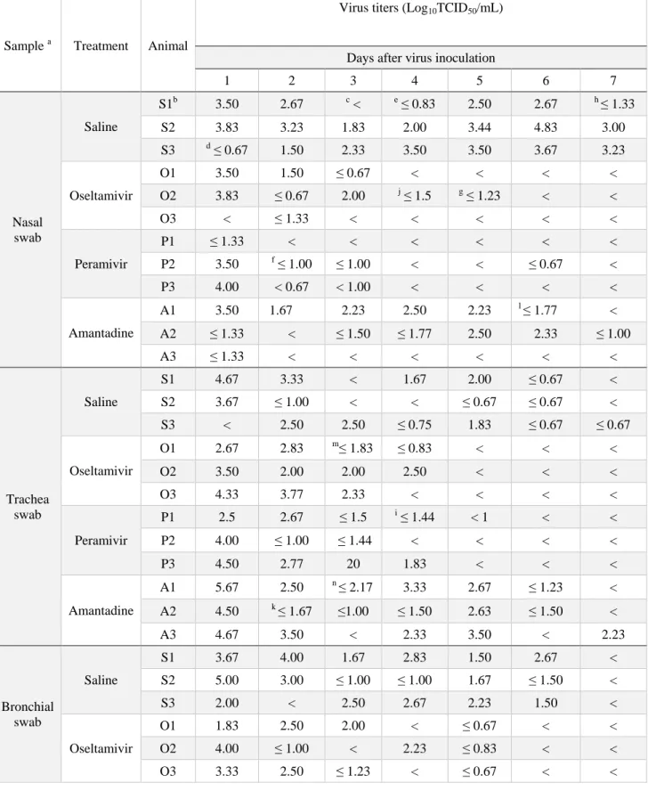
Table 1. Virus titers in swab samples of cynomolgus macaques infected with H5N6 and
517
treated with antiviral drugs.
518
Sample a Treatment Animal
Virus titers (Log10TCID50/mL)
Days after virus inoculation
1 2 3 4 5 6 7 Nasal swab Saline S1b 3.50 2.67 c < e ≤ 0.83 2.50 2.67 h ≤ 1.33 S2 3.83 3.23 1.83 2.00 3.44 4.83 3.00 S3 d ≤ 0.67 1.50 2.33 3.50 3.50 3.67 3.23 Oseltamivir O1 3.50 1.50 ≤ 0.67 < < < < O2 3.83 ≤ 0.67 2.00 j ≤ 1.5 g ≤ 1.23 < < O3 < ≤ 1.33 < < < < < Peramivir P1 ≤ 1.33 < < < < < < P2 3.50 f ≤ 1.00 ≤ 1.00 < < ≤ 0.67 < P3 4.00 < 0.67 < 1.00 < < < < Amantadine A1 3.50 1.67 2.23 2.50 2.23 l ≤ 1.77 < A2 ≤ 1.33 < ≤ 1.50 ≤ 1.77 2.50 2.33 ≤ 1.00 A3 ≤ 1.33 < < < < < < Trachea swab Saline S1 4.67 3.33 < 1.67 2.00 ≤ 0.67 < S2 3.67 ≤ 1.00 < < ≤ 0.67 ≤ 0.67 < S3 < 2.50 2.50 ≤ 0.75 1.83 ≤ 0.67 ≤ 0.67 Oseltamivir O1 2.67 2.83 m≤ 1.83 ≤ 0.83 < < < O2 3.50 2.00 2.00 2.50 < < < O3 4.33 3.77 2.33 < < < < Peramivir P1 2.5 2.67 ≤ 1.5 i ≤ 1.44 < 1 < < P2 4.00 ≤ 1.00 ≤ 1.44 < < < < P3 4.50 2.77 20 1.83 < < < Amantadine A1 5.67 2.50 n ≤ 2.17 3.33 2.67 ≤ 1.23 < A2 4.50 k ≤ 1.67 ≤1.00 ≤ 1.50 2.63 ≤ 1.50 < A3 4.67 3.50 < 2.33 3.50 < 2.23 Bronchial swab Saline S1 3.67 4.00 1.67 2.83 1.50 2.67 < S2 5.00 3.00 ≤ 1.00 ≤ 1.00 1.67 ≤ 1.50 < S3 2.00 < 2.50 2.67 2.23 1.50 < Oseltamivir O1 1.83 2.50 2.00 < ≤ 0.67 < < O2 4.00 ≤ 1.00 < 2.23 ≤ 0.83 < <
Peramivir P1 3.00 2.50 2.00 ≤ 1.50 ≤ 1.00 < < P2 4.33 3.50 ≤ 0.67 < < < < P3 4.67 3.00 < 1.38 < < < Amantadine A1 3.67 3.00 ≤ 1.00 ≤ 1.00 2.23 < < A2 4.50 ≤ 1.23 ≤ 1.77 < 1.50 3.50 o ≤ 2.25 < A3 4.00 2.33 ≤ 1.33 3.23 3.00 2.25 ≤ 1.00 519 a : Sampling organs. 520 b : Macaque identification. 521 c
<: No CPE-positive well in quadruplicate culture. A detection limit was 0.67 log10TCID50/mL.
522
d
≤ 0.67: One CPE-positive well in quadruplicate culture with the undiluted samples was 523
observed. 524
e ≤ 0.83: Two CPE-positive wells were observed in quadruplicate culture: one with undiluted
525
samples and one with 10-fold diluted sample. 526
f
≤ 1: Two CPE-positive wells in quadruplicate culture with undiluted sample were observed. 527
g ≤1.23: Three CPE-positive wells were observed in quadruplicate culture: two with undiluted
528
sample and one with 10-fold diluted sample. 529
h ≤1.33: Three CPE-positive wells in quadruplicate culture with undiluted sample were observed.
530
i ≤ 1.44: Four CPE-positive wells were observed in quadruplicate culture: two with undiluted
531
sample; one with 10-fold diluted sample and one with 100-fold diluted sample. 532
j ≤ 1.5: Four CPE-positive wells were observed in quadruplicate culture: two with undiluted
533
sample and two with 10-fold diluted sample. 534
k ≤ 1.67: Five CPE-positive wells were observed in quadruplicate culture: three with undiluted
535
sample; one with 10-fold diluted sample and one with 1000-fold diluted sample. 536
l ≤ 1.77: Five CPE-positive wells were observed in quadruplicate culture: three with undiluted
537
sample and two with 10-fold diluted sample. 538
m ≤ 1.83: Six CPE-positive wells were observed in quadruplicate culture: three with undiluted
539
sample; one with 10-fold diluted sample and two with 100-fold diluted sample. 540
n ≤ 2.17: Six CPE-positive wells were observed in quadruplicate culture: three with undiluted
541
sample; three with 10-fold diluted sample. 542
o ≤ 2.25: Seven CPE-positive wells were observed in quadruplicate culture: three with undiluted
Figure legends
545
Fig 1. Symptoms of cynomolgus macaques challenged with A/black swan/Akita/1/2016
546
(H5N6).
547
Cynomolgus macaques (n = 3) were inoculated with the virus on day 0. (a) Averages and 548
standard deviations of body temperature and (b) heart rate were determined by using a telemetry 549
probe system during the night (from 8:00 p.m. to 8:00 a.m.) and data were adjusted to day -1. (c, 550
d) Averages and standard deviations of food consumption and body weight are shown. (c) Food 551
consumption was estimated by the following formation: food consumption = [(number of pellets 552
given in the morning - number of pellets left at night)/number of pellets given in the morning] × 553
100 (%). (d) Body weight was monitored every day. Statistical differences among groups are 554
calculated with an ANOVA multi-comparison test. 555
556
Fig 2. Viral pneumonia in cynomolgus macaques challenged with A/black
557
swan/Akita/1/2016 (H5N6).
558
H&E staining of lung tissues collected 7 days after virus infection. Representative photos of 559
cynomolgus macaques treated with saline (a), oseltamivir (b), peramivir (c) and amantadine (d). 560
Black arrow heads point neutrophils. Bars, 50 μm. 561
562
Fig 3. Peripheral blood cell populations and cytokine/chemokine responses in cynomolgus
563
macaques challenged with A/black swan/Akita/1/2016 (H5N6).
564
(a-d) Concentrations in peripheral blood cells collected on the indicated days: (a) total leukocytes, 565
(b) granulocytes, (c) monocytes, and (d) lymphocytes. (e-h) Levels of cytokines/chemokines in 566
plasma after virus infection. The average values and standard deviations were shown. 567
568
Fig 4. Efficacy of antiviral drugs against A/black swan/Akita/1/2016 (H5N6) virus in vitro.
569
MDCK cells were infected with the virus at a multiplicity of infection (MOI) of 0.01 and 570
cultured with antiviral drugs of various concentrations: (a) oseltamivir, (b, d) peramivir, and (c) 571
amantadine. Sensitivity of the inoculum virus (a-c) and the virus recovered from a tonsil of 572
macaque P2 on day 7 (d) was examined. The supernatant of each well was collected at 24 h and 573
48 h after virus infection. Then virus titers in the supernatants were determined by the Reed 574
Muench method. Averages and standard deviations of three independent experiments were 575
shown in a-c. Averages and standard deviations of triplicate culture were shown in d. EC50
576
values were calculated by "Quest Graph™ EC50 Calculator." AAT Bioquest, Inc, 03 Feb. 2020, 577
https://www.aatbio.com/tools/ec50-calculator. The asterisks show significant differences in virus 578
titers with treatment at each antiviral drug concentration and without treatment (0 μg/mL) 579
(Student’s t-test, *: P < 0.05, **:P<0.01). 580