著者
INOUE Akitoshi, OHTA Shinichi, IMAI Yugo,
MURAKAMI Yoko, TOMOZAWA Yuki, SONODA Akinaga,
NITTA Norihisa
journal or
publication title
Minimally invasive therapy & allied
technologies : MITAT : official journal of the
Society for Minimally Invasive Therapy
page range
1-5
year
2020-02-26
URL
http://hdl.handle.net/10422/00012634
For Peer Review Only
Naturally shrunk visceral artery aneurysms by stenting for the superior mesenteric artery occlusion
Journal: Minimally Invasive Therapy & Allied Technologies Manuscript ID MITAT-2019-CR-0134.R1
Manuscript Type: Case Report Date Submitted by the
Author: n/a
Complete List of Authors: Inoue, Akitoshi; Shiga University of Medical Science, Department of Radiology
Ohta, Shinichi; Shiga University of Medical Science, Department of Radiology
Imai, Yugo; Shiga University of Medical Science, Department of Radiology
Murakami, Yoko; Shiga University of Medical Science, Department of Radiology
Tomozawa, Yuki; Shiga University of Medical Science, Department of Radiology
Sonoda, Akinaga; Shiga University of Medical Science, Department of Radiology
Nitta, Norihisa; Shiga University of Medical Science, Department of Radiology
Keywords: catheter interventions, endovascular therapy, image-guided procedures, interventional radiology, minimally invasive procedures, vascular intervention
For Peer Review Only
4 5 6 7 8 9 10 11 12 13 14 15 16 17 18 19 20 21 22 23 24 25 26 27 28 29 30 31 32 33 34 35 36 37 38 39 40 41 42 43 44 45 46 47 48 49 50 51 52 53 54 55For Peer Review Only
Naturally shrunk visceral artery aneurysms by stenting for the superior mesenteric artery occlusion
Akitoshi Inouea, Shinichi Ohtaa, Yugo Imaia, Yoko Murakamia, Yuki Tomozawaa, Akinaga
Sonodaa, Norihisa Nittaa
a. Department of Radiology, Shiga University of Medical Science, Shiga, Japan
Corresponding author
Akitoshi Inoue
Department of Radiology, Shiga University of Medical Science
Seta, Tsukinowa-cho, Otsu-city, Shiga 520-2192, Japan
Email: [email protected] 4 5 6 7 8 9 10 11 12 13 14 15 16 17 18 19 20 21 22 23 24 25 26 27 28 29 30 31 32 33 34 35 36 37 38 39 40 41 42 43 44 45 46 47 48 49 50 51 52 53
For Peer Review Only
Abstract
A 77-year-old female who experienced postprandial abdominal pain for four years was admitted to our institution presenting sudden and severe abdominal pain. Contrast-enhanced computed tomography (CECT) demonstrated complete short-segmented occlusion in the orifice of the superior mesenteric artery (SMA), and saccular aneurysms in the right hepatic artery and anterior superior pancreaticoduodenal artery. She was diagnosed with abdominal angina due to occlusion of the SMA. The SMA was recanalized by stenting, and a CECT scan confirmed naturally shrunk aneurysms after eight months. The patency of the SMA was maintained at five years after endovascular treatment.
Keywords
Abdominal angina, Chronic mesenteric ischemia, Superior mesenteric artery occlusion, Visceral artery aneurysm 4 5 6 7 8 9 10 11 12 13 14 15 16 17 18 19 20 21 22 23 24 25 26 27 28 29 30 31 32 33 34 35 36 37 38 39 40 41 42 43 44 45 46 47 48 49 50 51 52 53 54 55
For Peer Review Only
Introduction
The formation of certain visceral artery aneurysms (VAAs) is associated with wall shear stress due to abnormally increased blood flow from vascular stenosis or occlusion, especially for VAAs in the pancreaticoduodenal artery [1,2]. This pathology has been spontaneously improved after revascularization for extrinsic compression of the celiac artery to the median arcuate
ligament (MAL), which is a fibrous tissue located between two diaphragmatic crura [3,4]. At the moment, there is no article regarding aneurysm formation caused by the SMA occlusion and naturally shrunk VAAs after treatment of the SMA occlusion, therefore, we describe a case of naturally shrunk VAAs after endovascular treatment of the superior mesenteric artery (SMA) occlusion in a patient with abdominal angina.
Case Report
A 77-year-old female suffered from intermittent mild abdominal pain occurring
especially after meals for four years, and was diagnosed with abdominal angina according to an 4 5 6 7 8 9 10 11 12 13 14 15 16 17 18 19 20 21 22 23 24 25 26 27 28 29 30 31 32 33 34 35 36 37 38 39 40 41 42 43 44 45 46 47 48 49 50 51 52 53
For Peer Review Only
SMA stenosis detected by contrast-enhanced computed tomography (CECT). She hoped to avoid invasive treatment, and was followed up using CECT scans after her initial diagnosis. She was admitted to our institution due to sudden and severe abdominal pain, wherein CECT showed complete occlusion in the SMA (Fig.1A), and saccular aneurysms in the right hepatic artery (RHA) and anterior superior pancreaticoduodenal artery (ASPA) (Fig. 1B). Endovascular treatment was scheduled to recanalize the SMA to supply adequate blood flow to the small bowel before aneurysm embolization.
Before the procedure, 100 mg of aspirin and 300 mg of clopidogrel were administered. The SMA could not be reached from the aorta; therefore, a retrograde approach from the celiac artery was selected. First, 4-French (Fr) introducer sheath (Super sheath, length: 25 cm; Medikit, Tokyo, Japan) was inserted into the right femoral artery. Thereafter, a 4-Fr catheter (JC-1 type; Terumo Clinical Supply, Gifu, Japan) and a 0.035” guidewire (Radifocus, length: 150 cm, Terumo Clinical Supply) were inserted via the 4-Fr introducer sheath and placed in the celiac artery. A 6-Fr introducer sheath (Ansel sheath, shepherd hook type, length 55 cm, Cook Medical, 4 5 6 7 8 9 10 11 12 13 14 15 16 17 18 19 20 21 22 23 24 25 26 27 28 29 30 31 32 33 34 35 36 37 38 39 40 41 42 43 44 45 46 47 48 49 50 51 52 53 54 55
For Peer Review Only
Bloomington, IN, USA) was inserted under the guidance of the 4-Fr JC-1 type catheter and a 0.035” guidewire into the celiac artery, undergoing exchange with athe 4-Fr introducer sheath. Angiography via the celiac artery showed one saccular aneurysm within the RHA and one within the ASPA, and the orifice of the replaced RHA (Fig. 2A) and retrograde blood flow from the celiac artery to the SMA through the gastroduodenal artery (Fig.2B). A 4-Fr catheter (MP-1 type, Terumo Clinical Supply) through a 6-Fr introducer sheath was carried to the distal side of the SMA occlusion via the ASPA. A 0.035” guide wire (Radifocus; length: 300 cm,; Terumo Clinical Supply) was passed from the MP-1 type catheter through the occlusion and went to the aorta. A 5-Fr introducer sheath (Super Sheath, length: 25 cm; Medikit) was then inserted at the left femoral artery, and a 0.035” guidewire was caught in the aorta using an endovascular snare system (En Snare; shaft length: 120 cm, loop size: 12–20 mm, Merit Medical, South Jordan, UT, USA) which was inserted from a 5-Fr introducer sheath at the left femoral artery to establish pull-thorugh. At this point, a 4-Fr catheter (JC-1 type, Terumo Clinical Supply) was inserted into the distal side of the occlusion from the orifice of the SMA through a 5-Fr introducer sheath at the left femoral artery with a 0.035” guidewire. Next, a 0.014” guidewire was inserted via a 4-Fr 4 5 6 7 8 9 10 11 12 13 14 15 16 17 18 19 20 21 22 23 24 25 26 27 28 29 30 31 32 33 34 35 36 37 38 39 40 41 42 43 44 45 46 47 48 49 50 51 52 53
For Peer Review Only
catheter for exchange with the 0.035” guidewire. A balloon catheter (Aviator plus: balloon diameter: 4 mm, length: 20 mm, Cordis, Santa Clara, CA, U.S.A.) was carried to the occlusion point through a 0.014” guidewire, and was dilated before and after stent placement. A bare stent (PALMAZ Genesis: diameter 6 mm, length 15 mm, Santa Clara, Cordis, CA, U.S.A) was deployed, and patency of the SMA was confirmed by aortography.
The patient took 100 mg/day of aspirin and 75 mg/day of clopidogrel for three months. First follow-up CECT at eight months after stenting revealed that the SMA was still patent (Fig. 3A) and the aneurysms of the RHA and ASPA had shrunk (Fig. 3B). She received follow-up examinations for five years after the procedure with CECT imaging revealing gradually reduced diameters of both the aneurysms (Fig. 4 ).. No abdominal symptoms were detected, no
re-stenosis was required, and the aneurysms gradually decreased without rupture.
Discussion 4 5 6 7 8 9 10 11 12 13 14 15 16 17 18 19 20 21 22 23 24 25 26 27 28 29 30 31 32 33 34 35 36 37 38 39 40 41 42 43 44 45 46 47 48 49 50 51 52 53 54 55
For Peer Review Only
Abdominal angina is characterized by postprandial abdominal pain, diarrhea, vomiting, and weight loss due to the reduction of blood supply to the gastrointestinal tract, which is caused by slowly progressing stenosis. This stenosis occurs mainly in the SMA, but also in the celiac and inferior mesenteric artery without sufficient collateral blood flow [5]. The most common cause of stenosis is atherosclerosis, but fibromuscular dysplasia, Takayasu arteritis, systemic lupus erythematosus, and tumor invasion have also been reported in the literature [6–9].
Abdominal angina is diagnosed according to clinical symptoms and consistent imaging findings [10,11], and its treatment strategy which is applied in symptomatic patients with critical stenosis (>70%) to relieve gastrointestinal symptoms is categorized into open surgery or endovascular treatment. The average patency rate is reportedly 50% at two years after endovascular treatment [12] [10,11]. While the clinical success rate at three months is lower and the late recurrence rate is higher following endovascular treatment compared with open surgery, endovascular treatment is significantly associated with lower complication rates and shorter lengths of hospital stay [13]. Furthermore, endovascular treatment is advantageous for recurrence treatment because it is less invasive. 4 5 6 7 8 9 10 11 12 13 14 15 16 17 18 19 20 21 22 23 24 25 26 27 28 29 30 31 32 33 34 35 36 37 38 39 40 41 42 43 44 45 46 47 48 49 50 51 52 53
For Peer Review Only
Large, expanding, or inflammation-related VAAs generally present with a risk of rupture and require treatment because mortality from ruptured VAAs is high [14]. Indications of
treatment for hepatic artery aneurysms (HAAs) include symptomatic HAAs and asymptomatic HAAs caused by vasculitis and fibromuscular dysplasia and multiple HAAs. For
pancreaticoduodenal artery aneurysm (PDAAs), the mean size of ruptured PDAAs was reportedly 9 mm, although indications of treatment have not yet been determined owing to inadequate evidence [14]. Treatment of VAAs can be achieved by endovascular approaches, including coli packing with and without double-catheter, balloon-assist, and stent-assist techniques and isolation by embolization of the proximal and distal sides; however,
coil-embolization potentially has a risk of migration resulting in organ ischemia [15]. Vascular plugs and stent-graft therapy can be used to exclude VAAs; however, the rigidity and larger-bore of delivery systems can prevent to carry the device to aneurysms in peripheral or tortuous vessels [16]. The surgical approach also represents an alternative therapeutic modality for cases that are difficult for endovascular treatment [17]. In this case, we selected regulatory follow up for the patient’s VAAs, because she did not display signs of inflammation or aneurysm-related 4 5 6 7 8 9 10 11 12 13 14 15 16 17 18 19 20 21 22 23 24 25 26 27 28 29 30 31 32 33 34 35 36 37 38 39 40 41 42 43 44 45 46 47 48 49 50 51 52 53 54 55
For Peer Review Only
symptoms, her aneurysms were relatively small, and the time courses of her aneurysms were unknown. Endovascular treatment would be scheduled in the case of enlarged aneurysms; however, serial observation by CECT revealed that the aneurysms were shrinking.
Increased retrograde blood flow due to celiac artery stenosis may lead to aneurysms in the gastroduodenal artery [1]. A previous study using flow-sensitive four-dimensional magnetic resonance imaging in patients with pancreaticoduodenal artery aneurysms concomitant with celiac artery occlusion demonstrated a retrograde flow in the common hepatic and
gastroduodenal artery, and heterogeneous wall shear stress in the aneurysm [1518]. Wall shear stress is considered a factor contributing to the formation of aneurysms by atherosclerosis. Hemodynamic change in the pancreaticoduodenal arcade can change promptly following occlusion and recanalization of the artery [1619]. In a previously reported case, the posterior pancreaticoduodenal artery was enlarged and blood pressure in the common hepatic artery was maintained after embolization in a patient with the ASPA aneurysm consistent with celiac artery stenosis due to the MAL [2]. Furthermore, rapid development of a new aneurysm was also reported in the pancrecaticoduodenal artery after embolization for pancreaticoduodenal artery 4 5 6 7 8 9 10 11 12 13 14 15 16 17 18 19 20 21 22 23 24 25 26 27 28 29 30 31 32 33 34 35 36 37 38 39 40 41 42 43 44 45 46 47 48 49 50 51 52 53
For Peer Review Only
aneurysm [1720]. In this case, VAAs observed not only in the ASPA but also in the right hepatic artery. We consider that the blood flow of the replaced right hepatic artery decreased by the SMA stenosis, consequently, compensatory blood flow via intrahepatic anastomosis gave wall shear stress to the right hepatic artery. although aspirin may have facilitated the decrease of aneurysms via its anti-inflammatory effect.
Conversely, normalization of blood flow can reduce wall shear stress proceeding aneurysms. According to a previous report, a ruptured pancreaticoduodenal aneurysm was treated solely via bypass between the aorta and celiac trunk [3]. Additionally, two previous cases have demonstrated that stenting is a new strategy for addressing pancreaticoduodenal artery aneurysms associated with celiac artery stenosis by the MAL and atherosclerosis [4]. However, stenting is not suitable for extrinsic causes like MAL due to the risk of stent rupture, but it is appropriate for intrinsic causes like atherosclerosis.
Embolization and stenting in the visceral artery can change blood flow and exacerbate or improve VAAs associated with change of wall shear stress. It is essential to consider what change happen in blood flow by embolization or stenting before procedure. Notably, in patients 4 5 6 7 8 9 10 11 12 13 14 15 16 17 18 19 20 21 22 23 24 25 26 27 28 29 30 31 32 33 34 35 36 37 38 39 40 41 42 43 44 45 46 47 48 49 50 51 52 53 54 55
For Peer Review Only
with VAAs that coexist with vascular stenosis or occlusion, angioplasty with endovascular treatment prior to embolization for VAAs may treat the vascular stenosis or occlusion.
Acknowledgements
The authors of the present study would like to thank Makoto Yoshigoe (Department of Radiology, Shiga University of Medical Science) for creating volume rendering image of CT. The authors would like to thank Enago (www.enago.jp) for the English language review.
Compliance with Ethical Standards
Funding
The study was not supported by any funding.
Conflict of interest 4 5 6 7 8 9 10 11 12 13 14 15 16 17 18 19 20 21 22 23 24 25 26 27 28 29 30 31 32 33 34 35 36 37 38 39 40 41 42 43 44 45 46 47 48 49 50 51 52 53
For Peer Review Only
The authors declare that they have no conflict of interest.Ethical Statement
All procedures performed in studies involving human participants were in accordance with the ethical standards of the institutional and/or national research committee and with the 1964 Helsinki declaration and its later amendments or comparable ethical standards.
Informed consent
Our institutional review board did not require approval to report this case.
References 4 5 6 7 8 9 10 11 12 13 14 15 16 17 18 19 20 21 22 23 24 25 26 27 28 29 30 31 32 33 34 35 36 37 38 39 40 41 42 43 44 45 46 47 48 49 50 51 52 53 54 55
For Peer Review Only
1. Suzuki K, Tachi Y, Ito S, Maruyama K, Mori Y, Komada T, et al. Endovascular management of ruptured pancreaticoduodenal artery aneurysms associated with celiac axis stenosis.
Cardiovasc Intervent Radiol. 2008;31:1082–7.
2. Shibata E, Takao H, Amemiya S, Ohtomo K. Perioperative hemodynamic monitoring of common hepatic artery for endovascular embolization of a pancreaticoduodenal arcade aneurysm with celiac stenosis. Cardiovasc Intervent Radiol. 2017;40:465–9.
3. Salomon du Mont L, Lorandon F, Behr J, Leclerc B, Ducroux E, Rinckenbach S. Ruptured pancreaticoduodenal artery aneurysm due to a median arcuate ligament treated solely by revascularization of the celiac trunk. Ann Vasc Surg. 2017;43:310.e13–310.e16.
4. Tien YW, Kao HL, Wang HP. Celiac artery stenting: A new strategy for patients with pancreaticoduodenal artery aneurysm associated with stenosis of the celiac artery. J Gastroenterol. 2004;39:81–5.
5. Cademartiri F, Raaijmakers RH, Kuiper JW, van Dijk LC, Pattynama PM, Krstin GP. Multi-detector row CT angiography in patients with abdominal angina. Radiographics. 2004;24:969–84. 4 5 6 7 8 9 10 11 12 13 14 15 16 17 18 19 20 21 22 23 24 25 26 27 28 29 30 31 32 33 34 35 36 37 38 39 40 41 42 43 44 45 46 47 48 49 50 51 52 53
For Peer Review Only
6. Senadhi V. A rare cause of chronic mesenteric ischemia from fibromuscular dysplasia: A case report. J Med Case Rep. 2010;4:373.
7. Chaudhary SC, Gupta A, Himanshu D, Verma SP, Khanna R, Gupta DK. Abdominal angina: An unusual presentation of Takayasu’s arteritis. BMJ Case Rep. 2011;2011:bcr0220113900.
8. Matsuda M, Miyazaki D, Tojo K, Tazawa K, Shimojima Y, Kurozumi M,et al. Intestinal angina due to atherosclerosis in a 45-year-old systemic lupus erythematosus patient. Intern Med. 2010;49:2175–8.
9. Biolato M, Gabrieli ML, Parente A, Racco S, Costantini M, Bonomo L, et al. Abdominal angina due to recurrence of cancer of the papilla of Vater: A case report. J Med Case Rep. 2009;3:9314.
10. White CJ. Chronic mesenteric ischemia: diagnosis and management. Prog Cardiovasc Dis. 2011;54:36–40. 4 5 6 7 8 9 10 11 12 13 14 15 16 17 18 19 20 21 22 23 24 25 26 27 28 29 30 31 32 33 34 35 36 37 38 39 40 41 42 43 44 45 46 47 48 49 50 51 52 53 54 55
For Peer Review Only
11. Jaster A, Choudhery S, Ahn R, Sutphin P, Kalva S, Anderson M, et al. Anatomic and radiologic review of chronic mesenteric ischemia and its treatment. Clin Imaging. 2016;40:961– 9.
12. Sundermeyer A, Zapenko A, Moysidis T, Luther B, Kröger K. Endovascular treatment of chronic mesenteric ischaemia. Interv Med Appl Sci. 2014;6:118–24.
13. Barret M, Martineau C, Rahmi G, Pellerin O, Sapoval M, Alsac JM, et al. Chronic mesenteric ischemia: A rare cause of chronic abdominal pain. Am J Med. 2015;128:1363.e1– 1363.e8.
14. Hosn MA, Xu J, Sharafuddin M, Corson JD. Visceral artery aneurysms: decision making and treatment options in the new era of minimally invasive and endovascular surgery. Int J Angiol. 2019;28:11–6.
15. Tanigawa N, Kariya S, Kojima H, Tokuda T, Komemushi A, Sawada S. Transcatheter coil embolization of an aneurysm of an anomalous splenic artery: usefulness of double microcatheter method. Minim Invasive Ther Allied Technol. 2009;18:311–4.
4 5 6 7 8 9 10 11 12 13 14 15 16 17 18 19 20 21 22 23 24 25 26 27 28 29 30 31 32 33 34 35 36 37 38 39 40 41 42 43 44 45 46 47 48 49 50 51 52 53
For Peer Review Only
16. Chatani S, Inoue A, Ohta S, Takaki K, Sato S, Iwai T, et al. Transcatheter Arterial
Embolization for Postoperative Bleeding Following Abdominal Surgery. Cardiovasc Intervent Radiol. 2018;41:1346–55.
17. Xia T, Zhou JY, Mou YP, Xu XW, Zhou YC, Huang CJ, et al. Laparoscopic ligation of celiac trunk and splenic artery aneurysms with function preservation. Minim Invasive Ther Allied Technol. 2017;26:56–9.
1518. Mano Y, Takehara Y, Sakaguchi T, Alley MT, Isoda H, Shimizu T, et al. Hemodynamic assessment of celiaco-mesenteric anastomosis in patients with pancreaticoduodenal artery aneurysm concomitant with celiac artery occlusion using flow-sensitive four-dimensional magnetic resonance imaging. Eur J Vasc Endovasc Surg. 2013;46:321–8.
1619. Toriumi T, Shirasu T, Akai A, Ohashi Y, Furuya T, Nomura Y. Hemodynamic benefits of celiac artery release for ruptured right gastric artery aneurysm associated with median arcuate ligament syndrome: a case report. BMC Surg. 2017;17:116.
4 5 6 7 8 9 10 11 12 13 14 15 16 17 18 19 20 21 22 23 24 25 26 27 28 29 30 31 32 33 34 35 36 37 38 39 40 41 42 43 44 45 46 47 48 49 50 51 52 53 54 55
For Peer Review Only
1720. Hasegawa T, Seiji K, Ota H, Matsuura T, Satani N, Sato T, et al. Rapid development of new aneurysms in the adjacent pancreatic arcade arteries after urgent embolization of
pancreaticoduodenal artery aneurysms in cases with celiac stenosis. J Vasc Interv Radiol. 2018;29:1306–1308.e2.
Figures
Fig. 1 Contrast-enhanced computed tomography before endovascular treatment
A: Sagittal image of contrast-enhanced computed tomography at the arterial phase showing a complete occlusion at the orifice of the superior mesenteric artery (arrow). B: Volume rendering depicting aneurysms of 9.6 mm and 5.4 mm in diameter in the right hepatic artery (black arrow) and anterior superior pancreaticoduodenal artery (white arrow), respectively.
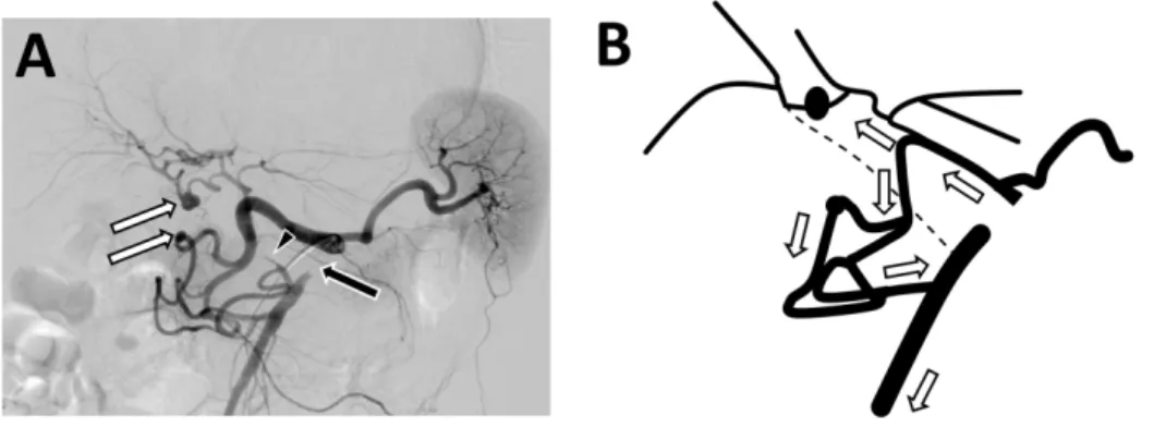
Fig. 2 Angiogram via the celiac artery 4 5 6 7 8 9 10 11 12 13 14 15 16 17 18 19 20 21 22 23 24 25 26 27 28 29 30 31 32 33 34 35 36 37 38 39 40 41 42 43 44 45 46 47 48 49 50 51 52 53
For Peer Review Only
A: Angiogram showing complete occlusion of the superior mesenteric artery (black arrow) associated with retrograde blood flow through the prominent pancreaticoduodenal arcade. Aneurysms are seen in the right hepatic and anterior superior pancreaticoduodenal art ery (white arrows). The orifice of the replaced right hepatic artery originated from the superior mesenteric artery (arrowhead). B: The white arrows in the schema shows the blood flow from the celiac artery to the proper hepatic artery anterogradely, and the superior mesenteric artery through the gastroduodenal artery retrogradely.
Fig. 3 Follow-up contrast-enhanced computed tomography at five years after endovascular treatment
A: Sagittal image of contrast-enhanced computed tomography at the arterial phase showing patency of the superior mesenteric artery (arrow). B: Volume rendering image demonstrating the shrunken aneurysms of 3.5 mm in the hepatic artery (black arrow) and of 2.8 mm in the superior anterior pancreaticoduodenal arteries (white arrow).
4 5 6 7 8 9 10 11 12 13 14 15 16 17 18 19 20 21 22 23 24 25 26 27 28 29 30 31 32 33 34 35 36 37 38 39 40 41 42 43 44 45 46 47 48 49 50 51 52 53 54 55
For Peer Review Only
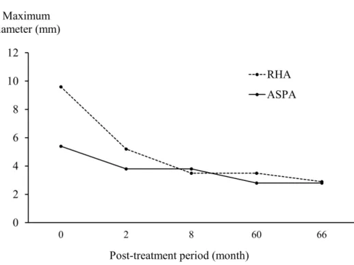
Fig. 4 The time course of the aneurysmal diameterRHA; Right hepatic artery, ASPA; superior anterior pancreaticoduodenal artery 4 5 6 7 8 9 10 11 12 13 14 15 16 17 18 19 20 21 22 23 24 25 26 27 28 29 30 31 32 33 34 35 36 37 38 39 40 41 42 43 44 45 46 47 48 49 50 51 52 53
For Peer Review Only
Fig. 1 Contrast-enhanced computed tomography before endovascular treatment
A: Sagittal image of contrast-enhanced computed tomography at the arterial phase showing a complete occlusion at the orifice of the superior mesenteric artery (arrow). B: Volume rendering depicting aneurysms
of 9.6 mm and 5.4 mm in diameter in the right hepatic artery (black arrow) and anterior superior pancreaticoduodenal artery (white arrow), respectively.
235x114mm (300 x 300 DPI) 4 5 6 7 8 9 10 11 12 13 14 15 16 17 18 19 20 21 22 23 24 25 26 27 28 29 30 31 32 33 34 35 36 37 38 39 40 41 42 43 44 45 46 47 48 49 50 51 52 53 54 55
For Peer Review Only
Fig. 2 Angiogram via the celiac artery
A: Angiogram showing complete occlusion of the superior mesenteric artery (black arrow) associated with retrograde blood flow through the prominent pancreaticoduodenal arcade. Aneurysms are seen in the right
hepatic and anterior superior pancreaticoduodenal art ery (white arrows). The orifice of the replaced right hepatic artery originated from the superior mesenteric artery (arrowhead). B: The white arrows in the schema shows the blood flow from the celiac artery to the proper hepatic artery anterogradely, and the
superior mesenteric artery through the gastroduodenal artery retrogradely. 254x190mm (300 x 300 DPI) 4 5 6 7 8 9 10 11 12 13 14 15 16 17 18 19 20 21 22 23 24 25 26 27 28 29 30 31 32 33 34 35 36 37 38 39 40 41 42 43 44 45 46 47 48 49 50 51 52 53
For Peer Review Only
Fig. 3 Follow-up contrast-enhanced computed tomography at five years after endovascular treatment A: Sagittal image of contrast-enhanced computed tomography at the arterial phase showing patency of the superior mesenteric artery (arrow). B: Volume rendering image demonstrating the shrunken aneurysms of
3.5 mm in the hepatic artery (black arrow) and of 2.8 mm in the superior anterior pancreaticoduodenal arteries (white arrow).
227x114mm (300 x 300 DPI) 4 5 6 7 8 9 10 11 12 13 14 15 16 17 18 19 20 21 22 23 24 25 26 27 28 29 30 31 32 33 34 35 36 37 38 39 40 41 42 43 44 45 46 47 48 49 50 51 52 53 54 55
For Peer Review Only
Fig. 4 The time course of the aneurysmal diameter
RHA; Right hepatic artery, ASPA; superior anterior pancreaticoduodenal artery 211x152mm (300 x 300 DPI) 4 5 6 7 8 9 10 11 12 13 14 15 16 17 18 19 20 21 22 23 24 25 26 27 28 29 30 31 32 33 34 35 36 37 38 39 40 41 42 43 44 45 46 47 48 49 50 51 52 53