Are our scintigraphic results useful as a clue
for interpreting kinetics of nuclear agents?
著者
佐藤 強志, 森田 康彦, 馬嶋 秀行
journal or
publication title
鹿児島大学歯学部紀要
volume
35
page range
21-28
year
2015
URL
http://hdl.handle.net/10232/26723
Are our scintigraphic results useful as a clue for interpreting kinetics of nuclear agents ?
Tsuyoshi Sato, Yasuhiko Morita*, Hideyuki Majima
Department of Maxillofacial Radiology, Kagoshima University Graduate School of Medical and Dental Sciences, 8-35-1 Sakuragaoka, Kagoshima 890-8544, Japan
*Department of Oral and Maxillofacial Radiology, Tokushima University Graduate School of Oral Sciences, 3-18-15 Kuramoto-cho, Tokushima 770-8504, Japan
ABSTRACT
Until about ten years ago, we have performed scintigraphy with 201-thallium chloride (201-TlCl) and 99m-Tc-hexakis-2-methoxy-isobutyl-isonitrile (99m-Tc-MIBI) for malignant tumors. In this article, we re-evaluated scintigraphic images retrospectively with a hope that the results might be a clue, even if it is small, for dentists to try to improve the accuracy of diagnosis of malignant tumors. From scintigraphy, we obtained the tumor retention index as a factor to estimate the uptake of radioactive agents in tumor cells. Moreover, we estimated transport proteins of Na+/K+-ATPase and
permeability-glycoprotein (P-gp) expressed on the cell membrane that might regulate the kinetic condition of radioactive agents. Among the tumor retention index, the transport protein and the histopathologic finding of tumors, there were relatively well correlations. The tumor retention index showed a difference clearly between malignant tumor and benign tumor. The transport protein revealed a distinct expression in accordance with the malignancy of tumor, and the uptake clearly depended upon the expression of transport protein.
Key words: Tumor scintigraphy, Malignant tumor, Transport protein, Gamma camera
1. INTRODUCTION
Until about ten years ago, not a few radioactive agents have been introduced for the purpose of diagnosing malignant tumors of the head and neck, for example 67-Ga (gallium), 201-Tl (thallium), 99m-Tc (technetium), 198-Au (aurum), 131-I (iodine) and so forth. However, these radioactive agents are now not popularly used as those times in routine examinations, because 18-F-fluoro-deoxy-glucose positron emission tomography (FDG-PET) is taking places of these radioactive agents. FDG-PET is a very superior method for malignant tumors.1 At the time
when FDG-PET has been introduced, we almost believed that most malignant tumors could be detected precisely and qualitatively with this method. However, this our expectation unfortunately ended with a fragile dream. This is not any all-purpose method. Even FDG-PET has some weak points. For example, FDG-PET is not able to distinguish malignant tumors from inflammatory lesions.2
This radioactive agent shows almost the same accumulation in both malignant tumors and inflammatory lesions depending on its high sensitivity and affinity both to tumors and inflammatory tissues. This weak point is also an eternal, essential problem among usual tumor scintigraphies for a long time. Many researchers have tried to resolve this problem for a long time, but this is left unresolved. Against this problem, we also did in spite of a small ability. We focused our eyes on transport proteins of radioactive agents as one of means of solving this problem. We performed evaluations concerning several subjects, for example an expression of transport proteins on cell membrane and a relation of transport proteins with accumulation. Among the results of our evaluations, we searched to pick up some factors that seemed to be helpful and useful for diagnosing malignant tumors, and we tried to find out a possibility of qualitative diagnosis of malignant tumors of the head and neck using the factors.3-8 In our scintigraphy for tumors, we
Tsuyoshi Sato, Yasuhiko Morita*, Hideyuki Majima 22
usually employed 201-thallium chloride (201-TlCl) and 99m-Tc-hexakis-2-methoxy-isobutyl-isonitrile (99m-Tc-MIBI) as radioactive agents. We selected a couple of factors that control and closely relate with the uptake of these radioactive agents. We evaluated the expression of Na+/K+-ATPase and permeability-glycoprotein (P-gp) on
the tumor cell membrane and the role of them as transport proteins in relation with both accumulation and washout of radioactive agents in tumor cells.
In this article, we re-evaluated retrospectively our results of tumor scintigraphy that we carried out. With this thing and that, most data used in this article were quoted from some of our previous reports on jurnals3-8 and modified to
some extent.
2. SCINTIGRAPHY FOR TUMORS
2-1. Clinical evaluation of scintigraphy with 201-Tl 201-Tl was first used to evaluate the viability of the myocardium. After a while, this agent was introduced for the examination of malignant tumors of the head and neck.9,10 In this section, we evaluated the usefulness of
201-Tl for malignant tumors of the head and neck.
(Methods and Materials of Scintigraphy with 201-Tl) We used 85 patients with a malignant tumor of the head and neck (squamous cell carcinoma) and 10 patients with a benign tumor (7 with pleomorphic adenoma and 3 with Warthin’s tumor).
Intravenous injection of 74MBq of 201-Tl was performed. An early dynamic scan (for 5 min immediately after injection), a delayed dynamic scan and a spot scan (at 2.5 hrs after injection) were carried out using a gamma camera. From the dynamic scan, 2-second scans were obtained continuously. A single 2-second scan constituted a frame data. Two regions of interest (ROI) on each frame covering both tumor and control areas were used to estimate the uptake of 201-Tl (Figure 1). Early and delayed retention indexes were calculated from the results of each dynamic scan. The early retention index was the ratio of count of tumor to count of control in the early dynamic scan. The delayed retention index was the ratio of count of tumor to count of control in the delayed dynamic scan. From these two indexes, the tumor retention index was calculated; the ratio of the delayed retention index to the early retention index (Figure 2). We used this tumor
B
A
Figure 1: Two regions of interest (ROI) on a frame image covered the tumor area (A) and the symmetrical region (B: control region).
Figure 2: Two curves showed radioactive count after injection in the tumor and control areas. In the dynamic scintigraphy, the early retention index was A/a and the delayed retention index was B/b. The tumor retention index was (delayed retention index) / (early retention index).
Radioactive count Time Tumor Control
A
a
B
Early dynamic scintigraphy Spot imageb
Delayed dynamic scintigraphyretention index for the evaluation of scintigraphy. The tumor retention index was compared with the histopathologic type and tissue differentiation.
(Results: Tumor retention index of 201-Tl, Histopathologic type and Tissue differentiation)
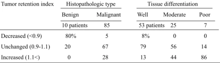
Tumor retention indexes varied widely ranging from 0.76 to 1.46 in patients. In the histopathologic type, the tumor retention indexes ranged from 0.76 to 0.93 (average was 0.82) in the benign group, and 0.78 to 1.46 (1.04) in the malignant group, respectively. In the tissue differentiation, tumor retention indexes ranged from 0.78 to 1.24 (average was 1.03) in the well group, from 0.91 to 1.42 (1.09) in the moderate group, and from 1.05 to 1.46 (1.24) in the poor group, respectively. We classified these tumor retention indexes into three groups: decreased (<0.9), unchanged (0.9-1.1), and increased (1.1<). The increased tumor retention index means that the washout of 201-Tl from tumor is delayed or the washout function is lost. On the other hand, the decreased tumor retention index indicates that the washout of 201-Tl is fast. As for histopathologic type, 80% of patients in the benign group belonged to the decreased tumor retention index group and no patient showed the increased. On the other hand, 28% and 67% of patients in the malignant group were included in the increased and unchanged groups. Only 5% of patients indicated the decreased. As for the tissue differentiation, 86% of patients in the poor group were included in the increased group and no patient showed the decreased. On the other hand, only 13% of patients in the well group belonged to the increased group (Table 1). These results showed that 201-Tl once taken up in malignant tumors had a tendency to remain.
2-2. Accumulation of 201-Tl and Na+/K+-ATPase expression
It was reported that the expression of Na+/K+-ATPase on
cell membrane was one of the most important factors concerning the accumulation mechanism of 201-Tl in malignant tumors.11 However, the role of Na+/K+-ATPase
on the uptake mechanism of 201-Tl is not clearly understood, and there are few reports on tumors of the head and neck. In this section, we evaluated the role of Na+/K+
-ATPase expression on 201-Tl scintigraphy of malignant tumors of the head and neck.
(Methods and Materials of Immunohistochemistry for Na+/K+-ATPase expression)
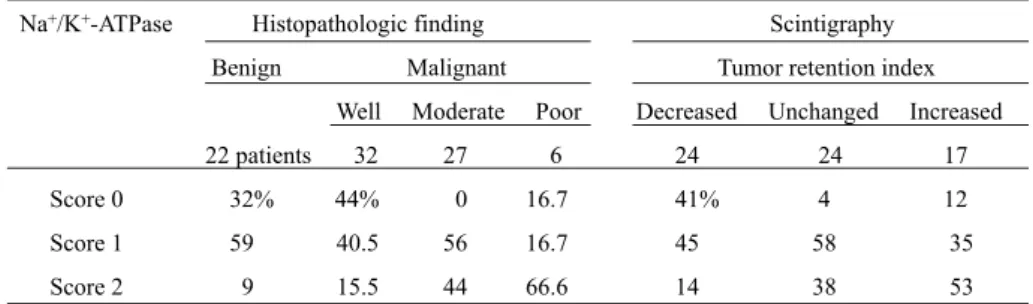
Sixty-five patients with malignant tumor of the head and neck (squamous cell carcinoma) and 22 patients with benign tumor were used.
Immunohistochemical staining was performed with tumor samples. Briefly, sections of tumors were treated with sodium citrate buffer, heated for the antigen retrieval, and then treated hydrogen peroxide for 10 min to block endogenous peroxidase activity. Sections were incubated with the primary and secondary antibody. After incubation, the sections were washed with Tris buffer saline, reacted with avidin-biotinylated-peroxidase complex, and stained with diaminobendizine.12 Na+/K+-ATPase expression was
graded into score 0 (stained under 5%), score 1 (from 5 to 49%), or score 2 (over 50%)13 with reference to
histopathologic findings in malignant tumors of the head and neck.
(Results: Na+/K+-ATPase expression, Histopathologic
finding and Tumor retention index)
As for the Na+/K+-ATPase expression and histopathologic
finding, patients of the benign group showed score 0 (32%), Table 1. Tumor retention index of 201-Tl, Histopathologic type and Tissue differentiation
Tumor retention index Histopathologic type Tissue differentiation Benign Malignant Well Moderate Poor 10 patients 85 53 patients 25 7 Decreased (<0.9) 80% 5 8% 0 0
Unchanged (0.9-1.1) 20 67 79 56 14
Tsuyoshi Sato, Yasuhiko Morita*, Hideyuki Majima 24
score 1 (59%) and score 2 (9%). In the malignant group, scores 0, 1 and 2 were shown in 44%, 40.5% and 15.5% in the well group, respectively. Patients of the moderate group showed score 1 (56%) and score 2 (44%). In patients of the poor group, scores 0, 1 and 2 were shown in 16.7%, 16.7% and 66.6%, respectively. As for the Na+/K+-ATPase
expression and tumor retention index, patients of the decreased group showed scores 0, 1, and 2 in 41%, 45% and 14%, respectively. Patients of the unchanged group revealed score 0 (4%), score 1 (58%) and score 2 (38%), respectively. In patients of the increased group, scores 0, 1 and 2 were observed in 12%, 35% and 53%, respectively (Table 2). The frequency of score 2 was elevated according as the tumor retention index became large. These results indicated that Na+/K+-ATPase expression was typical in
malignant tumors and played the role of uptake of 201-Tl. 2-3. Clinical evaluation of scintigraphy with 99m-Tc-MIBI
This scintigraphic agent has been widely used to evaluate the viability of the myocardium, and the accumulation of this agent in malignant tumors has been also reported.14 In
this section, we evaluated the usefulness of 99m-Tc-MIBI scintigraphy for the diagnosis of malignant tumor of the head and neck.
(Methods and Materials of Scintigraphy with 99m-Tc-MIBI) Nineteen patients with squamous cell carcinoma of the head and neck were used. The method of scintigraphy was almost the same as that of 201-Tl. Scintigraphy was performed with an intravenous injection of 600MBq of 99m-Tc-MIBI.15 The tumor retention index was compared
with the tissue differentiation.
(Results: Tumor retention index of 99m-Tc-MIBI and Tissue differentiation)
Retention indexes ranged from 1.1 to 3.1 in the early dynamic scan, and averages were 1.03 (well group), 1.8 (moderate) and 1.65 (poor). In the delayed dynamic scan, retention indexes ranged from 1.0 to 2.9, and averages were 1.1 (well group), 1.48 (moderate) and 1.27 (poor). From these retention indexes, tumor retention indexes were calculated. Tumor retention indexes ranged from 0.70 to 1.0, and averages were 0.91 (well group), 0.93 (moderate) and 0.79 (poor), respectively. Then, we classified grades of tumor retention indexes into >0.9 (slightly decreased), 0.9 to 0.8 (intermediately decreased) and 0.8> (severely decreased). Most of tumor retention indexes were under 1.0. We could find a decreasing tendency of tumor retention indexes from the early dynamic scan to delayed dynamic Table 2. Na+/K+-ATPase expression, Histopathologic finding and Tumor retention index
Na+/K+-ATPase Histopathologic ifnding Scinitgraphy
Benign Malignant Tumor retention index Well Moderate Poor Decreased Unchanged Increased
22 patients 32 27 6 24 24 17
Score 0 32% 44% 0 16.7 41% 4 12
Score 1 59 40.5 56 16.7 45 58 35
Score 2 9 15.5 44 66.6 14 38 53
Table 3. Tumor retention index of 99m-Tc-MIBI and Tissue differentiation n o it a it n e r e ff i d e u s s i T x e d n i n o it n e t e r r o m u T
Well Moderate Poor 7 patients 8 4 Slightly decreased (>0.9) 71% 12.5 0 Intermediately decreased (0.9-0.8) 29 50 50 Severely decreased (0.8>) 0 37.5 50 % decrease from early to delayed (average) 9% 17.8 21
scan in malignant tumors of the head and neck. All patients in the well group belonged to the slightly or intermediately decreased indexes. On the other hand, 50% of patients in the poor group showed the severely decreased index. The " % decreases from the early to delayed tumor retention index" were ranged from 0% to 30%, and the average of poor group was 21% (Table 3). These results revealed that 99m-Tc-MIBI once taken up in malignant tumors was discharged from tumors gradually, and this was opposite to 201-Tl.
2-4. Accumulation of 99m-Tc-MIBI and P-gp expression 99m-Tc-MIBI once accumulated is discharged gradually from tumors. This washout of 99m-Tc-MIBI from tumors is recognized with a tumor retention index, which is considered to depend on the expression of P-gp in tumor cell membrane.14,16 Pg-p is observed on the cell membrane
of both normal and tumor cells, and the expression is more distinct in malignant tumor cells.7 However, there are few
reports concerning the role of P-gp on Tc-99m-MIBI scintigraphy in malignant tumor of the head and neck. In this section, we evaluated immunohistochemically the level and role of P-gp in malignant tumors.
(Methods and Materials of Immunohistochemistry for P-gp expression)
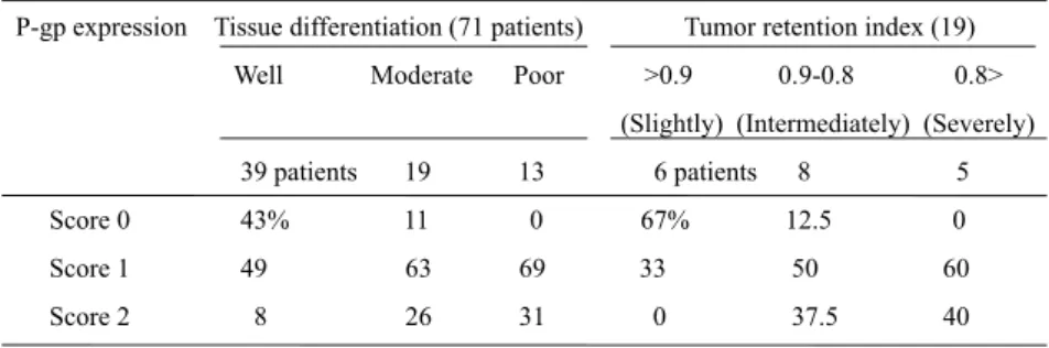
One group of 19 patients underwent both 99m-Tc-MIBI scintigraphy and an immunohistochemical examination. Moreover, another group of 71 patients underwent an immunohistochemical examination of P-gp expression.
Samples of malignant tumor were treated in citrate buffer to retrieve the antibody activity. They were incubated with H2O2, horse serum and a primary monoclonal antibody of
JSB-1. They were incubated with secondary antibody
solution, diaminobenzidine, H2O2, and peroxidase substrate
solution. Finally, the nuclei were counter-stained with hematoxylin. In addition to these samples, we used three other tissue sections for the control of negative, a moderately positive and a severely positive stains.16 We
classified grades of staining of P-gp expression into score 0 (less than 5% of tumor cells), score 1 (5-50%) and score 2 (over 50%).17 We compared P-gp expression with the tissue
differentiation and the tumor retention index in malignant tumors of the head and neck.
(Results: P-gp expression, Tissue differentiation and Tumor retention index)
With respect to the P-gp expression and tissue differentiation in 71 patients, 43% and 49% of patients in the well group showed score 0 and 1. On the other hand, most patients in the poor group showed score 1 and score 2. No patient showed score 0. As for the P-gp expression and tumor retention index in 19 patients, 67% of patients in the slightly decreased group showed score 0, and no patient showed score 2. On the other had, 40% of patients in the severely decreased group showed score 2, and no patient showed score 0 (Table 4). These results indicated that P-gp expression was distinct in patients of low differentiation group and showed a well correlation with the discharge of 99m-Tc-MIBI.
2-5. Comparison of 201-Tl with 99m-Tc-MIBI
99m-Tc-MIBI and 201-Tl had each different uptake mechanism. 99m-Tc-MIBI accumulated distinctly in malignant tumors in the early phase, but the accumulation became less intense in the late phase. 201-Tl also accumulated in malignant tumors in the early phase, but the accumulation in the delayed phase of malignant tumors did Table 4: P-gp expression, Tissue differentiation and Tumor retention index
P-gp expression Tissue differentiation (71 patients) Tumor retention index (19) Well Moderate Poor >0.9 0.9-0.8 0.8>
(Slightly) (Intermediately) (Severely) 39 patients 19 13 6 patients 8 5 Score 0 43% 11 0 67% 12.5 0 Score 1 49 63 69 33 50 60 Score 2 8 26 31 0 37.5 40
Tsuyoshi Sato, Yasuhiko Morita*, Hideyuki Majima 26
not show any distinct decrease. In this section, we compared and evaluated the usefulness of 201-Tl and 99m-Tc-MIBI for the diagnosis of malignant tumors of the head and neck.
(Results: Diagnostic reliability of 201-Tl with 99m-Tc-MIBI) The true positive, false positive, false negative, true negative, sensitivity, specificity and accuracy of the two scintigraphic agents are shown (Table 5). The sensitivity, specificity and accuracy of 201-Tl scintigraphy were 82.9%, 80.1% and 82.7%, respectively. On the other hand, the sensitivity and accuracy were 68.4% and 68.4% in 99m-Tc-MIBI scintigraphy. Thus, 201-Tl is a little superior to 99m-Tc-MIBI as an agent for malignant tumors of the head and neck.
3. DISCUSSION
We made re-evaluation of some of our previous reports3-8
on scintigraphy for malignant tumors and lymph node metastasis. There were some clues to find a solution to problems in scintigraphy. The results in this article indicated a possible hint to make a qualitative diagnosis of malignant tumors or to differentiate malignant tumors from inflammatory lesions. For example, tumor retention indexes showed different tendencies between malignant lesions and benign lesions including inflammatory changes, or the level of transport proteins on cell membrane have a possible clue to reveal grades of tissue differentiation of tumors like tumor markers.
Scintigraphy for tumors with 201-Tl and 99m-Tc-MIBI: Both 201-Tl and 99m-Tc-MIBI are now rarely used for diagnosis of malignant tumors of the head and neck8
because FDG-PET has been widely introduced for the same purpose.1 However, 201-Tl and 99m-Tc-MIBI have some
advantages to FDG-PET, for example, transport proteins ( Na+/K+-ATPase for 201-Tl and P-gp for 99m-Tc-MIBI)
were helpful for qualitative diagnosis and have a possibility to become factors like tumor markers. In addition, 201-Tl and 99m-Tc-MIBI are not so expensive. In this article, we re-evaluated retrospectively the usefulness of 201-Tl and 99m-Tc-MIBI for a diagnosis of tumors of the head and neck. We could obtain important information from dynamic scintigraphy. In the early phase, both 201-Tl and 99m-Tc-MIBI accumulated well in viable tumor cells,9,15 although
they have physical differences. Tl+ has physical effects
similar to K+ and is taken up actively because it has an ion
radius similar to K+, and malignant tumors need a large
amount of K+.18,19 On the other hand, 99m-Tc-MIBI
accumulated in tumor cells by plasma membrane potentials.6 With respect to the accumulation mechanism in
the delayed phase, we performed some evaluations and obtained some useful results. 99m-Tc-MIBI first reached tumor cells through the tumor vascular system and was taken into tumor cells by plasma membrane potentials. Next, the accumulated 99m-Tc-MIBI was discharged from tumor cells by P-gp expressed on the cell membrane which was well known as a responsible protein in the multi-drug resistance.20 On the other hand, 201-Tl was first brought to
tumor cells like 99m-Tc-MIBI, and the accumulation in tumor cells was increased by the active transportation with Na+/K+-ATPase expressed on the cell membrane.21 In our
investigation, the accumulation of 201-Tl in the delayed phase correlated well with Na+/K+-ATPase.5 As for the
relationship with the tumor retention index, the tissue differentiation and tumor retention index showed an evident correlation. This suggested that tumor retention indexes correlated with transport proteins. Tomura and co-workers22
reported a tendency that the tumor retention index of malignant tumors decreased in 99m-Tc-MIBI scintigraphy. They reported an about 30% decrease. On the other hand, Tonami and co-workers23 reported a decreased tumor
retention index of 4.6% to 6% in benign tumors, and demonstrated an increase of more than 20% in malignant tumors in 201-Tl scintigraphy. Thus, the tumor retention index decreased with 99m-Tc-MIBI and increased with 201-Tl when tumors were malignant.24 In this article, we
showed the usefulness of 99m-Tc-MIBI and 201-Tl, especially we showed that the tumor retention index Table 5. Diagnostic reliability of 201-Tl and 99m-Tc-MIBI
201-Tl 99m-Tc-MIBI True positive 77% 68 False positive 1 0 False negative 16 32 True negative 5 0 Sensitivity 82.9% 68.4 Specificity 80.1 -Accuracy 82.7 68.4
showed a good correlation with the grade of tumor malignancy, and that the accumulation chiefly depended on transport proteins of Na+/K+-ATPase and P-gp.5,25,26
4. SUMMARY
At the present time that 201-Tl and 99m-Tc-MIBI became not to be used popularly in comparison with FDG-PET, we do not expect that our previous results are useful or helpful to the routine dental practice directly. However, FDG-PET is recently found to have a problem in diagnosis of malignant tumors, for example FDG-PET accumulates both in malignant tumors and inflammatory lesions. This is just the problem that we also tried to resolve until now. Therefore, we hope that even a small part of our results shown in this article could be a clue or hint for dentists to try to find out a solution of problem, if it is a very small help.
5. REFERENCES
1. Gambihir SS, Crernin J, Schwinner J, et al. A tabulated summary of the FDG PET literature. J Nucl Med 2001;42:1-93.
2. Shreve PD, Anzai Y, Wahl Rl. Pitfalls in oncologic diagnosis with FDG-PET imaging physiologic and benign variants. Radiographics 1999;19:61-77.
3. Sato T, Indo H, Kawabata Y, Iwashita Y, Morita Y, Noikura T, Sugihara K, Mimura T. Dynamic scintigraphy with thallium-201 chloride (Tl-201) for the diagnosis of tumors of the head and neck. Oral Surg Oral Med Oral Pathol Oral Radiol Endod 2001;92:228-235.
4. Sato T, Kawabata Y, Indo H, Suenaga S, Kawano K, Iwashita Y, Morita Y, Majima H, Sugihara K, Mimura T. Scintigraphic evaluation of patients with malignant tumor of the head and neck by thallium-201-chloride (Tl-201) scintigraphy. Oral Science International 2005;2:8-16.
5. Sato T, Indo H, Kawabata Y, Kobayashi T, Suenaga S, Iwashita Y, Nitta T, Sugihara K, Majima H. Thallium-201 chloride (Tl-201) accumulation and Na+/
K+-ATPase expression in tumours of the head and neck.
Dentomaxillofacial Radiology 2005;34:212-217. 6. Sato T, Kawabata Y, Saigo Y, Iwashita Y, Suenaga S,
Indo H, Hamahira S, Kawano K, Nitta T, Morita Y, Majima H, Sugihara K. Interpretation of scintigraphic
findings of oral malignant tumours with a new scanning agent of technetium-99m-hexakis-2-methoxy-isobutyl-isonitrile (Tc-99m-MIBI). Dentomaxillofacial Radiology 2006;35:24-29.
7. Sato T, Kawabata Y, Nitta T, Saigo Y, Iwashita Y, Suenaga S, Hamahira S, Indo H, Morita Y, Majima H, Sugihara K. Expression of permeability-glycoprotein (P-gp) and uptake of technetium-99m-hexakis-2-methoxy-isobutyl-isonitrile (99Tcm-MIBI) in malignant
tumour of the head and neck. Dentomaxillofacial Radiology 2005;34:274-278.
8. Sato T, Kawabata Y, Kobayashi Y, Suenaga S, Indo H, Kawano K, Iwashita Y, Morita Y, Majima H. Scintigraphy for interpretation of malignant tumours of the head and neck: comparison of technetium-99m-hexakis-2-methoxy-isobutyl-isonitrile (Tc-MIBI) and thallium-201-chloride (Tl-201). Dentomaxillofacial Radiology 2005;34:268-273.
9. Hisada K, Tonami N, Miyamae Y, Yamazaki T, Maeda T, et al. Clinical evaluation of tumor imaging with 201-Tl chloride. Radiology 1978;129:497-500.
10. Kostakoglu L, Uysal U, Özyar E, Hayran M, Uzal D, Demirkazik FB, Kars A, Atahan L, Bekdik CF. Monitoring response to therapy with thallium-201 and technetium-99m-sestamibi SPECT in nasopharyngeal carcinoma. J Nucl Med 1997;38:1009-1014.
11. Takekawa H, Itoh K, Abe S, Ogura S, Isobe H, Furudate M, et al. Thallium-201 uptake, histopathological d i f f e r e n t i a t i o n a n d N a - K - AT P a s e i n l u n g adenocarcinoma. Jnucl Med 1996;37:955-958.
12. Abbott A, Ball WJ. The epitope for the inhibitory antibody M7-PB-E9 contains Ser-646 and Asp-652 of the sheep Na+,K+-ATPase alpha–subunit. Biochemistry
1993;32:3511-8.
13. Espineda C, Seligson DB, James Ball W Jr, Rao J, Palotie A, Horvath S, Huang Y, Shi T, Rajasekaran AK. Analysis of the Na, K-ATPase alpha– and beta–subunit expression profiles of bladder cancer using tissue microarrays. Cancer 2003;97:1859-68.
14. Kao CH, Tsai SC, Wang JJ, Ho YJ, Ho ST, Changlai SP. Technetium-99m-sestamethoxyisobutylisonitrile scan as a predictor of chemotherapy response in malignant lymphomas compared with P-glycoprotein expression, multidrug resistance-related protein expression and other prognosis factors. Br J Haematol
2001;113:369-Tsuyoshi Sato, Yasuhiko Morita*, Hideyuki Majima 28
74.
15. Taki J, Sumiya H, Tsuchiya H, Tomita K, Nonomura A, Tonami N. Evaluating benign and malignant bone and soft tissue lesions with technetium-99m-MIBI scintigraphy. J Nucl Med 1997;38:501-506.
16. Thiebaut F, Tsuruo T, Hamada H, Gottesman MM, Pastan I, Willingham, M. C. Cellular localization of the multidrug-resistance gene product P-glycoprotein in normal human tissues. Proc Natl Acad Sci USA. 1987;84:7735-8.
17. Yamashita K, Yonezawa S, Tanaka S, Shirahama H, Sakoda K, Imai K, Xing PX, McKenzie Ian FC, Hilkens J, Kim YS, Sato E. Immunohistochemical study of mucin carbohydrates and core proteins in hepatolithiasis and cholangiocarcinoma. Int J Cancer 1993;55:83-91. 18. Kasarov LB, Friedman H. Enhanced Na+-K+-activated
adenosine triphosphatase activity in transformed fibroblasts. Cancer Res 1974;34:1862-5.
19. Orihashi N, Suga K, Yoneshiro S, Fujita T, Ohno Y, Arita T, et al. Evaluation of Tl-201 SPECT in differential diagnosis of benign and malignant lesions of the chest. Yamaguchi Med J 1992;41:274-83 (in Japanese).
20. Hendrikse NH, Franssen EJ, Van-der Graaf WT, Meijer C, Piers DA, Vaalburg W, de Vries EG. 99mTc-sestamibi is a substrate for P-glycoprotein and the multidrug resistance-associated protein. Br J Cancer 1998;77:353-8.
21. Schweil AM, Mckillop JH, Milroy R, Wilson R, Abdel-Dayem HM, Omar YT. Mechanism of 201-Tl uptake in tumors. Eur J Nucl Med 1989;15:376-379.
22. Tomura N, Hirano H, Watanabe O, Kato K, Watarai J, Sasaki K, Tamura K. Evaluation of single photon emission tomography of tumors in the head and neck with technetium-99m MIBI. Kaku Igaku 1997;34: 471-479 (in Japanese).
23. Tonami N, Shuke N, Yokoyama K, Seki H, Takayama T, Kinuya S, et al. Thallium-201 single photon emission computed tomography in the evaluation of suspected lung cancer. J Nucl Med 1989;30:997-1004.
24. Yamamoto Y, Kawasaki Y, Nishiyama Y, Fukunaga K, Satoh K, Takashima H, et al. Comparative evaluation of 99m-Tc-MIBI (hexakis 2-methoxy isobutyl isonitrile) and 201Tl-chloride in primary lung cancer. Kaku Igaku 1996;33:501-11 (in Japanese).
25. Kostakoglu L. Elahi N, Kiratli P, Ruacan S, Sayek I, Baltali E, Sungur A, Hayran M, Bekdik CF. Clinical validation of the influence of p-glycoprotein on technetium-99m-sestamibi uptake in malignant tumors.
J Nucl Med 1997;38:1003-1008.
26. Rabkin D, Chhieng DC, Miller MB, Jennings T, Feustel P, Steiniger J, Parnes SM. P-glycoprotein expression in squamous cell carcinoma of the tongue base.
LARYNGOSCOPE 1995;105:1294 –1299.
LEGENDS
Figure 1: Two regions of interest (ROI) on a frame image covered the tumor area (A) and the symmetrical region (B: control region).
Figure 2: Two curves showed radioactive count after injection in the tumor and control areas. In the dynamic scintigraphy, the early retention index was A/a and the delayed retention index was B/b. The tumor retention index was (delayed retention index) / (early retention index).