32
* Department of Rehabilitation, Faculty of Health Science and Technology, Kawasaki University of Medical Welfare, Kurashiki, Okayama 701-0193, Japan
E-Mail: ishida@mw.kawasaki-m.ac.jp 1. Introduction
Lumbar stability and low back stabilizing exercises have emerged as popular topics related to optimal athletic/occupational performance and to the rehabilitation of painful backs. The objective of such exercise is to enhance the function of the critical trunk muscles in a way that spares the spine from damage [1]. Ultrasound imaging was introduced in rehabilitation to evaluate muscle morphology and function in persons with musculoskeletal disorders such as low back pain [2]. Most rehabilitative ultrasound imaging research has focused on transversus abdominis (TrA) and lumbar multifidus (LM) muscles, because dysfunction of these muscles has been linked to low back pain (LBP) [3-6]. Muscle morphology refers to the shape, size, and structure of a muscle and may be important in rehabilitation as an indication of muscle atrophy and/ or hypertrophy. The ability of ultrasound imaging to quantify muscle size depends on its reliability and validity. Using ultrasound to quantify muscle size is relatively straightforward and relies on its ability to
Influence of Inward Pressure Applied by
the Transducer on Trunk Muscle
Thickness during Ultrasound Imaging
Hiroshi ISHIDA
*and Susumu WATANABE
*(Accepted Oct. 22, 2013)
Key words: transversus abdominis, internal oblique, external oblique, lumbar multifidus, ultrasound imaging
Abstract
The purpose of this study was to clarify the change in trunk muscle thickness induced by different inward pressures applied by the transducer during ultrasound imaging. Eleven healthy male volunteers participated in the study. The thickness of the right transversus abdominis (TrA), internal oblique (IO), external oblique (EO), and lumbar multifidus (LM) muscles were measured by ultrasound imaging in the following two conditions, with inward pressures of approximately 0.1 N and 2.0 N. The mean difference between the 0.1 N and 2.0 N conditions was less than the minimal detectable change (MDC) of the 0.1 N and 2.0 N conditions in the TrA and IO muscles. The mean difference between the 0.1 N and 2.0 N conditions was greater than the MDC of the 0.1 N and 2.0 N conditions in the EO and LM muscles. Sensitivity to pressure is different among the trunk muscles. Therefore, maintaining consistent transducer-induced inward pressure is required to clarify the minimal changes of muscle thickness induced by the intervention in EO and LM muscles. However, this is not so in the case of the TrA and IO muscles.
measure muscle thickness and cross-sectional area. Despite the excellent intraclass correlation coefficient (ICC) values reported to date, further investigation is required to determine whether methods can be used to reduce measurement errors [7]. Diligent attention to steadying the position, orientation, and inward pressure of the transducer is required during ultrasound imaging [8]. A recent study indicated that inward pressures of the transducer during ultrasound imaging decreased the thickness of the TrA, internal oblique (IO), and external oblique (EO) muscles [9]. However, little is known about the influence of inward pressure of the transducer on LM muscle thickness during ultrasound imaging. Therefore, the purpose of this study was to clarify the changes in TrA, IO, EO, and LM muscle thickness induced by different inward pressures applied by the transducer during ultrasound imaging.
2. Materials and methods
2.1. Participants
Eleven healthy male volunteers participated in this study. Their age, height, and weight (mean ± standard deviation) were 21.5 ± 2.9 years, 172.9 ± 5.4 cm, and 66.5 ± 8.7 kg, respectively. Informed consent was obtained from all participants.
2.2. Procedure
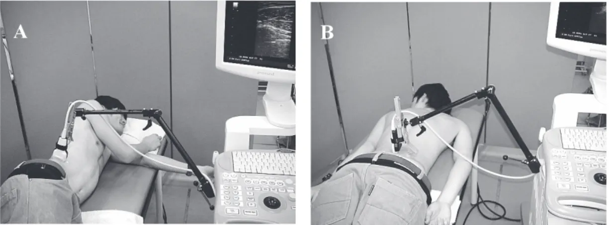
The same experimenter was responsible for collecting all ultrasound imaging data. B-mode ultrasound (SSD-3500SX; Aloka Co. Ltd., Japan) with a 10 MHz transducer was used for collecting ultrasound imaging with muscle thickness of TrA, IO, EO and LM. For collecting ultrasound imaging of the lateral abdominal muscles, the subject was positioned in the side-lying position (Fig. 1). Gel was interposed between the transducer and the skin. The transducer was then placed transversely on the right side of the body, with its center positioned 25 mm anterior to the midaxillary line and at the midpoint between the inferior rib and iliac crest [10]. A custom-made holder was designed to enable hands-free application of the ultrasound transducer, which could maintain the inward pressure of approximately 2.0 N applied by the weight of the transducer and holder and allow the use of a linear-motion guide, the THK Miniature Linear Guide (RSR7; THK Co. Ltd., Japan) (Fig. 2) [9]. The location of the transducer holder was fixed. Therefore, after fixation, we could repeat testing under different instrument conditions (different inward pressures) using the same position and orientation of the transducer holder [9]. The inward pressure of approximately 0.1 N was applied by holding the transducer holder manually to avoid contact between the transducer and skin (Fig. 3). We measured the force output of the transducer holder using a force plate (P08-1713; Kyowa Electronic Instruments Co. Ltd., Japan). For collecting ultrasound imaging of the LM muscle, the subject was positioned in the prone position, with a towel placed under the abdomen to minimize lumbar lordosis
Fig. 1 (A) Measurement of ultrasound imaging for the lateral abdominal muscles (B) The lumbar multifidus muscle
Fig. 2 Custom-made holder
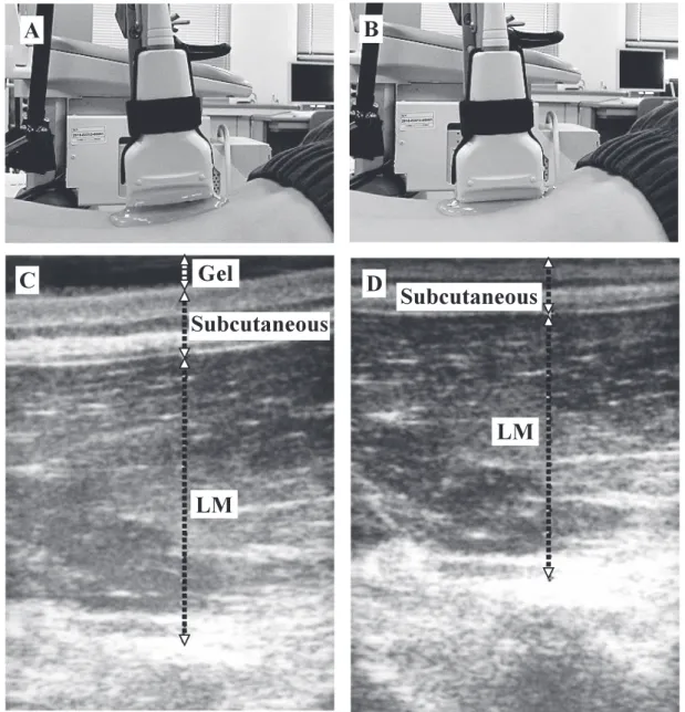
Fig. 3 Measurement of ultrasound imaging of the lumbar multifidus muscle (A) The 0.1 N condition
(B) The 2.0 N condition
(C) Ultrasound imaging of lumbar multifidus (LM) muscle in 0.1 N condition (D) Ultrasound imaging of LM muscle in 2.0 N condition
(Fig. 1). The transducer was placed longitudinally along the spine with the mid-point over the L4 spinous process. It was moved laterally and angled slightly medially until the L4/5 zygapophyseal joint could be identified [11]. Two images were collected at 0.1 N and 2.0 N, and this was repeated three times. The two conditions were performed in random order. The thickness of each of the abdominal muscles (TrA, IO, and EO muscles) was measured at the center line of the image. The thickness of the LM muscle was measured above the L4/5 zygapophyseal joint. The average values of the three trials for each condition were used for analysis. The absolute changes from the 0.1 N condition were calculated as 0.1 N condition – 2.0 N condition (mm) for the TrA, IO, EO, and LM muscles.
2.3. Statistical analysis
SPSS 16.0J for Windows was used for statistical analyses. Intrarater reliability of ultrasound imaging measurement was examined by calculating the ICC values. The standard error of measurement (SEM = SDpool × 1-ICC ) and the minimal detectable change (MDC) for a 95% confidence interval (MDC = SEM × 1.96 × 2 ) were calculated for each ultrasound measurement taken [11]. The paired t-test was used to determine differences between 0.1 N and 2.0 N conditions. Values were considered statistically significant at p < 0.05.
3. Results
The intra-examiner reliabilities are listed in Table 1. The thicknesses of abdominal and LM muscles are shown in Table 2. Significant differences were observed in all parameters (Table 2). The mean difference between the 0.1 N and 2.0 N conditions were less than the MDC of the 0.1 N and 2.0 N conditions in the TrA and IO muscles. The mean difference between the 0.1 N and 2.0 N conditions were greater than the MDC of the 0.1 N and 2.0 N conditions in the EO and LM muscles.
√ √
Table 1 Intra-examiner reliability
ICC (1, 3) SEM (mm) MDC (mm) TrA 0.1 N 0.99 0.1 0.4 2.0 N 0.99 0.1 0.4 IO 0.1 N 0.99 0.4 1.2 2.0 N 0.99 0.4 1.2 EO 0.1 N 0.98 0.4 1.0 2.0 N 0.99 0.3 0.7 LM 0.1 N 0.99 0.4 1.1 2.0 N 0.99 0.4 1.1
ICC : intraclass correlation coefficients SEM : standard error of measurement MDC : minimal detectable change TrA : transversus abdominis IO : internal oblique EO : external oblique LM : lumbar multifidus
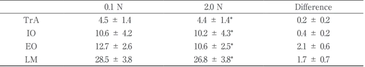
Table 2 Thicknesses of abdominal and LM muscles (mm)
0.1 N 2.0 N Difference
TrA 4.5 ± 1.4 4.4 ± 1.4* 0.2 ± 0.2
IO 10.6 ± 4.2 10.2 ± 4.3* 0.4 ± 0.2
EO 12.7 ± 2.6 10.6 ± 2.5* 2.1 ± 0.6
LM 28.5 ± 3.8 26.8 ± 3.8* 1.7 ± 0.7
TrA: transversus abdominis IO : internal oblique EO : external oblique LM : lumbar multifidus
4. Discussion
We investigated the changes in the TrA, IO, EO, and LM muscle thickness induced by differing inward pressures applied by the transducer during ultrasound imaging. In this study, the difference in magnitude produced by the forces under different conditions is meaningless because the mean difference between the 0.1 N and 2.0 N conditions were less than the MDC of the 0.1 N and 2.0 N conditions in the TrA and IO muscles. The difference in magnitude produced by the forces under different conditions is meaningful because the mean difference between the 0.1 N and 2.0 N conditions were greater than the MDC of the 0.1 N and 2.0 N conditions in the EO and LM muscles. Sensitivity of pressure is different among the trunk muscles. Inward pressure of 2.0 N is an acceptable pressure to measure the thickness of TrA and IO muscles. Inward pressure of 2.0 N is not an acceptable pressure to measure the thickness of EO and LM muscles. These data suggest that elasticity of tissues between the target muscle and the transducer might influence pressure applied to the target muscle during ultrasound imaging [12]. More thickness changes occurred in the EO and LM muscles than the others because tissue thickness between the EO and LM muscles and the transducer was less than in the other tests. Possible causes of lower observed changes in the TrA and IO muscles might be the existence of the EO between the TrA and IO and the transducer. The LM muscle was sensitive to the pressure, because there was reaction force from the spine under the LM. Elasticity of tissues under the target muscle might influence pressure on the target muscle during ultrasound imaging. For example, Akbari et al. [13] found that eight weeks of motor control exercise resulted in an increase of 1.1 mm in LM muscle thickness, and that general exercise resulted in an increase of 0.4 mm at rest in individuals with LBP. Different inward pressures applied by the transducer during ultrasound imaging can induce these changes. Therefore, maintaining consistent transducer-induced inward pressure is required to clarify the minimal changes of muscle thickness induced by the intervention in EO and LM muscles. However, this is not so in the case of the TrA and IO muscles.
The limitation of this study is the fact that only healthy young subjects were included in the analysis. Wallwork et al. [4] demonstrated that LBP is related to a decrease in the thickness of the LM muscle. As measurement of muscle hardness may be influenced by muscle thickness [12]. Furthermore, the influence of inward pressure of the transducer on atrophic LM muscle remains unknown. Therefore, we plan to clarify the changes in LM muscle thickness induced by different inward pressures applied by the transducer during ultrasound imaging in the weak or deconditioned LM muscles of middle-aged and elderly individuals with LBP.
References
1. Kavcic N, Grenier S, McGill SM: Quantifying tissue loads and spine stability while performing commonly prescribed low back stabilization exercises. Spine 29: 2319-2329, 2004.
2. Teyhen DS: Rehabilitative ultrasound imaging: the roadmap ahead. J Orthop Sports Phys Ther 37: 431-433, 2007.
3. Hungerford B, Gilleard W, Hodges P: Evidence of altered lumbopelvic muscle recruitment in the presence of sacroiliac joint pain. Spine 28: 1593-1600, 2003.
4. Wallwork TL, Stanton WR, Freke M, Hides JA: The effect of chronic low back pain on size and contraction of the lumbar multifidus muscle. Man Ther 14: 496-500, 2009.
5. Yoshihara K, Shirai Y, Nakayama Y, Uesaka S: Histochemical changes in the multifidus muscle in patients with lumbar intervertebral disc herniation. Spine 26: 622-626, 2001.
6. Zhao WP, Kawaguchi Y, Matsui H, Kanamori Y, Kimura T: Histochemistry and morphology of the multifidus muscle in lumbar disc herniation: comparative study between diseased and normal sides.
Spine 25: 2191-2199, 2000.
7. Whittaker JL, Teyhen DS, Elliott JM, Cook K, Langevin HM, Dahl HH, Stokes M: Rehabilitative ultrasound imaging: understanding the technology and its applications. J Orthop Sports Phys Ther 37:
434-449, 2007.
8. Teyhen DS, Gill NW, Whittaker JL, Cook K, Langevin HM, Dahl HH, Stokes M: Rehabilitative ultrasound imaging of the abdominal muscles. J Orthop Sports Phys Ther 37: 450-466, 2007.
9. Ishida H, Watanabe S: Influence of inward pressure of the transducer on lateral abdominal muscle thickness during ultrasound imaging. J Orthop Sports Phys Ther 42: 815-818, 2012.
10. Mannion AF, Pulkovski N, Gubler D, Gorelick M, O’Riordan D, Loupas T, Schenk P, Gerber H, Sprott H: Muscle thickness changes during abdominal hollowing: an assessment of between-day measurement error in controls and patients with chronic low back pain. Eur Spine J 17: 494-501, 2008.
11. Kiesel KB, Underwood FB, Mattacola CG, Nitz AJ, Malone TR: A comparison of select trunk muscle thickness change between subjects with low back pain classified in the treatment-based classification system and asymptomatic controls. J Orthop Sports Phys Ther 37: 596-607, 2007.
12. Muraki S, Fukuda O, Fukumoto K: Validity of muscle strength evaluation by elasticity measuring instruments using ultrasound signal. Descente Sports Sci 30: 105-113, 2009 (in Japanese).
13. Akbari A, Khorashadizadeh S, Abdi G: The effect of motor control exercise versus general exercise on lumbar local stabilixing muscles thickness: randomized controlled trial of patients with chronic low back pain. J Back Musculoskelet Rehabil 21: 105-112, 2008.