Research Article
Preventive Effects of a Kampo Medicine, Kakkonto, on
Inflammatory Responses via the Suppression of
Extracellular Signal-Regulated Kinase Phosphorylation in
Lipopolysaccharide-Treated Human Gingival Fibroblasts
Hiroyuki Kitamura,
1Hiroko Urano,
2and Toshiaki Ara
1,31Department of Hard Tissue Research, Graduate School of Oral Medicine, Matsumoto Dental University, Shiojiri, Nagano 399-0781, Japan
2Institute for Oral Science, Graduate School of Oral Medicine, Matsumoto Dental University, Shiojiri, Nagano 399-0781, Japan 3Department of Pharmacology, Matsumoto Dental University, 1780 Gobara, Hirooka, Shiojiri, Nagano 399-0781, Japan
Correspondence should be addressed to Toshiaki Ara; ara t@po.mdu.ac.jp Received 30 November 2013; Accepted 8 January 2014; Published 18 February 2014 Academic Editors: A. Pittaluga and E. M. Urbanska
Copyright © 2014 Hiroyuki Kitamura et al. This is an open access article distributed under the Creative Commons Attribution License, which permits unrestricted use, distribution, and reproduction in any medium, provided the original work is properly cited.
Periodontal disease is accompanied by inflammation of the gingiva and destruction of periodontal tissues, leading to alveolar bone loss in severe clinical cases. The chemical mediator prostaglandin E2(PGE2) and cytokines such as interleukin- (IL-)6 and IL-8 have been known to play important roles in inflammatory responses and tissue degradation. In the present study, we investigated the effects of a kampo medicine, kakkonto (TJ-1), on the production of prostaglandin E2(PGE2), IL-6, and IL-8 by human gingival fibroblasts (HGFs) treated with lipopolysaccharide (LPS) from Porphyromonas gingivalis. Kakkonto concentration dependently suppressed LPS-induced PGE2production but did not alter basal PGE2levels. In contrast, kakkonto significantly increased LPS-induced IL-6 and IL-8 production. Kakkonto decreased cyclooxygenase- (COX-)1 activity to approximately 70% at 1 mg/mL but did not affect COX-2 activity. Kakkonto did not affect cytoplasmic phospholipase A2(cPLA2), annexin1, or LPS-induced COX-2 expression. Kakkonto suppressed LPS-induced extracellular signal-regulated kinase (ERK) phosphorylation, which is known to lead to ERK activation and cPLA2phosphorylation. These results suggest that kakkonto decreased PGE2production by inhibition of ERK phosphorylation which leads to inhibition of cPLA2phosphorylation and its activation. Therefore, kakkonto may be useful to improve gingival inflammation in periodontal disease.
1. Introduction
Periodontal disease is accompanied by inflammation of the gingiva and destruction of periodontal tissues, leading to
alveolar bone loss in severe clinical cases. Prostaglandin E2
(PGE2), interleukin- (IL-)6, and IL-8 are known to play
important roles in inflammatory responses and tissue
degra-dation. PGE2 has several functions in vasodilation, the
enhancement of vascular permeability and pain, and the induction of osteoclastogenesis and is believed to play impor-tant roles in inflammatory responses and alveolar bone resorption in periodontal disease [1]. IL-6 has the ability
to induce osteoclastogenesis [2,3]. IL-8 acts as a
chemoat-tractant for neutrophils [4] that produce proteases such as cathepsin, elastase, and matrix metalloproteinase- (MMP-)8, leading to tissue degradation.
Recently, we reported that several kampo medicines, shosaikoto [5], hangeshashinto [6], and orento [7],
sup-press lipopolysaccharide- (LPS-) induced PGE2 production
by HGFs and suggested that these kampo medicines have anti-inflammatory effects in periodontal disease. Another kampo medicine, kakkonto (TJ-1), has been clinically used for various diseases such as the common cold, coryza, the initial stage of febrile diseases, and inflammatory diseases. There Volume 2014, Article ID 784019, 7 pages
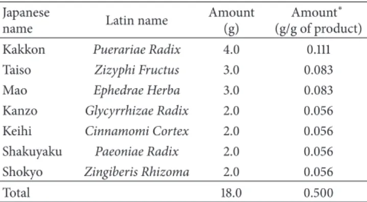
Table 1: Ingredients of the kakkonto formula. Japanese
name Latin name
Amount (g)
Amount∗ (g/g of product) Kakkon Puerariae Radix 4.0 0.111 Taiso Zizyphi Fructus 3.0 0.083
Mao Ephedrae Herba 3.0 0.083
Kanzo Glycyrrhizae Radix 2.0 0.056 Keihi Cinnamomi Cortex 2.0 0.056 Shakuyaku Paeoniae Radix 2.0 0.056 Shokyo Zingiberis Rhizoma 2.0 0.056
Total 18.0 0.500
∗7.5 g of kakkonto product contains 3.75 g of a dried extract of the mixed
crude drugs.
are several reports that kakkonto shows antiallergic effects [8,9] and antiviral effects [10–13] in animal and in vitro exper-imental models. For anti-inflammatory effects, kakkonto has
been reported to decrease PGE2 production in cultured
rabbit astrocytes [14]. Therefore, we considered the
possi-bility that kakkonto decreases PGE2 production by human
gingival fibroblasts (HGFs) and has anti-inflammatory effects with respect to periodontal disease. However, the anti-inflammatory effects of kakkonto are not adequately under-stood.
HGFs are the most prominent cells in periodontal tissue. Moreover, LPS-treated HGFs produce inflammatory
chemi-cal mediators such as PGE2and inflammatory cytokines such
as IL-6 and IL-8 [2,15, 16]. Moreover, because HGFs have
sustained production of PGE2[17], IL-6, and IL-8 [18] in the
presence of LPS, these mediators and cytokines in periodon-tal tissues are thought to be derived from HGFs. Therefore, we believe that examining the effects of drugs on HGFs, as well as on monocytes and macrophages, is important in the study of periodontal disease. In the present study, we
examined the effect of kakkonto on LPS-induced PGE2,
IL-6, and IL-8 production using this in vitro model.
2. Materials and Methods
2.1. Reagents. Kakkonto was purchased from Tsumura & Co. (Tokyo, Japan; lot number: D23122), and its components are
listed in Table 1. Kakkonto was suspended in Dulbecco’s
modified Eagle’s medium (D-MEM, Sigma, St. Louis, MO, USA) containing 10% heat-inactivated fetal calf serum, 100 units/mL penicillin, and 100 mg/mL streptomycin (culture
medium) and was rotated at 4∘C overnight. Then, the
sus-pension was centrifuged and the supernatant was filtrated
through a 0.45𝜇m-pore membrane. Phorbol 12-myristate
13-acetate (PMA) purchased from Sigma. Other reagents were purchased from Nacalai tesque (Kyoto, Japan). LPS from Porphyromonas gingivalis 381 was provided by Professor Nobuhiro Hanada (School of Dental Medicine, Tsurumi University, Japan).
2.2. Cells. HGFs were prepared as described previously [6]. In brief, HGFs were prepared from free gingiva during the
extraction of an impacted tooth with the informed consent of the subjects who consulted Matsumoto Dental University Hospital. The free gingival tissues were cut into pieces and seeded onto 24-well plates (AGC Techno Glass Co., Chiba,
Japan). HGFs were maintained in culture medium at 37∘C in
a humidified atmosphere of 5% CO. For passage, HGFs were trypsinized, suspended, and plated into new cultures in a 1 : 3 dilution ratio. HGFs were used between the 10th and 15th passages in the assays. This study was approved by the Ethical Committee of Matsumoto Dental University (number 0063). 2.3. Cell Viability. The numbers of cells were measured using WST-8 (Cell Counting Kit-8; Dojindo, Kumamoto, Japan) according to the manufacturer’s instructions. In brief, HGFs (10,000 cells/well) were seeded in 96-well plates (AGC Techno Glass Co., Chiba, Japan) and incubated in
serum-containing medium at 37∘C overnight. Then, the cells were
treated with various concentrations of kakkonto (0, 0.5, 1, 2, 5, and 10 mg/mL) in the absence or presence of LPS (10 ng/mL)
for 24 h (200𝜇L each well) in quadruplicate for each sample.
Then, the media were removed by aspiration and the cells
were treated with 100𝜇L of mixture of WST-8 with culture
medium for 2 h at 37∘C in CO incubator. Optical density
was measured (measured wavelength at 450 nm and reference wavelength at 655 nm) using an iMark microplate reader (Bio-Rad, Hercules, CA, USA), and the mean background value was subtracted from each value. Data is represented as
means± SD (𝑛 = 4).
2.4. Enzyme-Linked Immunosorbent Assay (ELISA). HGFs (10,000 cells/well) were seeded in 96-well plates and
incu-bated in serum-containing medium at 37∘C overnight. Then,
the cells were treated with various concentrations of kakkonto (0, 0.01, 0.03, 0.1, 0.3, and 1 mg/mL) in the absence or presence
of LPS (10 ng/mL) for 24 h (200𝜇L each well) in triplicate for
each sample. After the culture supernatants were collected, viable cell numbers were measured using WST-8 as described
above. The concentrations of PGE2, IL-6, and IL-8 in the
culture supernatants were measured by ELISA according to
the manufacturer’s instructions (PGE2, Cayman Chemical,
Ann Arbor, MI, USA; IL-6 and IL-8, Biosource International Inc., Camarillo, CA, USA) and were adjusted by the number of viable cells. Data are represented as ng or pg per 10,000 cells
(mean± SD, 𝑛 = 3).
2.5. Cyclooxygenase Activity. The effects of kakkonto on the activities of cyclooxygenase (COX)-1 and COX-2 were analyzed using a COX inhibitor screening assay kit (Cayman Chemical, Ann Arbor, MI, USA) according to the manu-facturer’s instructions. COX activities were evaluated by the measurement of prostaglandin produced from arachidonic acid by COX-1 or COX-2. These values were normalized to a relative value of 100% for cells without LPS or kakkonto
treatments, and are represented as means± SD (𝑛 = 3).
2.6. Western Blotting. HGFs were cultured in 60 mm dishes and treated with combinations of LPS and kakkonto for the indicated times. Then, cells were washed twice with Tris-buffered saline, transferred into microcentrifuge tubes,
0 0.5 1 2 5 10 Kakkonto (mg/mL) 0 20 40 60 80 100 120 V ia bili ty (% o f co nt ro l) ∗∗∗ ∗∗∗ ∗∗ ∗∗∗ ∗∗∗ # ### ### ###
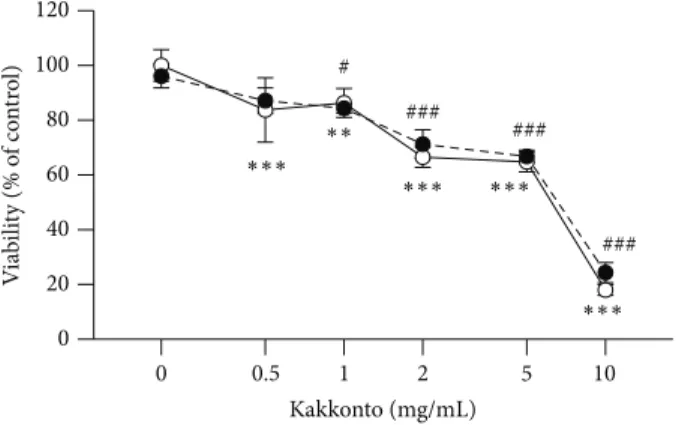
Figure 1: Effects of kakkonto on HGFs viability. The effect of kakkonto on the viability of HGFs at 24 h. HGFs were plated in 96-well microplates at 10,000 cells/mL, and media containing LPS and kakkonto were added. Cell numbers were evaluated by WST-8 at 24 h. The optical density (OD) was normalized to a relative value of 100% for cells without LPS or kakkonto treatments and is represented as means± SD (𝑛 = 4). Open circles, treatment without LPS; closed circles, treatment with 10 ng/mL of LPS.∗∗𝑃 < 0.01 and∗∗∗𝑃 < 0.001 (without kakkonto versus with kakkonto
in the absence of LPS). #𝑃 < 0.01 and##𝑃 < 0.001 (without kakkonto versus with kakkonto in the presence of LPS).𝑃 values were calculated by pairwise comparisons and corrected with the Holm method (16 null hypotheses).
and centrifuged at 6,000×g for 5 min at 4∘C. Supernatants
were aspirated and cells were lysed on ice in lysis buffer (50 mM Tris-HCl, pH 7.4, 1% Nonidet P-40, 0.25% sodium deoxycholate, 150 mM NaCl, 1 mM ethyleneglycol bis(2-aminoethylether) tetraacetic acid (EGTA), 1 mM sodium orthovanadate, 10 mM sodium fluoride, 1 mM
phenylmethyl-sulfonyl fluoride, 10𝜇g/mL aprotinin, 5 𝜇g/mL leupeptin, and
1𝜇g/mL pepstatin) for 30 min at 4∘C. Then, samples were
cen-trifuged at 12,000×gfor 15 min at 4∘C, and supernatants were
collected. The protein concentration was measured using a BCA Protein Assay Reagent Kit (Pierce Chemical Co., Rock-ford, IL, USA).
The samples (10𝜇g of protein) were fractionated in a
polyacrylamide gel under reducing conditions and trans-ferred onto a polyvinylidene difluoride (PVDF) membrane (Hybond-P; GE Healthcare, Uppsala, Sweden). The mem-branes were blocked with 5% ovalbumin for 1 h at room temperature and incubated with primary antibody for an additional 1 h. The membranes were further incubated with horseradish peroxidase-conjugated secondary antibodies for 1 h at room temperature. Protein bands were visualized with an ECL Kit (GE Healthcare).
Antibodies against COX-2 (sc-1745, 1 : 500 dilution),
cyto-plasmic phospholipase A2 (cPLA2, sc-438, 1 : 200 dilution),
annexin1 (sc-11387, 1 : 1,000 dilution), and actin (sc-1616, 1 : 1,000 dilution), which detects a broad range of actin isoforms, were purchased from Santa Cruz Biotechnology (Santa Cruz, CA, USA). Antibodies against extracellular signal-regulated kinase (ERK; p44/42 MAP kinase antibody, 1 : 1,000 dilution) and phosphorylated ERK (Phospho-p44/42 MAPK (Thr202/Tyr204) (E10) monoclonal antibody, 1 : 2,000 dilution) were from Cell Signaling Technology (Danvers,
MA, USA). Horseradish peroxidase-conjugated anti-goat IgG (sc-2020, 1 : 20,000 dilution) was from Santa Cruz, and anti-rabbit IgG (1 : 20,000 dilution) and anti-mouse IgG (1 : 20,000 dilution) were from DakoCytomation (Glostrup, Denmark). 2.7. Statistical Analysis. Differences between groups were evaluated by the two-tailed pairwise comparison test with a pooled variance, followed by correction with the Holm method (total 16 null hypotheses; 5 null hypotheses without kakkonto versus with kakkonto in the absence of LPS, 5 null hypotheses without kakkonto versus with kakkonto in the presence of LPS, and 6 null hypotheses without LPS versus
with LPS) (Figures1and2). Differences between the control
group and experimental groups were evaluated by a
two-tailed Dunnett’s test (Figure3).
All computations were performed with the statistical pro-gram R (http://www.r-project.org/). Dunnett’s test was per-formed using the “glht” function in the “multcomp” package.
Values with𝑃 < 0.05 were considered significantly different.
3. Results
3.1. Effects of Kakkonto on HGFs Viability. First, we examined the effect of kakkonto on HGFs viability. The viability of HGFs was approximately 90% at up to 1 mg/mL of kakkonto for a 24 h treatment in the absence or presence of LPS
(Figure1). The viabilities were approximately 70% and 20% at
5 mg/mL and 10 mg/mL of kakkonto, respectively (Figure1).
Therefore, we used kakkonto at the concentrations of up to 1 mg/mL in further experiments.
3.2. Effects of Kakkonto on PGE2, IL-6, and IL-8 Production. We examined whether kakkonto affects the production of
PGE2and inflammatory cytokines (IL-6 and IL-8) by HGFs.
Because kakkonto affects cell viability, the concentrations of
PGE2, IL-6, and IL-8 needed to be adjusted according to
viable cell number.
When HGFs were treated with 10 ng/mL of LPS, HGFs
produced large amounts of PGE2, IL-6, and IL-8.
Indometh-acin decreased LPS-induced PGE2 production in a
concen-tration-dependent manner but slightly decreased LPS-induced IL-6 and IL-8 production (data not shown).
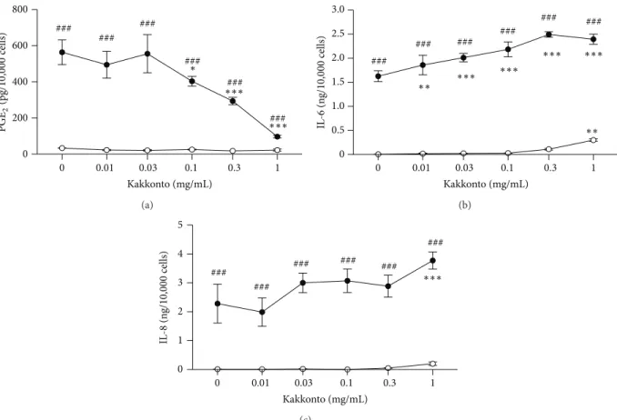
Kakkonto significantly decreased PGE2production in a
con-centration-dependent manner (Figure2(a)). In the absence
of LPS, kakkonto had no effect on PGE2 production
(Fig-ure2(a)). In contrast, kakkonto increased LPS-induced IL-6
and IL-8 production (Figures2(b)and2(c)). In the absence of
LPS, up to 0.1 mg/mL of kakkonto did not affect 6 and IL-8 production, but above 0.3 mg/mL of kakkonto, their
con-centrations were increased (Figures 2(b)and2(c)). Similar
results were obtained using human skin fibroblast TIG-103 cells (data not shown).
3.3. Effects of Kakkonto on COX Activities. Because PGE2 production is regulated by COX enzymes and suppressed by acid NSAIDs such as aspirin and diclofenac sodium, which inhibit COX activities, we examined whether kakkonto inhibits COX-1 and COX-2 activities. Kakkonto decreased
0 0.01 0.03 0.1 0.3 1 Kakkonto (mg/mL) 0 200 400 600 800 ∗∗∗ ∗∗∗ ### ### ### ### ### ### ∗ PGE 2 (pg/ 10 ,000 cells) (a) 0 0.01 0.03 0.1 0.3 1 Kakkonto (mg/mL) 0 0.5 1.0 1.5 2.0 2.5 3.0 IL -6 (n g/10,000 cells) ∗∗∗ ∗∗∗ ∗∗∗ ∗∗∗ ∗∗ ∗∗ ### ### ### ### ### ### (b) 0 0.01 0.03 0.1 0.3 1 Kakkonto (mg/mL) 0 1 2 3 4 5 IL -8 (n g/10,000 cells) ∗∗∗ ### ### ### ### ### ### (c)
Figure 2: Effects of kakkonto on the production of PGE2, IL-6, and IL-8. HGFs were treated with combinations of LPS (0 and 10 ng/mL) and kakkonto (0, 0.01, 0.3, 0.1, 0.3, and 1 mg/mL) for 24 h. Concentrations of PGE2(a), IL-6 (b), and IL-8 (c) were measured by ELISA, adjusted by cell number, and expressed as per 10,000 cells (mean± SD, 𝑛 = 3). Open circles, treatment without LPS; closed circles, treatment with 10 ng/mL of LPS.∗∗𝑃 < 0.01 and∗∗∗𝑃 < 0.001 (without kakkonto versus with kakkonto).##𝑃 < 0.001 (without LPS versus with LPS). 𝑃 values were calculated by pairwise comparisons and corrected with the Holm method (16 null hypotheses).
0 0.1 0.3 1 Kakkonto (mg/mL) 0 50 100 150 C O X-1 ac tivi ty (% o f co nt ro l) ∗ (a) Kakkonto (mg/mL) 0 0.1 0.3 1 0 50 100 150 C O X-2 ac tivi ty (% o f co nt ro l) (b)
Figure 3: Effects of kakkonto on COX activities. COX activities were evaluated by measurement of prostaglandin produced from arachidonic acid by COX-1 or COX-2. These values were normalized to a relative value of 100% for cells without LPS or kakkonto treatments and are represented as means± SD (𝑛 = 3).∗𝑃 < 0.05 (Dunnett’s test).
LPS (ng/mL) Kakkonto (mg/mL) COX-2 Annexin1 Actin 10 0 0 0.1 1 0 0.1 1 cPLA2
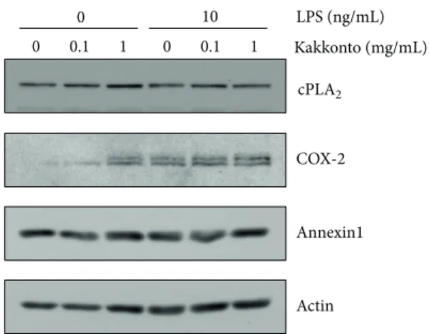
Figure 4: Effects of kakkonto on cPLA2, COX-2, and annexin1 expressions. HGFs were treated with a combination of LPS (0 or 10 ng/mL) and kakkonto (0, 0.01, or 1 mg/mL) for 8 h, and protein levels were examined by western blotting.
COX-1 activity to approximately 70% at 1 mg/mL but did not
affect COX-2 activity (Figure3).
3.4. Effects of Kakkonto on Molecular Expression in the Arachi-donic Acid Cascade. We examined whether kakkonto affects the expression of molecules in the arachidonic acid cascade.
cPLA2 is the most upstream enzyme in the arachidonic
acid cascade and releases arachidonic acid from plasma
membranes. Kakkonto did not alter cPLA2 expression in
the absence or presence of LPS (Figure4). COX-2 was not
detected in the absence of LPS. Treatment with kakkonto alone increased COX-2 expression. However, kakkonto
did not alter LPS-induced COX-2 expression (Figure 4).
Annexin1, also named lipocortin1, is an anti-inflammatory
mediator produced by glucocorticoids that inhibit cPLA2
activity [19,20]. However, neither LPS nor kakkonto showed
an effect on annexin1 expression (Figure4).
3.5. Effects of Kakkonto on ERK Phosphorylation. cPLA2 is reported to be directly phosphorylated at Ser505 by ERK,
resulting in cPLA2activation [21,22]. Therefore, we examined
whether kakkonto suppresses LPS-induced ERK phospho-rylation. ERK phosphorylation was enhanced at 0.5 h after LPS treatment and thereafter was attenuated. One mg/mL of kakkonto suppressed LPS-induced ERK phosphorylation at
0.5 h to 2 h (Figure5).
4. Discussion
In the present study, we examined the effect of kakkonto
on LPS-induced PGE2, IL-6, and IL-8 production by
HGFs. Kakkonto concentration dependently decreased
LPS-induced PGE2production but did not affect PGE2
produc-tion without LPS treatment, similar to shosaikoto, hange-shashinto, and orento [5–7]. Moreover, kakkonto suppressed LPS-induced ERK phosphorylation. In contrast, kakkonto increased LPS-induced IL-6 and IL-8 production. It is widely
known that PGE2 leads to inflammatory responses such
pERK 0 0.5 1 2 0.5 1 2 0.5 LPS Kakkonto PMA (h) ERK Actin LPS +
Figure 5: Effects of kakkonto on LPS-induced ERK phosphoryla-tion. HGFs were untreated (0 h), treated with LPS (10 ng/mL), or treated with both LPS and kakkonto (1 mg/mL) for 0.5, 1, and 2 h. As a positive control, HGFs were treated with 1𝜇M of PMA for 0.5 h. Western blotting was performed using antiphosphorylated ERK or anti-ERK antibodies. pERK: phosphorylated ERK. Upper band indicates ERK1 (p44 MAPK) and lower band indicates ERK2 (p42 MAPK).
as vasodilation, enhanced vascular permeability, and pain generation [1]. Acid non-steroidal anti-inflammatory drugs NSAIDs show anti-inflammatory effects by suppression of
PGE2production, even though they do not affect 6 and
IL-8 production. Our findings showing that kakkonto decreases
LPS-induced PGE2 production suggest that kakkonto also
has anti-inflammatory effects in periodontal disease and that
its effects are mainly mediated by suppression of PGE2
production even though kakkonto increased LPS-induced IL-6 and IL-8 production.
Our results showed that kakkonto suppressed LPS-induced ERK phosphorylation in HGFs. Previously, we demonstrated that orento inhibits LPS-induced ERK
phos-phorylation and cPLA2activation, leading to the suppression
of PGE2production in HGFs [7]. Therefore, we consider that
kakkonto decreased LPS-induced PGE2production through
the suppression of ERK phosphorylation in HGFs.
Although kakkonto increased COX-2 expression in the
absence of LPS, kakkonto did not alter PGE2production. We
consider a likely reason to be the suppression of cPLA2
acti-vation through the inhibition of ERK phosphorylation and/or the suppression of COX-1 activity. However, the components that induce COX-2 expression remain unknown.
Our results showed that kakkonto increased LPS-induced IL-6 and IL-8 production by HGFs. Previously, we reported that the activation of the protein kinase A (PKA) pathway by adrenaline or aminophylline increases LPS-induced IL-6 and IL-8 production in HGFs [23] and that H-89, a PKA inhibitor, decreases LPS-induced IL-6 and IL-8 production [23,24]. Therefore, kakkonto may activate the PKA pathway. In general, steroidal anti-inflammatory drugs (SAIDs)
suppress the expression of cPLA2, COX-2, and inflammatory
cytokines (such as IL-6 and IL-8) and induce the expression of
annexin1. However, kakkonto did not affect cPLA2, annexin1,
IL-8 production. This therefore suggests that the mechanism
by which kakkonto decreases PGE2 production is different
from that of SAIDs.
Many studies have demonstrated that NSAID administra-tion prevents gingival inflammaadministra-tion [25] and several clinical
studies have indicated that the concentration of PGE2 in
gingival crevicular fluid (GCF) is increased in periodontal disease [26] and is decreased by oral administration or
mouthwash with NSAIDs [27, 28]. Considering that both
NSAIDs and kakkonto suppress PGE2 production, it is
possible that administration of kakkonto also decreases the
PGE2concentration in GCF and results in the improvement
of gingival inflammation. Therefore, kakkonto may be useful for the improvement of gingival inflammation in periodon-tal disease. Importantly, kakkonto did not affect the basal
level of PGE2, although kakkonto decreased COX-1 activity
to approximately 70%. Because PGE2 produced by COX-1
protects gastric mucosa, these results suggest that kakkonto may cause minimal gastrointestinal dysfunction.
5. Conclusion
We demonstrated that kakkonto suppresses LPS-induced
ERK phosphorylation, resulting in the suppression cPLA2
activation and further PGE2 production by HGFs. These
results suggest that kakkonto is clinically useful for the improvement of inflammatory responses in periodontal dis-ease.
Ethical Approval
This study was approved by the Ethical Committee of Mat-sumoto Dental University (no. 0063).
Conflict of Interests
The authors have no conflict of interests to disclose.
Acknowledgments
The authors thank Professor Nobuo Yoshinari (Department of Periodontology) for HGFs preparation. The study was aided by funding from the Nagano Society for the Promotion of Science and a Scientific Research Special Grant from Matsumoto Dental University.
References
[1] K. Noguchi and I. Ishikawa, “The roles of cyclooxygenase-2 and prostaglandin E2in periodontal disease,” Periodontology 2000, vol. 43, no. 1, pp. 85–101, 2007.
[2] P. M. Bartold and D. R. Haynes, “Interleukin-6 production by human gingival fibroblasts,” Journal of Periodontal Research, vol. 26, no. 4, pp. 339–345, 1991.
[3] H. Takada, J. Mihara, I. Morisaki, and S. Hamada, “Induction of interleukin-1 and -6 in human gingival fibroblast cultures stimulated with Bacteroides lipopolysaccharides,” Infection and
Immunity, vol. 59, no. 1, pp. 295–301, 1991.
[4] H. Okada and S. Murakami, “Cytokine expression in periodon-tal health and disease,” Critical Reviews in Oral Biology and
Medicine, vol. 9, no. 3, pp. 248–266, 1998.
[5] T. Ara, Y. Maeda, Y. Fujinami, Y. Imamura, T. Hattori, and P. L. Wang, “Preventive effects of a Kampo medicine, Shosaikoto, on inflammatory responses in LPS-treated human gingival fibroblasts,” Biological and Pharmaceutical Bulletin, vol. 31, no. 6, pp. 1141–1144, 2008.
[6] Y. Nakazono, T. Ara, Y. Fujinami, T. Hattori, and P. L. Wang, “Preventive effects of a kampo medicine, hangeshashinto on inflammatory responses in lipopolysaccharide-treated human gingival fibroblasts,” Journal of Hard Tissue Biology, vol. 19, no. 1, pp. 43–50, 2010.
[7] T. Ara, K. Honjo, Y. Fujinami, T. Hattori, Y. Imamura, and P. L. Wang, “Preventive effects of a kampo medicine, orento on inflammatory responses in lipopolysaccharide treated human gingival fibroblasts,” Biological and Pharmaceutical Bulletin, vol. 33, no. 4, pp. 611–616, 2010.
[8] Y. Ozaki, “Studies on antiinflammatory effect of Japanese ori-ental medicines (Kampo medicines) used to treat inflammatory diseases,” Biological and Pharmaceutical Bulletin, vol. 18, no. 4, pp. 559–562, 1995.
[9] T. Yamamoto, K. Fujiwara, M. Yoshida et al., “Therapeutic effect of kakkonto in a mouse model of food allergy with gas-trointestinal symptoms,” International Archives of Allergy and
Immunology, vol. 148, no. 3, pp. 175–185, 2009.
[10] K. Nagasaka, M. Kurokawa, M. Imakita, K. Terasawa, and K. Shiraki, “Efficacy of Kakkon-to, a traditional herb medicine, in herpes simplex virus type 1 infection in mice,” Journal of Medical
Virology, vol. 46, no. 1, pp. 28–34, 1995.
[11] M. Kurokawa, M. Tsurita, J. Brown, Y. Fukuda, and K. Shiraki, “Effect of interleukin-12 level augmented by Kakkon-to, a herbal medicine, on the early stage of influenza infection in mice,”
Antiviral Research, vol. 56, no. 2, pp. 183–188, 2002.
[12] M. S. Wu, H. R. Yen, C. W. Chang et al., “Mechanism of action of the suppression of influenza virus replication by Ko-Ken Tang through inhibition of the phosphatidylinositol 3-kinase/Akt signaling pathway and viral RNP nuclear export,” Journal of
Ethnopharmacology, vol. 134, no. 3, pp. 614–623, 2011.
[13] J. S. Chang, K. C. Wang, D. E. Shieh, F. F. Hsu, and L. C. Chiang, “Ge-Gen-Tang has anti-viral activity against human respiratory syncytial virus in human respiratory tract cell lines,” Journal of
Ethnopharmacology, vol. 139, no. 1, pp. 305–310, 2012.
[14] M. Kutsuwa, N. Nakahata, M. Kubo, K. Hayashi, and Y. Ohizumi, “A comparative study of Kakkon-to and Keishi-to on prostaglandin E2release from rabbit astrocytes,” Phytomedicine, vol. 5, no. 4, pp. 275–282, 1998.
[15] H. J. Sismey-Durrant and R. M. Hopps, “Effect of lipopolysac-charide from Porphyromonas gingivalis on prostaglandin E2and interleukin-1-𝛽 release from rat periosteal and human gingival fibroblasts in vitro,” Oral Microbiology and Immunology, vol. 6, no. 6, pp. 378–380, 1991.
[16] M. Tamura, M. Tokuda, S. Nagaoka, and H. Takada, “Lipo-polysaccharides of Bacteroides intermedius (Prevotella
inter-media) and Bacteroides (Porphyromonas) gingivalis induce
interleukin-8 gene expression in human gingival fibroblast cultures,” Infection and Immunity, vol. 60, no. 11, pp. 4932–4937, 1992.
[17] T. Ara, Y. Fujinami, Y. Imamura, and P. L. Wang, “Lipopol-ysaccharide-treated human gingival fibroblasts continuously produce PGE2,” Journal of Hard Tissue Biology, vol. 17, no. 3, pp. 121–124, 2008.
[18] T. Ara, K. Kurata, K. Hirai et al., “Human gingival fibroblasts are critical in sustaining inflammation in periodontal disease,”
Journal of Periodontal Research, vol. 44, no. 1, pp. 21–27, 2009.
[19] C. Gupta, M. Katsumata, A. S. Goldman, R. Herold, and R. Pid-dington, “Glucocorticoid-induced phospholipase A2 -inhibi-tory proteins mediate glucocorticoid teratogenicity in vitro,”
Proceedings of the National Academy of Sciences of the United States of America, vol. 81, no. 4 I, pp. 1140–1143, 1984.
[20] B. P. Wallner, R. J. Mattaliano, C. Hession et al., “Cloning and expression of human lipocortin, a phospholipase A2inhibitor with potential anti-inflammatory activity,” Nature, vol. 320, no. 6057, pp. 77–81, 1986.
[21] L. L. Lin, M. Wartmann, A. Y. Lin, J. L. Knopf, A. Seth, and R. J. Davis, “cPLA2is phosphorylated and activated by MAP kinase,”
Cell, vol. 72, no. 2, pp. 269–278, 1993.
[22] M. A. Gij´on, D. M. Spencer, A. L. Kaiser, and C. C. Leslie, “Role of phosphorylation sites and the C2 domain in regulation of cytosolic phospholipase A2,” Journal of Cell Biology, vol. 145, no. 6, pp. 1219–1232, 1999.
[23] T. Ara, Y. Fujinami, H. Urano, K. Hirai, T. Hattori, and H. Miyazawa, “Protein kinase A enhances lipopolysaccharide-induced IL-6, IL-8, and PGE2production by human gingival fibroblasts,” Journal of Negative Results in BioMedicine, vol. 11, article 10, 2012.
[24] A. Kamemoto, T. Ara, T. Hattori, Y. Fujinami, Y. Imamura, and P. L. Wang, “Macrolide antibiotics like azithromycin increase lipopolysaccharide-induced IL-8 production by human gingival fibroblasts,” European Journal of Medical Research, vol. 14, no. 7, pp. 309–314, 2009.
[25] G. E. Salvi and N. P. Lang, “Host response modulation in the management of periodontal diseases,” Journal of Clinical
Peri-odontology, vol. 32, no. 6, pp. 108–129, 2005.
[26] S. Offenbacher, D. H. Farr, and J. M. Goodson, “Measurement of prostaglandin E in crevicular fluid,” Journal of Clinical
Periodon-tology, vol. 8, no. 4, pp. 359–367, 1981.
[27] M. M. Abramson, L. F. Wolff, S. Offenbacher, D. M. Aeppli, N. D. Hardie, and H. M. Friedman, “Flurbiprofen effect on gin-gival crevicular fluid prostaglandin and thromboxane levels in humans,” Journal of Periodontal Research, vol. 27, no. 5, pp. 539– 543, 1992.
[28] M. K. Jeffcoat, M. S. Reddy, S. Haigh et al., “A comparison of topical ketorolac, systemic flurbiprofen, and placebo for the inhibition of bone loss in adult periodontitis,” Journal of