学位論文 博士(医学)甲
Targeted massively parallel
sequencing and histological
assessment of skeletal muscles for
the molecular diagnosis of
inherited muscle disorders
遺伝性筋疾患の分子診断における
骨格筋の組織学的検討を伴う
ターゲットシーケンスの有用性
西川 敦子
山梨大学
ORIGINAL ARTICLE
Targeted massively parallel sequencing and
histological assessment of skeletal muscles for the
molecular diagnosis of inherited muscle disorders
Atsuko Nishikawa,
1,2Satomi Mitsuhashi,
1,3Naomasa Miyata,
3Ichizo Nishino
1,3 ▸ Additional material ispublished online only. To view please visit the journal online (http://dx.doi.org/10.1136/ jmedgenet-2016-104073). 1
Department of Neuromuscular Research, National Institute of Neuroscience, National Center of Neurology and Psychiatry, Tokyo, Japan
2Department of Education, Interdisciplinary Graduate School of Medicine and Engineering, University of Yamanashi, Yamanashi, Japan 3
Department of Clinical Development, Medical Genome Center, National Center of Neurology and Psychiatry, Tokyo, Japan
Correspondence to Dr Satomi Mitsuhashi, Department of Neuromuscular Research, National Institute of Neuroscience, National Center of Neurology and Psychiatry, 4-1-1 Ogawahigashi-cho, Kodaira, Tokyo 187-8502, Japan; [email protected] AN and SM contributed equally. Received 2 June 2016 Revised 28 July 2016 Accepted 8 August 2016 To cite: Nishikawa A, Mitsuhashi S, Miyata N, et al. J Med Genet Published Online First: [ please include Day Month Year] doi:10.1136/jmedgenet-2016-104073
ABSTRACT
Background Inherited skeletal muscle diseases are genetically heterogeneous diseases caused by mutations in more than 150 genes. This has made it challenging to establish a high-throughput screening method for identifying causative gene mutations in clinical practice. Aim In the present study, we developed a useful method for screening gene mutations associated with the pathogenesis of skeletal muscle diseases.
Methods We established four target gene panels, each covering all exonic andflanking regions of genes involved in the pathogenesis of the following muscle diseases: (1) muscular dystrophy (MD), (2) congenital myopathy/congenital myasthenic syndrome, (3) metabolic myopathy and (4) myopathy with protein aggregations/ rimmed vacuoles. We assigned one panel to each patient based on the results of clinical and histological analyses of biopsied muscle samples and performed high-throughput sequencing by using Ion PGM next-generation sequencer. We also performed protein analysis to confirm defective proteins in patients with major muscular dystrophies. Further, we performed muscle-derived cDNA analysis to identify splice-site mutations.
Results We identified possible causative gene mutations in 33% of patients (62/188) included in this study. Our results showed that the MD panel was the most useful, with a diagnostic rate of 46.2%. Conclusions Thus, we developed a high-throughput sequencing technique for diagnosing inherited muscle diseases. The use of this technique along with
histological and protein analyses may be useful and cost-effective for screening mutations in patients with inherited skeletal muscle diseases.
INTRODUCTION
Inherited skeletal muscle diseases, including muscu-lar dystrophy (MD), congenital myopathy (CMP), metabolic myopathy (MM), distal myopathy and myofibrillar myopathy (MFM), are heterogeneous diseases. Until now, most muscle diseases have been categorised according to their histological presenta-tion and clinical phenotypes. Since 1978, our laboratory, which is a part of a referral hospital, has been providing nationwide histological diagnoses for patients with muscle diseases in Japan. Until now, we have diagnosed muscle diseases in ∼15 000 biopsied muscle samples. Approximately 50% of patients with muscle diseases have inher-ited muscle diseases. However, genetic diagnosis is not always possible because of the diversity of
disease-causing genes (∼150 genes) and because of the large size of some muscle genes such as NEB and TTN. Thus far, we have only performed routine gene sequencing of small genes such as ACTA1, CAPN3, SIL1, GNE and MTM1. We have also performed gene sequencing of some large genes such as RYR1; however, this was not per-formed for routine diagnosis but was perper-formed as a part of a sporadic study. Recent advances in next-generation sequencing have prompted us to use this technology for gene sequencing along with routine histological analysis for disease diagnosis.
MDs are categorised based on their clinical and histological presentation. Clinically, MDs are cate-gorised based on the presence of progressive muscle weakness with high creatine kinase levels. Histologically, MDs are categorised based on the presence of necrotic and regenerating musclefibres, consequential endomysial fibrosis and fat tissue infiltration.1 2 Different types of MDs, including limb-girdle muscular dystrophy (LGMD), congeni-tal muscular dystrophy (CMD), Emery–Dreifuss muscular dystrophy (EDMD), Ullrich CMD and Bethlem myopathy, are categorised according to their clinical phenotypes.3–7
Histological presentation of CMP is important for its diagnosis. CMPs are characterised by hypo-tonia along with various abnormalities in facial development at birth because of congenital muscle weakness. CMPs are subdivided into different types such as nemaline myopathy, central core disease, myotubular myopathy and CMP with fibre-type disproportion (CFTD)8–11 based on their histo-logical characteristics. For example, nemaline myopathy is diagnosed based on the presence of nemaline bodies.12 13 Congenital myasthenic syn-drome (CMS) is caused by an abnormality in neuromuscular junctions. Some patients with CMS may show phenotypes similar to those of patients with CMP.14 15
MMs are characterised by heterogeneous clinical symptoms such as muscle weakness, exercise intolerance or rhabdomyolysis.16–20 Commonly, MMs are caused by defects in enzymes involved in glycogen or lipid metabolism, as evidenced by glycogen or lipid accumulation in biopsied muscle samples.
Myopathy with protein aggregation/rimmed vacuoles is a heterogeneous disease. One example is MFM, which is characterised by the presence of myofibrillar disorganisation and accumulation of protein aggregates in muscle tissue with various
Nishikawa A, et al. J Med Genet 2016;0:1–11. doi:10.1136/jmedgenet-2016-104073 1
Methods
JMG Online First, published on September 6, 2016 as 10.1136/jmedgenet-2016-104073
Copyright Article author (or their employer) 2016. Produced by BMJ Publishing Group Ltd under licence.
group.bmj.com on September 25, 2016 - Published by
http://jmg.bmj.com/ Downloaded from
clinical phenotypes.21 22 Therefore, histological analysis of muscle samples is important for diagnosing muscle diseases.
Until now, >150 genes have been identified to be associated with the pathogenesis of inherited muscle disorders.23However, these genes need to be sequenced for performing accurate molecular diagnosis. High-throughput screening of causative gene mutations has been increasingly performed because of the ever-expanding availability of next-generation sequencers. Whole-exome sequencing (WES) allows the screening of various known neuromuscular disease-related gene mutations and can potentially identify new causative genes. For example, screening of LGMD genes by performing WES has been useful for detect-ing candidate causative mutations in 40% of sporadic patients examined.24 Targeted sequencing of genes involved in the pathogenesis of muscle diseases may be beneficial because it is time-effective and cost-effective, can be performed in small-sized laboratories or hospitals and provides high coverage of genes of interest. However, establishment of a comprehensive diagnostic system for screening different patients with myop-athies in a diagnostic setting is challenging. In the present study, we divided patients with inherited muscle diseases into four groups based on the histological characteristics of their biopsied muscle samples and screened gene mutations in these patients. Our results showed that mutation screening by using a targeted gene panel along with histological analysis was an efficient and feasible method for diagnosing inherited muscle diseases in the clinical setting.
METHODS Patients
The study included 188 sporadic patients who were suspected of having inherited muscle diseases based on their clinical and muscle histopathological analyses but who did not undergo molecular diagnosis. All the patients were unrelated sporadic cases. Biopsied muscle and peripheral blood samples obtained from these patients were sent to our laboratory for diagnostic evaluation between 2014 and 2015. Patients with suspected mitochondrial disease were excluded. All clinical information and samples used for diagnostic purpose in this study were col-lected after obtaining written informed consent from the patients.
Histochemical analysis
Skeletal muscle samples were obtained from the patients by per-forming an open surgery. The samples were snap-frozen in liquid nitrogen; cut into 10μm-thick sections by using standard procedures and analysed by performing routine histochemical staining procedures, including H&E staining, modified Gomori trichrome staining, NADH-tetrazolium reductase staining, suc-cinate dehydrogenase staining, cytochrome c oxidase staining, periodic acid-Schiff (PAS) staining, phosphofructokinase stain-ing, myosin ATPase stainstain-ing, acid phosphatase and alkaline phosphatase staining, non-specific esterase staining, acetylcholi-nesterase staining, Congo red staining, myoadenylate deaminase staining, menadione-linked alpha-glycerophosphate dehydrogen-ase staining and Oil red O staining (figure 1).
Immunohistochemical analysis
Immunohistochemical analysis of proteins associated with the pathogenesis of MDs was performed for patients who were sus-pected of having MDs. Immunohistochemical analysis was per-formed using mouse monoclonal antibodies against dystrophin C-terminus (NCL-DYS2), dystrophin rod (NCL-DYS1), dys-trophin N-terminus (NCL-DYS3),α-sarcoglycan (NCL-a-SALC),
β-sarcoglycan (NCL-b-SALC), γ-sarcoglycan (NCL-g-SALC), δ-sarcoglycan (NCL-d-SALC), β-dystroglycan (NCL-b-DG), utrophin (NCL-DRP2), dysferlin (NCL-Hamlet), emerin (NCL-EMERIN) (all from Novocastra Lab); merosin M-chain (MAB1922; CHEMICON International); glycosylated α-dystroglycan (VIA4-1; Upstate); caveolin 3 (C38320; Transduction Lab) and collagen type VI (63175; ICN Biomedicals). Immunofluorescence staining of collagen types IV and VI was performed as described previously25by using rabbit anti-collagen IV (ab6586; Abcam) and mouse anti-collagen VI (VI-26; Abnova) antibodies.
Gene selection and primer design
Multiple primer sets covering exonic and exon–intron border regions (+30 to−30) of genes associated with the pathogenesis of MDs, CMP/CMS, MM and MFM/rimmed vacuolar myop-athy (see online supplementary tables S1–S4) were designed using Ion AmpliSeq Designer software (Thermo Fisher Scientific). These genes were selected using the 2013 version of the gene table of monogenic neuromuscular disorders.26 Genes associated with the pathogenesis of channelopathies were included in the MM panel because we rarely encounter patients with this disease type. Target gene numbers for the MD, CMP, MM and MFM panels were 65, 41, 45 and 36, respectively, and target gene sizes were 502, 352, 422 and 242 kb, respectively. Coverage rates of the targets (exons andflanking regions) were 96.8%, 97.2%, 97.8% and 96.7%, respectively.
Ion PGM sequencing and data analysis
Genomic DNA was isolated from peripheral blood lymphocytes by using standard techniques. Target region was enriched using Ion AmpliSeq Library Kit 2.0 (Thermo Fisher Scientific). Emulsion PCR was performed using Ion PGM IC 200 Kit (Thermo Fisher Scientific). Samples were loaded onto Ion 318 Chip by using Ion Chef (Thermo Fisher Scientific) and were sequenced using Ion PGM (Thermo Fisher Scientific), according to the manufacturer’s protocol. Single nucleotide changes, dele-tions and microinserdele-tions were reported and were annotated using National Center for Biotechnology Information (NCBI) and University of California Santa Cruz (UCSC) reference sequences and were compared using online genome databases such as National Heart, Lung, and Blood Institute (NHLBI) exome sequencing project (ESP) with ∼6500 exomes, 1000 Genomes Project, dbSNP138, Human Genetic Variation Database (HGVD) for Japanese genetic variants and Exome Aggregation Consortium. Wefiltered variants with an allele fre-quency of <0.01 in these databases. Human genome reference used for these analyses was hg19. Identified candidate mutations were validated by performing Sanger sequencing with ABI Prism 3130 DNA Analyzer (Applied Biosystems). Nomenclature of the variants was confirmed using Mutalyzer, and prediction of disease-causing mutations was assessed using MutationTaster. All transcripts used in this study are presented in online supple-mentary tables S1–S4.
cDNA analysis
Analysis of muscle-derived cDNA was performed for patients with splice-site mutations for whom biopsied muscle samples were still available after performing histochemical and protein analyses. Total RNA was extracted from the frozen skeletal muscle samples by using TRIzol Reagent (Thermo Fisher Scientific) and RNeasy Mini Kit (QIAquick Gel Extraction Kit (QIAGEN)), and cDNA was synthesised using Oligo(dT) 15 Primer (Promega) and SuperScript IV Reverse Transcriptase
2 Nishikawa A, et al. J Med Genet 2016;0:1–11. doi:10.1136/jmedgenet-2016-104073
Methods
group.bmj.com on September 25, 2016 - Published by
http://jmg.bmj.com/ Downloaded from
(Thermo Fisher Scientific). Primers against regions flanking splice-site mutations were designed using Primer3 (http:// bioinfo.ut.ee/primer3-0.4.0/). The synthesised cDNA was ampli-fied using PCR Master Mix (Promega). PCR products obtained were extracted from agarose gel by using QIAquick Gel Extraction Kit (QIAGEN) and were sequenced directly or cloned into pCR4 vector by using TOPO-TA Cloning Kit for sequencing (Thermo Fisher Scientific) to identify the effect of these mutations. Primers used in this study are listed in online supplementary table S5.
Identification of pathogenic variants
Likely pathogenic variants were defined based on the following criteria: (1) clinical presentation and/or abnormal muscle
histopathology consistent with the disease category; (2) identi fi-cation of the variant at least once in patients with the same disease phenotype or categorisation of the variant as ‘patho-genic’ by ClinVar according to the recommendation of American College of Medical Genetics and Genomics (ACMG)27and/or (3) the presence of the variant as a null muta-tion in recessive genes, identification of the variant through a protein study (eg, identification of a defect in the encoded protein by performing immunohistochemical analysis; online supplementary figure S1) or identification of the variant as a truncating splice-site mutation based on cDNA analysis.
We used the results of prediction analysis obtained using MutationTaster, a prediction software for determining patho-genicity, as a reference. However, we did not take these results Figure 1 Muscular gene panels. Each
panel includes 65, 41, 45 and 36 genes associated with the pathogenesis of muscular dystrophy (MD), congenital myopathy (CMP)/congenital myasthenic syndrome (CMS), metabolic myopathy (MM) and myopathy with protein aggregation/rimmed vacuole (MFM), respectively, and covers 96.8%, 97.2%, 97.8% and 96.7% exons andflanking regions, respectively. AchE,
acetylcholinesterase staining; ACP, acid phosphatase staining; ALP, alkaline phosphatase staining; COX, cytochrome c oxidase staining; MAG, menadione-linked alpha-glycerophosphate dehydrogenase staining; mGT, modified Gomori trichrome staining; NADH, NADH-tetrazolium reductase staining; NSE, non-specific esterase staining; ORO, Oil red O staining; PAS, periodic acid-Schiff staining; PFK,
phosphofructokinase staining; SDH, succinate dehydrogenase staining.
Nishikawa A, et al. J Med Genet 2016;0:1–11. doi:10.1136/jmedgenet-2016-104073 3
Methods
group.bmj.comon September 25, 2016 - Published by http://jmg.bmj.com/
into account because the scores may not have been accurate. Patients who had variants with unknown pathogenicity were categorised as undiagnosed.
RESULTS MD panel
We enrolled 65 patients with suspected MDs based on the results of their clinical and muscle histopathological analyses. Immunohistochemical analyses of proteins associated with the pathogenesis of MDs were performed for all patients included in this group. The results of immunohistochemical analysis showed that these proteins were present in normal muscle samples (see online supplementaryfigure S1) but were absent or were stained abnormally in diseased muscle samples. The average coverage (>20 reads) in the MD panel was 98.0%.
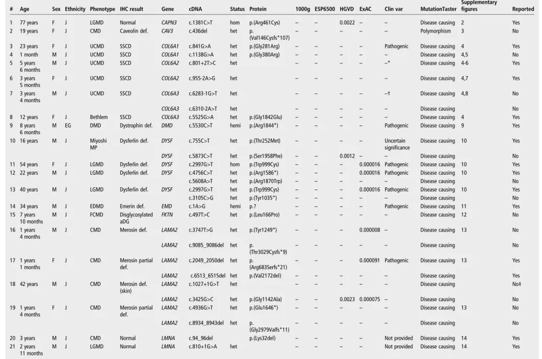
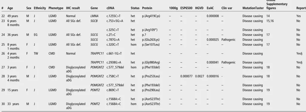
Likely causative gene mutations were identified in CAPN3 (1), CAV3 (2), COL6A1 (3,4), COL6A2 (5,6), COL6A3 (7,8), DMD (9), DYSF (10–13), EMD (14), FKTN (15), LAMA2 (16–19), LMNA (20–22), SGCB (23), SGCG (24,25), TRAPPC11 (26), POMGNT2 (27, 28) and POMT2 (29, 30) in 30 patients with MDs (figure 2andtable 1, the numbers in parentheses indicate the numbers of patients mentioned intable 1and online supple-mentaryfigures S2–S19). Clinical findings of all the patients were consistent with the detected genotype. Relevant protein defects or dislocations were confirmed based on the results of immunohisto-chemical analysis (see online supplementaryfigure S1), which sup-ported the genetic diagnosis in all patients, except in patients with mutations inCAPN3, LMNA and TRAPPC11. In addition, we per-formed cDNA analysis for patients 5–7 who harboured splice-site mutations in genes encoding collagen type VI and confirmed the presence of aberrant splicing that disrupted repetitive glycine resi-dues in a triple-helical region, which is pathogenic in either gene (see online supplementaryfigures S6–S8).
Two patients had unreported LMNA variants whose pheno-types were consistent with those associated with laminopathy (see online supplementary table S5). However, these mutations did not meet our criteria. One patient with laminin alpha-2 de fi-ciency and one patient with sarcoglycan gamma deficiency, as determined by performing immunohistochemical analysis, did not have any pathogenic variants inLAMA2 or SGCG (data not shown).
CMP panel
We enrolled 65 patients with CMP. One patient was suspected as having CMS according to the results of electrophysiological analysis. Muscle biopsy was not performed for this patient because muscles of patients with CMS usually show non-specific changes and are not very useful for diagnosis. The average coverage (>20 reads) in the CMP panel was 98.0%.
We prioritised variants in genes that were consistent with clin-ical and histologclin-ical phenotypes of reported patients. We identi-fied causative gene mutations in ACTA1 (31), CHRNE (32), KLHL40 (33, 34), NEB (35), MTM1 (36–38), RYR1 (39–45), TPM2 (46) and TPM3 (47) in 17 patients (figure 2,table 2and online supplementary figures S20–S26). Patient 36 had an intronic mutation in MTM1 (c.1261-10A>G). Analysis of cDNA obtained from this patient confirmed the presence of an aberrant splicing that was reported previously28(see online sup-plementaryfigure S23).
We identified variants in DNM2, RYR1 and NEB in 13 patients, which was consistent with their respective phenotypes. However, we could not describe these variants as pathogenic because they have not been reported previously or have been reported for a different phenotype (such as malignant hyperthermia). These variants are listed in online supplementary table S5.
MM panel
We enrolled 10 patients with suspected MM. One patient was diagnosed with glycogen storage disease based on glycogen accumulation, as determined by performing PAS staining (48). Two patients ( patients 49 and 50) were diagnosed with glycogen phosphorylase deficiency based on the results of routine histo-chemical staining (see online supplementary figures S26–S28), and three patients were diagnosed with lipid storage myopathy. The remaining four patients showed non-specific clinical and histological phenotypes but were suspected of having MM, with two patients having myalgia, one patient having rhabdomyolysis and one patient having cyclic vomiting. cDNA analysis was per-formed for patient 48 to confirm the presence of aberrant spli-cing, which was similar to that reported in a patient with the same mutation29 (see online supplementary figure S27). The average coverage (>20 reads) in the MM panel was 98.9%.
We detected causative mutations (table 3) in AGL (48) and PYGM (49,50) in three patients with glycogenosis (figure 2and table 3) and did not detect any variants in other patients, sug-gesting the heterogeneous nature of the clinical phenotype. Myopathy with protein aggregation/rimmed vacuole panel (MFM panel)
We enrolled 48 patients who showed protein aggregation, rimmed vacuoles and/or myofibrillar disorganisation in biopsied muscle samples (table 4) and identified probable causative gene mutations in DNAJB6 (51), GNE (52–55), MYH2 (56), MYOT (57),SEPN1 (58), TTN (59, 60) and VCP (61, 62) in 12 patients (figure 2, table 4 and online supplementary figures S29–S34). The average coverage (>20 reads) in the MFM panel was 98.9%.
Patient 58 had one fibre with a rimmed vacuole; however, nicotinamide adenine dinucleotide (NADH) staining detected multi-minicores in this patient (see online supplementaryfigure S32). The clinical phenotype and muscle histopathological pres-entation, except for the rimmed vacuole, in this patient were consistent with a multi-minicore disease associated withSEPN1 mutations. Patients 59 and 60 had common Japanese variants in TTN, which are associated with hereditary myopathy with early Figure 2 The total diagnostic yield was 33.0%. The diagnostic yield
of the muscular dystrophy (MD) panel was 46.2%, congenital myopathy (CMP)/congenital myasthenic syndrome (CMS) panel was 26.2%, metabolic myopathy (MM) panel was 30.0% and myopathy with protein aggregation/rimmed vacuole (MFM) panel was 25.0%. Determined: likely pathogenic variants; undetermined: variants that did not meet our criteria for likely pathogenic variants.
4 Nishikawa A, et al. J Med Genet 2016;0:1–11. doi:10.1136/jmedgenet-2016-104073
Methods
group.bmj.com on September 25, 2016 - Published by
http://jmg.bmj.com/ Downloaded from
Table 1 Pathogenic variants identified using the MD panel
# Age Sex Ethnicity Phenotype IHC result Gene cDNA Status Protein 1000g ESP6500 HGVD ExAC Clin var MutationTaster Online Supplementary
figures Reported
1 77 years F J LGMD Normal CAPN3 c.1381C>T hom p.(Arg461Cys) – – 0.0022 – – Disease causing 2 Yes
2 19 years F J CMD Caveolin def. CAV3 c.436del het p.
(Val146Cysfs*107)
– – – – – Polymorphism 3 No
3 23 years F J UCMD SSCD COL6A1 c.841G>A het p.(Gly281Arg) – – – – Pathogenic Disease causing 4 Yes
4 1 month M J UCMD SSCD COL6A1 c.1138G>A het p.(Gly380Arg) – – – – – Disease causing 4,5 No
5 5 years 6 months
M J UCMD SSCD COL6A2 c.801+2T>C het – – – – –* Disease causing 4-6 Yes
6 3 years 5 months
F J UCMD SSCD COL6A2 c.955-2A>G het – – – – – Disease causing 4,7 Yes
7 3 years 4 months
M J UCMD SSCD COL6A3 c.6283-1G>T het – – – – –† Disease causing 4,8 No
COL6A3 c.6310-2A>T het – – – – – Disease causing No
8 12 years F J Bethlem SSCD COL6A3 c.5525G>A het p.(Gly1842Glu) – – – – – Disease causing 4 Yes
9 8 years 6 months
M EG DMD Dystrophin def. DMD c.5530C>T hemi p.(Arg1844*) – – – – Pathogenic Disease causing 9 Yes
10 16 years M J Miyoshi MP
Dysferlin def. DYSF c.755C>T het p.(Thr252Met) – – – – Uncertain significance
Disease causing 10 Yes
DYSF c.5873C>T het p.(Ser1958Phe) – – 0.0012 – – Disease causing No
11 54 years F J LGMD Dysferlin def. DYSF c.2997G>T hom p.(Trp999Cys) – – – 0.000016 Pathogenic Disease causing 10 Yes 12 22 years M J LGMD Dysferlin def. DYSF c.4756C>T het p.(Arg1586*) – – – 0.000016 Pathogenic Disease causing 10 Yes
c.5608A>T het p.(Arg1870Trp) – – – – – Disease causing No
13 40 years M J LGMD Dysferlin def. DYSF c.2997G>T het p.(Trp999Cys) – – – 0.000016 Pathogenic Disease causing 10 Yes
c.3105C>G het p.(Tyr1035*) – – – – – Disease causing No
14 34 years M J EDMD Emerin def. EMD c.1A>G hemi p.? – – – – Pathogenic Disease causing 11 Yes
15 7 years 10 months
M J FCMD Disglycosylated aDG
FKTN c.497T>C het p.(Leu166Pro) – – – – – Disease causing 12 No
16 1 years 4 months
M J CMD Merosin def. LAMA2 c.3747T>G het p.(Tyr1249*) – – – 0.000008 – Disease causing 13 No
LAMA2 c.9085_9086del het p.
(Thr3029Cysfs*9) – – – – – Disease causing No 17 1 years 1 months F J CMD Merosin partial def.
LAMA2 c.2049_2050del het p.
(Arg683Serfs*21)
– – – 0.000091 Pathogenic Disease causing 13 Yes
LAMA2 c.6513_6515del het p.(Val2172del) – – – – – Disease causing Yes
18 42 years M J CMD Merosin def. (skin)
LAMA2 c.1027+1G>T het – – – – – Disease causing No‡
LAMA2 c.3425G>C het p.(Gly1142Ala) – – 0.0023 0.000075 – Disease causing No
19 1 years 4 months
F J CMD Merosin partial def.
LAMA2 c.4936G>T het p.(Glu1646*) – – – – – Disease causing 13 No
LAMA2 c.8934_8943del het p.
(Gly2979Valfs*11)
– – – – – Disease causing No
20 3 years M J CMD Normal LMNA c.94_96del p.(Lys32del) – – – – Not provided Disease causing 14 Yes
21 2 years 11 months
M J LGMD Normal LMNA c.810+1G>A het – – – – Not provided Disease causing 14 Yes
Continued Nishika w a A, et al . J Med Genet 2016; 0 :1 – 11. doi:10.1136 /jmedgenet-2 016-104073 5
Methods
group.bmj.com on September 25, 2016 - Published by http://jmg.bmj.com/ Downloaded fromTable 1 Continued
# Age Sex Ethnicity Phenotype IHC result Gene cDNA Status Protein 1000g ESP6500 HGVD ExAC Clin var MutationTaster Online Supplementary
figures Reported
22 49 years M J LGMD Normal LMNA c.1255C>T het p.(Arg419Cys) – – – 0.000008 – Disease causing 14 Yes
23 6 years 8 months
M J LGMD All SGs def. SGCB c.753+5G>A het – – – – – Disease causing 15,16 No
c.325C>T het p.(Arg109*) – – – – – Disease causing No
24 36 years M EG LGMD All SGs def. SGCG c.2T>C het p.? – – – – – Disease causing 17 No
SGCG c.787G>A het p.(Glu263Lys) – – – 0.000025 Pathogenic Disease causing No
25 8 years 1 months
F J LGMD All SGs def. SGCG c.320C>T hom p.(Ser107Leu) – – – – – Disease causing 17 No
26 4 years 2 months
F TW CMD Normal TRAPPC11 c.661-1G>T het – – – – – Disease causing Yes§
TRAPPC11 c.2938G>A het p.(Gly980Arg) – – – 0.000041 Pathogenic Disease causing Yes§ 27 3 years F J CMD Disglycosylated
aDG POMGNT2 c.577_579del
hom p.(Phe193del) – – – – – Disease causing 18 No
28 3 years 4 months
M J LGMD Disglycosylated
aDG POMGNT2 c.758C>T
het p.(Pro253Leu) – 0.000077 0.0027 0.000016 - Disease causing 18 No
POMGNT2 c.577_579del het p.(Phe193del) – – – – – Disease causing No
29 15 years F J LGMD Disglycosylated aDG
POMT2 c.869C>T het p.(Pro290Leu) – – – – – Disease causing 19 No
c.1568A>C het p.(Asn523Thr) – – – – – Disease causing No
30 33 years M J LGMD Disglycosylated aDG
POMT2 c.1568A>C hom p.(Asn523Thr) – – – – – Disease causing 19 No
p.? means protein is unknown according to hgvs nomenclature recommendation. *c.801+1 is pathogenic.
†c.6283-2 is likely pathogenic. ‡c.1027+3A>G has been reported. §This case.
aDG, alpha dystroglycan; Bethlem, Bethlem myopathy; CMD, congenital muscular dystrophy; def., deficiency, as determined by performing immunohistochemical staining; DMD, Duchenne muscular dystrophy; EDMD, Emery–Dreifuss muscular dystrophy; EG, Egyptian; ExAC, Exome Aggregation Consortium; FCMD, Fukuyama congenital muscular dystrophy; IHC, immunohistochemistry; J, Japanese; LGMD, limb-girdle muscular dystrophy; MD, muscular dystrophy; SGs, sarcoglycans; SSCD, sarcolemma-specific collagen deficiency; TW, Taiwanese; UCMD, Ullrich congenital muscular dystrophy.
6 Nishika w a A, et al . J Med Genet 2016; 0 :1 – 11. doi:10.1136/jm edgenet-20 16-104073
Methods
group.bmj.com on September 25, 2016 - Published by http://jmg.bmj.com/ Downloaded fromTable 2 Pathogenic variants identified using the CMP/CMS panel
# Age Sex Ethnicity Phenotype Gene cDNA Status Protein 1000g ESP6500 HGVD ExAC Clin var MutationTaster
Online supplementary
figures Reported
31 3 months F J NM ACTA1 c.282C>A het p.(Asn94Lys) – – – – – Disease causing 20 Yes
32 18 years M EG CMS CHRNE c.1181_1187dup hom p.(Glu396Aspfs*3) – – – – – Disease causing No
33 2 months M J NM KLHL40 c.1405G>T het p.(Gly469Cys) – – – 0.000033 Pathogenic Disease causing 21 Yes
KLHL40 c.1582G>A het p.(Glu528Lys) – – – 0.000067 Pathogenic Disease causing Yes
34 5 months M J NM KLHL40 c.1405G>T p.(Gly469Cys) – – – 0.000033 Pathogenic Disease causing 21 Yes
KLHL40 c.1582G>A p.(Glu528Lys) – – – 0.000067 Pathogenic Disease causing Yes
35 9 months F J NM NEB c.24681C>G hom p.(Tyr8227*) – – – – – Disease causing 21 No
36 1 year M ? MTM MTM1 c.1261-10A>G hemi – – Pathogenic Polymorphism 22,23 Yes
37 11 months M J MTM MTM1 c.1497G>A hemi p.(Trp499*) – – Pathogenic Disease causing 22 No
38 1 years 6 months
M J MTM MTM1 c.1536dup hemi p.(Phe513Leufs*4) – – – Disease causing 22 No
39 8 years F J CCD RYR1 c.131G>A het p.(Arg44His) – 0.000078 – 0.000010 Uncertain
significance
Disease causing 24 Yes
RYR1 c.7635G>C het p.(Glu2545Asp) – – – 0.000009 Pathogenic Disease causing Yes
40 67 years F J CCD RYR1 c.14378T>C het p.(Leu4793Pro) – – – – Pathogenic Disease causing 24 Yes
41 4 years M J CCD RYR1 c.14581C>T het p.(Arg4861Cys) – – – – Pathogenic Disease causing 24 Yes
42 4 years 10 months
M J CCD RYR1 c.14740A>G het p.(Arg4914Gly) – – – – Pathogenic Disease causing 24 Yes
43 30 years F J CCD RYR1 c.14741G>T het p.(Arg4914Met) – – – – –* Disease causing 24 Yes
44 3 years 1 months
F J CCD RYR1 c.14590T>G het p.(Tyr4864Asp) – – – – –† Disease causing 24 Yes
45 7 months F J CFTD RYR1 c.1001G>T het p.(Gly334Val) – – – – Uncertain
significance
Disease causing 25 No
RYR1 c.1186_1187inv het p.(Glu396Ser) – – – – Uncertain
significance
Disease causing No
RYR1 c.4071_4072del het p.
(Gly1359Hisfs*16)
– – – – Pathogenic Disease causing No
RYR1 c.4717C>A het p.(Pro1573Thr) – – – – – Disease causing Yes
46 70 years F J NM TPM2 c.428T>C het p.(Leu143Pro) – – – – – Disease causing 21 Yes
47 9 years 5 months
F J CFTD TPM3 c.502C>G het p.(Arg168Gly) – – – – Pathogenic Disease causing 25 Yes
*Arg >Thr and Gly are reported to be pathogenic. †Tyr >Cys is reported to be pathogenic.
CCD, central core disease; CFTD, congenital myopathy with fibre-type disproportion; CMP, congenital myopathy; CMS, congenital myasthenic syndrome; EG, Egyptian; ExAC, Exome Aggregation Consortium; J, Japanese; MTM, myotubular myopathy; NM, nemaline myopathy. Nishika w a A, et al . J Med Genet 2016; 0 :1 – 11. doi:10.1136 /jmedgenet-2 016-104073 7
Methods
group.bmj.com on September 25, 2016 - Published by http://jmg.bmj.com/ Downloaded fromrespiratory failure (HMERF). These patients have not yet devel-oped respiratory failure; however, their other symptoms and the results of muscle histopathological analysis are consistent with HMERF.
Summary of the four panels
The overall diagnostic yield was 33.0% in 188 patients. The rates for detecting the most likely causative gene mutations by the MD, CMP, MM and MFM panels were 46.2%, 26.2%, 30.0% and 25.0%, respectively (figure 2).
DISCUSSION
In the present study, we developed four targeted gene sequen-cing panels by using the Ion Torrent sequensequen-cing system and assessed their unbiased diagnostic yield in combination with histological and protein analyses. We separated genes into the four panels rather than combining them into a single large panel to mainly achieve cost efficiency and time efficiency. This approach also reduced labour required for interpreting data but might have overlooked known genes associated with unexpected phenotypes. However, it is essential to improve these panels because some novel muscle disease-related genes were identified after the development of these panels. The rate of genetic diag-nosis varied for each panel, with the MD panel having the highest diagnostic rate (46.2%), which was comparable with or higher than that reported in a previous study involving WES.24 MD is a heterogeneous inherited muscle disease. The most prevalent forms of MD in both children and adults in Japan are dystrophinopathy, which is caused by DMD mutations; myo-tonic dystrophy, which is caused by CTG expansion in the 30 untranslated region (UTR) inDMPK; facioscapulohumeral MD, which is caused by the contraction of the D4Z4 repeat (a 3.3 kb macrosatellite repeat in 4q35) and Fukuyama CMD, which is caused by a retrotransposonal 3 kb insertion in the 30UTR of FKTN. These diseases are usually clinically distinguishable and can be diagnosed in local hospitals by performing multiplex ligation-dependent probe amplification, PCR or Southern blot-ting before their evaluation at our laboratory. In the present study, most of the prevalent MDs were excluded at the routine clinical testing level. Patients with other MDs are usually cate-gorised clinically based on the presence of LGMD, CMD or EDMD. Known gene mutations in patients with these diseases are mostly caused by single nucleotide variants or small inser-tions and deleinser-tions. Therefore, next-generation sequencing is a powerful tool to detect these mutations. Among 62 genes exam-ined for MD, 66% causes the disease in a recessive manner, sug-gesting the presence of a loss-of-function mechanism. Therefore, immunohistochemical analysis to detect the loss of a protein is an effective method to diagnose these recessive dis-eases.30 31 In the present study, 25 patients yielded abnormal results for immunohistochemical analysis, and their diagnosis was confirmed by performing molecular analysis. These results suggest that immunohistochemical analysis is very helpful for improving the precision of genetic diagnosis and should be used routinely together with genetic testing. In one patient with laminin alpha-2 deficiency and one patient with sarcoglycan gamma deficiency, which were detected by performing immuno-histochemical analysis, we could not detect any pathogenic var-iants in LAMA2 or SGCG. However, the coverage of the gene was 96.9% and 91.6%, respectively, in MD panel. This might be because of the limitation of protein analysis or technical issues associated with this method (ie, variants outside of the target region, such as promoter region; variants in homopoly-mers or the presence of copy number variations). Therefore,
Table 3 Pa thogenic variants identified using the MM panel # Age Se x Ethnicity Phenotype Pr otein study Gene cDNA Sta tus Pr otein 1000g ESP6500 HGVD ExA C Clin var Muta tionT as ter
Online supplementary figur
es R eported 48 47 F J Glycogenosis type III AGL c.1735+1G>T hom 0.0005 –– 0.000016 Pa thogenic Disease causing 26, 27 Y es 49 57 M J McArdle disease Phosphorylase ↓ PYGM c.1531delG hom p.(Asp511Thrfs*28) –– – – – Disease causing 28 Y es 50 13 M J McArdle disease Phosphorylase ↓ PYGM c.2128_2130del hom p.(Phe710del ) –– – – Pa thogenic Disease causing 28 Y es ExA C, Exome Aggr ega tion C onsortium; J, Japanese; MM, metabolic my opa thy .
8 Nishikawa A, et al. J Med Genet 2016;0:1–11. doi:10.1136/jmedgenet-2016-104073
Methods
group.bmj.com on September 25, 2016 - Published by
http://jmg.bmj.com/ Downloaded from
Table 4 Pathogenic variants identified using the MFM panel
# Age Sex Ethnicity Phenotype Gene cDNA Status Protein 1000g ESP6500 HGVD ExAC Clin var MutationTaster
Online supplementary
figures Reported
51 57 years M J AVM DNAJB6 c.279C>G het p.(Phe93Lys) – – – – – Disease causing 29 Yes
52 42 years F J DMRV GNE c.1807G>C het p.(Val603Leu) 0.0005 – 0.0043 0.00002 – Disease causing 30 Yes
c.620A>T het p.(Asp207Val) 0.0009 – 0.0018 0.00004 – Disease causing Yes
53 29 years M J DMRV GNE c.188_197dup het p.(Glu66Aspfs2) – – – – – Disease causing 30 Yes
c.1807G>C het p.(Val603Leu) 0.0005 – 0.0043 0.00002 – Disease causing Yes
54 41 years F J DMRV GNE c.620A>T het p.(Asp207Val) 0.0009 – 0.0018 0.00004 – Disease causing 30 Yes
c.1807G>C het p.(Val603Leu) 0.0005 – 0.0043 0.00002 – Disease causing Yes
55 30 years F J DMRV GNE c.131G>C hom p.(Cys44Ser) – – – – – Disease causing 30 Yes
56 38 years M J Distal myopathy MYH2 c.2414T>C het p.(Val805Ala) 0.0018 – 0.0093 0.00081 - Disease causing 31 Yes
57 61 years M J MFM MYOT c.179C>G het p.(Ser60Cys) – – – – Pathogenic Disease causing 13 Yes
58 41 years M J AVM_scoliosis_resp
failure SEPN1
c.565C>T het p.(Arg189) – – – 0.00001 – Disease causing 32 No
c.1574T>G het p.(Met525Arg) – – 0.0037 0.00006 – Disease causing Yes
59 54 years M J MFM TTN c.95135G>A het p.(Cys31712Tyr) – – – – – Disease causing 33 Yes
60 46 years F J HMERF TTN c.95136T>G het p.(Cys31712Tyr) – – – – – Disease causing 33 Yes
61 44 years M J Myopathy VCP c.463C>T het p.(Arg155Cys) – – – – Pathogenic Disease causing 34 Yes
62 45 years M J Distal myopathy VCP c.476G>A het p.(Arg159His) – – – – Pathogenic Disease causing 34 Yes
AVM, autophagic vacuolar myopathy; DMRV, distal myopathy with rimmed vacuole; ExAC, Exome Aggregation Consortium; HMERF, hereditary myopathy with early respiratory failure; J, Japanese; MFM, myofibrillar myopathy.
Nishika w a A, et al . J Med Genet 2016; 0 :1 – 11. doi:10.1136 /jmedgenet-2 016-104073 9
Methods
group.bmj.com on September 25, 2016 - Published by http://jmg.bmj.com/ Downloaded fromfurther analysis such as Sanger sequencing of all target exonic regions or WES may be necessary to detect these mutations. Immunohistochemical analysis is not useful in patients with some dominant gene mutations, such as patients with mutations in LMNA. Two unreported LMNA variants were identified in patients with EDMD included in our study. We could not deter-mine their pathogenicity despite the consistency with clinical and pathologicalfindings. Therefore, we included these patients in the undiagnosed category. It is necessary to include more patients in disease variant databases to check for shared muta-tions. Family analysis will be helpful to determine the pathogen-icity of these variants. However, only sporadic cases were analysed in the present study.
Several patients with nemaline myopathy had variants in NEB; however, we could not determine whether these variants were pathogenic because most variants were unreported and the coverage ofNEB was low due to a repeat region with high hom-ology, suggesting the presence of other mutations in this region.32 Adoption of a next generation sequencing approach for NEB is challenging for identifying variants in this region. Transcript analysis might be useful for identifying these variants. We also identified several rare variants in RYR1 in patients with CFTD or CMP with type 1 fibre predominance. Mutations in RYR1 are associated with these phenotypes;33 34 however, it is unclear whether these mutations are pathogenic because there is no suitable in vitro or in vivo analytical method to prove their pathogenicity. Interestingly, patients 6 and 7 shared the same phenotype and the same rare variant inNEB; moreover, patients 12 and 13 shared the same rare variant in RYR1. Thus, collec-tion of variant data from diseased and healthy subjects is important for determining the pathogenicity of these variants.
MM is caused by defects in enzymes involved in glycogen and lipid metabolism. In Japan, glycogen storage disease is mainly diagnosed by performing biochemical assessment of these enzymes. Therefore, such samples are rarely sent to our laboratory for genetic diagnosis. Future studies involving more patients are necessary to evaluate the diagnostic yield of the MM panel and to obtain a mutation spectrum of this disease.
Some patients with myopathies showfibres with marked accu-mulation of protein aggregates or rimmed vacuoles, which is a marker of autophagy. These highly heterogeneous diseases include MFM, VCP myopathy, GNE myopathy, TTN myopathy, oculopharyngeal distal myopathy, oculopharyngeal MD and autophagic vacuolar myopathy.22 35–40MFM has been described only recently, and not many patients with MFM have been reported until now.41 42Many unknown causative genes may be involved in the pathogenesis of this disease. Therefore, it is
increasingly important to accumulate genotype–phenotype spec-trum of these relatively new rare diseases.
In conclusion, our genetic diagnosis technique combined with histological, mRNA and protein analyses is useful and efficient for screening pathogenic variants and for performing molecular diagnosis of patients with muscle diseases. The results of this study further emphasise the importance of developing a com-prehensive disease mutation database and identifying multidi-mensional phenotypes from clinical, histological and molecular studies.
Acknowledgements The authors thank Mami Arai, Ayumi Oda, Kaoru Tatezawa, Chikako Miyazaki, Keiko Ishikawa, Chizuru Sumino and Kazu Iwasawa for providing technical support.
Contributors AN and SM: study concept, analysis and interpretation of the data, drafting/revising the manuscript: NM and IN: analysis and interpretation of the data, drafting/revising the manuscript.
Funding This study was supported partly by a grant for Research on Rare and Intractable Diseases (H26-Itaku (Nan)-Ippan-081) from the Japan Agency for Medical Research and Development, AMED and by Intramural Research Grants 26-8, 26-7 for Neurological and Psychiatric Disorders from the National Center of Neurology and Psychiatry.
Competing interests None declared.
Ethics approval This study was approved by the ethics committee of the National Center of Neurology and Psychiatry.
Patient consent Obtained.
Provenance and peer review Not commissioned; externally peer reviewed.
REFERENCES
1 Mitsuhashi S, Kang PB. Update on the genetics of limb girdle muscular dystrophy.
Semin Pediatr Neurol2012;19:211–18.
2 Engel AG, Franzini-Armstrong C. Myology. 3rd edn. USA: McGRAW-Hill, 2004:691–747.
3 Wicklund MP, Kissel JT. The limb-girdle muscular dystrophies.Neurol Clin
2014;32:729–49, ix.
4 Kobayashi O, Hayashi Y, Arahata K, Ozawa E, Nonaka I. Congenital muscular dystrophy: clinical and pathologic study of 50 patients with the classical (occidental) merosin-positive form.Neurology1996;46:815–18.
5 Bonne G, Quijano-Roy S. Emery-Dreifuss muscular dystrophy, laminopathies, and other nuclear envelopathies.Handb Clin Neurol2013;113:1367–76.
6 Nonaka I, Une Y, Ishihara T, Miyoshino S, Nakashima T, Sugita H. A clinical and histological study of Ullrich’s disease (congenital atonic-sclerotic muscular dystrophy).Neuropediatrics1981;12:197–208.
7 Bushby KM, Collins J, Hicks D. Collagen type VI myopathies.Adv Exp Med Biol
2014;802:185–99.
8 Sewry CA, Jimenez-Mallebrera C, Muntoni F. Congenital myopathies.Curr Opin Neurol2008;21:569–75.
9 Laing NG. Congenital myopathies.Curr Opin Neurol2007;20:583–9.
10 North KN, Wang CH, Clarke N, Jungbluth H, Vainzof M, Dowling JJ, Amburgey K, Quijano-Roy S, Beggs AH, Sewry C, Laing NG, Bönnemann CG, International Standard of Care Committee for Congenital Myopathies. Approach to the diagnosis of congenital myopathies.Neuromuscul Disord2014;24:97–116.
11 Wang CH, Dowling JJ, North K, Schroth MK, Sejersen T, Shapiro F, Bellini J, Weiss H, Guillet M, Amburgey K, Apkon S, Bertini E, Bonnemann C, Clarke N, Connolly AM, Estournet-Mathiaud B, Fitzgerald D, Florence JM, Gee R, Gurgel-Giannetti J, Glanzman AM, Hofmeister B, Jungbluth H, Koumbourlis AC, Laing NG, Main M, Morrison LA, Munns C, Rose K, Schuler PM, Sewry C, Storhaug K, Vainzof M, Yuan N. Consensus statement on standard of care for congenital myopathies.J Child Neurol2012;27:363–82.
12 Ilkovski B, Cooper ST, Nowak K, Ryan MM, Yang N, Schnell C, Durling HJ, Roddick LG, Wilkinson I, Kornberg AJ, Collins KJ, Wallace G, Gunning P, Hardeman EC, Laing NG, North KN. Nemaline myopathy caused by mutations in the muscle alpha-skeletal-actin gene.Am J Hum Genet2001;68:1333–43.
13 Ryan MM, Ilkovski B, Strickland CD, Schnell C, Sanoudou D, Midgett C, Houston R, Muirhead D, Dennett X, Shield LK, De Girolami U, Iannaccone ST, Laing NG, North KN, Beggs AH. Clinical course correlates poorly with muscle pathology in nemaline myopathy.Neurology2003;60:665–73.
14 Engel AG, Shen XM, Selcen D, Sine SM. Congenital myasthenic syndromes: pathogenesis, diagnosis, and treatment.Lancet Neurol2015;14:420–34. 15 Kinali M, Beeson D, Pitt MC, Jungbluth H, Simonds AK, Aloysius A, Cockerill H,
Davis T, Palace J, Manzur AY, Jimenez-Mallebrera C, Sewry C, Muntoni F, Robb SA.
Web resources
▸ The URLs for data presented are as follows: ▸ Ensembl, http://www.ensembl.org/index.html ▸ OMIM, http://www.omim.org/ ▸ 1000 Genomes, http://www.1000genomes.org/ ▸ ESP6500, http://evs.gs.washington.edu/EVS/ ▸ dbSNP138, http://www.ncbi.nlm.nih.gov/projects/SNP/ ▸ HGVD, http://www.genome.med.kyoto-u.ac.jp/SnpDB/ ▸ Exome Aggregation Consortium (ExAC), http://exac.
broadinstitute.org/
▸ MutationTaster: http://www.mutationtaster.org ▸ Mutalyzer: http://www.mutalyzer.nl
10 Nishikawa A, et al. J Med Genet 2016;0:1–11. doi:10.1136/jmedgenet-2016-104073
Methods
group.bmj.com on September 25, 2016 - Published by
http://jmg.bmj.com/ Downloaded from
Congenital myasthenic syndromes in childhood: diagnostic and management challenges.J Neuroimmunol2008;201–202:6–12.
16 Sharp LJ, Haller RG. Metabolic and mitochondrial myopathies.Neurol Clin
2014;32:777–99.
17 Angelini C. Spectrum of metabolic myopathies.Biochim Biophys Acta
2015;1852:615–21.
18 Zutt R, van der Kooi AJ, Linthorst GE, Wanders RJ, de Visser M. Rhabdomyolysis: review of the literature.Neuromuscul Disord2014;24:651–9.
19 Tobon A. Metabolic myopathies.Continuum (Minneap Minn)2013;19(Muscle Disease):1571–97.
20 Quinlivan R, Jungbluth H. Myopathic causes of exercise intolerance with rhabdomyolysis.Dev Med Child Neurol2012;54:886–91.
21 Claeys KG, Fardeau M. Myofibrillar myopathies.Handb Clin Neurol
2013;113:1337–42.
22 Selcen D. Myofibrillar myopathies.Neuromuscul Disord2011;21:161–71. 23 Kaplan J-C, Hamroun D. The 2016 version of the gene table of monogenic
neuromuscular disorders (nuclear genome).Neuromuscul Disord2015;25:991–20. 24 Ghaoui R, Cooper ST, Lek M, Jones K, Corbett A, Reddel SW, Needham M, Liang
C, Waddell LB, Nicholson G, O’Grady G, Kaur S, Ong R, Davis M, Sue CM, Laing NG, North KN, MacArthur DG, Clarke NF. Use of whole-exome sequencing for diagnosis of limb-girdle muscular dystrophy: outcomes and lessons learned.JAMA Neurol2015;72:1424–32.
25 Yonekawa T, Nishino I. Ullrich congenital muscular dystrophy: clinicopathological features, natural history and pathomechanism(s).J Neurol Neurosurg Psychiatr
2015;86:280–7.
26 Kaplan JC, Hamroun D. The 2013 version of the gene table of monogenic neuromuscular disorders (nuclear genome).Neuromuscul Disord2012;22:1108–35. 27 Green RC, Berg JS, Grody WW, Kalia SS, Korf BR, Martin CL, McGuire AL,
Nussbaum RL, O’Daniel JM, Ormond KE, Rehm HL, Watson MS, Williams MS, Biesecker LG, American College of Medical Genetics and Genomics. ACMG recommendations for reporting of incidentalfindings in clinical exome and genome sequencing.Genet Med2013;15:565–74.
28 de Gouyon BM, Zhao W, Laporte J, Mandel JL, Metzenberg A, Herman GE. Characterization of mutations in the myotubularin gene in twenty six patients with X-linked myotubular myopathy.Hum Mol Genet1997;6:1499–504.
29 Okubo M, Aoyama Y, Murase T. A novel donor splice site mutation in the glycogen debranching enzyme gene is associated with glycogen storage disease type III.
Biochem Biophys Res Commun1996;224:493–9.
30 Sewry CA. Muscular dystrophies: an update on pathology and diagnosis.Acta Neuropathol2010;120:343–58.
31 Costanza L, Moggio M. Muscular dystrophies: histology, immunohistochemistry, molecular genetics and management.Curr Pharm Des2010;16:978–87.
32 Donner K, Sandbacka M, Lehtokari VL, Wallgren-Pettersson C, Pelin K. Complete genomic structure of the human nebulin gene and identification of alternatively spliced transcripts.Eur J Hum Genet2004;12:744–51.
33 Clarke NF, Waddell LB, Cooper ST, Perry M, Smith RL, Kornberg AJ, Muntoni F, Lillis S, Straub V, Bushby K, Guglieri M, King MD, Farrell MA, Marty I, Lunardi J, Monnier N, North KN. Recessive mutations in RYR1 are a common cause of congenitalfiber type disproportion.Hum Mutat2010;31:E1544–50. 34 Rocha J, Taipa R, Melo Pires M, Oliveira J, Santos R, Santos M. Ryanodine
myopathies without central cores—clinical, histopathologic, and genetic description of three cases.Pediatr Neurol2014;51:275–8.
35 Watts GD, Wymer J, Kovach MJ, Mehta SG, Mumm S, Darvish D, Pestronk A, Whyte MP, Kimonis VE. Inclusion body myopathy associated with Paget disease of bone and frontotemporal dementia is caused by mutant valosin-containing protein.
Nat Genet2004;36:377–81.
36 Eisenberg I, Avidan N, Potikha T, Hochner H, Chen M, Olender T, Barash M, Shemesh M, Sadeh M, Grabov-Nardini G, Shmilevich I, Friedmann A, Karpati G, Bradley WG, Baumbach L, Lancet D, Asher EB, Beckmann JS, Argov Z, Mitrani-Rosenbaum S. The UDP-N-acetylglucosamine 2-epimerase/
N-acetylmannosamine kinase gene is mutated in recessive hereditary inclusion body myopathy.Nat Genet2001;29:83–7.
37 Pfeffer G, Elliott HR, Griffin H, Barresi R, Miller J, Marsh J, Evilä A, Vihola A, Hackman P, Straub V, Dick DJ, Horvath R, Santibanez-Koref M, Udd B, Chinnery PF. Titin mutation segregates with hereditary myopathy with early respiratory failure.
Brain2012;135:1695–713.
38 Tomé FM, Fardeau M. Nuclear inclusions in oculopharyngeal dystrophy.Acta Neuropathol1980;49:85–7.
39 van der Sluijs BM, ter Laak HJ, Scheffer H, van der Maarel SM, van Engelen BG. Autosomal recessive oculopharyngodistal myopathy: a distinct phenotypical, histological, and genetic entity.J Neurol Neurosurg Psychiatr2004;75: 1499–501.
40 Nishino I. Autophagic vacuolar myopathy.Semin Pediatr Neurol2006; 13:90–5.
41 Nakano S, Engel AG, Waclawik AJ, Emslie-Smith AM, Busis NA. Myofibrillar myopathy with abnormal foci of desmin positivity. I. Light and electron microscopy analysis of 10 cases.J Neuropathol Exp Neurol1996;55:549–62.
42 De Bleecker JL, Engel AG, Ertl BB. Myofibrillar myopathy with abnormal foci of desmin positivity. II. Immunocytochemical analysis reveals accumulation of multiple other proteins. J Neuropathol Exp Neurol 1996;55:563–77.
Nishikawa A, et al. J Med Genet 2016;0:1–11. doi:10.1136/jmedgenet-2016-104073 11
Methods
group.bmj.comon September 25, 2016 - Published by http://jmg.bmj.com/
muscle disorders
for the molecular diagnosis of inherited
histological assessment of skeletal muscles
Targeted massively parallel sequencing and
Nishino
Atsuko Nishikawa, Satomi Mitsuhashi, Naomasa Miyata and Ichizo
published online September 6, 2016
J Med Genet
http://jmg.bmj.com/content/early/2016/09/06/jmedgenet-2016-104073
Updated information and services can be found at:
These include:
References
#BIBL
http://jmg.bmj.com/content/early/2016/09/06/jmedgenet-2016-104073
This article cites 41 articles, 5 of which you can access for free at:
service
Email alerting
box at the top right corner of the online article.
Receive free email alerts when new articles cite this article. Sign up in the
Collections
Topic
Articles on similar topics can be found in the following collections
(1244)
Molecular genetics
(625)
Epidemiology
(254)
Neuromuscular disease
(145)
Muscle disease
Notes
http://group.bmj.com/group/rights-licensing/permissions
To request permissions go to:
http://journals.bmj.com/cgi/reprintform
To order reprints go to:
http://group.bmj.com/subscribe/
To subscribe to BMJ go to:
group.bmj.com on September 25, 2016 - Published by http://jmg.bmj.com/ Downloaded fromSupplemental Table 1. MD panel
gene symbol ENST# NM# Example of the phenotype inheritance OMIM
ACVR1 ENST00000263640 NM_001105.4 Fibrodysplasia ossificans progressiva AD MIM135100 AGRN ENST00000379370 NM_198576.3 Myasthenic syndrome AR MIM615120 ALG13 ENST00000394780 NM_001099922.2 Congenital disorder of glycosylation XLR MIM300884
ANO5 ENST00000324559 NM_213599.2 LGMD2L AR MIM611307
B3GALNT2 ENST00000366600 NM_152490.2 alpha-dystroglycanopathy AR MIM615181 B3GNT1 ENST00000311181 NM_006876.2 alpha-dystroglycanopathy AR MIM615287
CAPN3 ENST00000397163 NM_000070.2 LGMD2A AR MIM253600
CAV3 ENST00000343849 NM_033337.2 LGMD1C AD MIM607801
CHKB ENST00000406938 NM_005198.4 Megaconial myopathy AR MIM602541 COL12A1 ENST00000322507 NM_004370.5 Ullrich/Bethlem myopathy AD/AR MIM616470/616471
COL6A1 ENST00000361866 NM_001848.2 Ullrich/Bethlem myopathy AD/AR MIM254090/158810 COL6A2 ENST00000300527 NM_001849.3 Ullrich/Bethlem myopathy AD/AR MIM254090/158810 COL6A3 ENST00000295550 NM_004369.3 Ullrich/Bethlem myopathy AD/AR MIM254090/158810
DAG1 ENST00000545947 NM_001177634.2 Dystroglycanopathy AR MIM616538 DES ENST00000373960 NM_001927.3 Myofibrillar myopathy/LGMD2R AD/AR MIM601419/615325 DMD ENST00000357033 NM_004006.2 Duchenne/Becker Muscular Dystrophy XLR MIM310200/300376
DNAJB6 ENST00000262177 NM_058246.3 LGMD1E AD MIM603511
DOK7 ENST00000389653 NM_173660.4 Myasthenia, limb girdle AR MIM254300 DOLK ENST00000372586 NM_014908.3 Congenital disorder of glycosylation AR MIM610768 DPAGT1 ENST00000409993 NM_001382.3 Congenital disorder of glycosylation/Myathenic syndrome AR MIM608093/614750
DPM1 ENST00000371584 NM_003859.1 Congenital disorder of glycosylation AR MIM608799 DPM2 ENST00000373110 NM_003863.3 Congenital disorder of glycosylation AR MIM615042 DPM3 ENST00000368399 NM_018973.3 Congenital disorder of glycosylation AR MIM612937
DYSF ENST00000258104 NM_003494.3 LGMD2B AR MIM253601
EMD ENST00000369842 NM_000117.2 Emery-Dreifuss muscular dystrophy XLR MIM310300 FAT1 ENST00000441802 NM_005245.3 Facioscapulohumeral muscular dystrophy-like myopathy? AD
FHL1 ENST00000394155 NM_001159702.2 Reducing body myopathy XLD MIM300717 FKRP ENST00000318584 NM_024301.4 alpha-dystroglycanopathy AR MIM613153 FKTN ENST00000602661 NM_001079802.1 alpha-dystroglycanopathy AR MIM253800
FLNC ENST00000325888 NM_001458.4 MFM AD/AR MIM609524
GFPT1 ENST00000357308 NM_001244710.1 Congenital myathenia AR MIM610542
GMPPB ENST00000308375 alpha-dystroglycanopathy AR MIM615350
ISPD ENST00000407010 NM_001101426.3 alpha-dystroglycanopathy AR MIM614643 ITGA7 ENST00000257880 XM_005268841.1 Congenital muscular dystrophy AR MIM613204 KLHL9 ENST00000359039 NM_018847.2 Distal myopathy? AD
LAMA2 ENST00000421865 NM_000426.3 Congenital muscular dystrophy AR MIM607855 LARGE ENST00000354992 NM_004737.4 alpha-dystroglycanopathy AR MIM613154
LMNA ENST00000368300 NM_170707.3 LGMD1B AD MIM159001
MEGF10 ENST00000274473 NM_032446.2 EMARDD AR MIM614399
MICU1 ENST00000398761 NM_006077.3 Myopathy with extrapyramidal signs AR MIM615673
MYOT ENST00000239926 NM_006790.2 LGMD1A/MFM AD MIM609200
PLEC1 ENST00000322810 NM_201380.3 Muscular dystrophy with epidermolysis bullosa simplex/LGMD2Q AR MIM226670 POMGNT1 ENST00000371992 NM_001243766.1 alpha-dystroglycanopathy AR MIM253280 POMGNT2 ENST00000344697 NM_032806.5 alpha-dystroglycanopathy AR MIM614830 POMT1 ENST00000372228 NM_007171.3 alpha-dystroglycanopathy AR MIM236670 POMT2 ENST00000261534 NM_013382.5 alpha-dystroglycanopathy AR MIM613150 PTRF ENST00000357037 NM_012232.5 Muscular dystrphy and lipodystrophy AR MIM613327
SGCA ENST00000262018 NM_000023.2 LGMD2D AR MIM608099
SGCB ENST00000381431 NM_000232.4 LGMD2E AR MIM604286
SGCD ENST00000337851 NM_000337.5 LGMD2F AR MIM601287
SGCG ENST00000218867 NM_000231.2 LGMD2C AR MIM253700
POMK ENST00000331373 NM_032237 alpha-dystroglycanopathy AR MIM615249 SMCHD1 ENST00000320876 NM_015295.2 Facioscapulohumeral muscular dystrophy type2 AD MIM158901 STIM1 ENST00000300737 NM_003156.3 Tubular aggregate myopathy AD MIM160565 SYNE1 ENST00000367255 NM_182961.3 Emery-Dreifuss muscular dystrophy AD MIM612998 SYNE2 ENST00000358025 NM_182914.2 Emery-Dreifuss muscular dystrophy AD MIM612999
TCAP ENST00000309889 NM_003673.3 LGMD2G AR MIM601954
TMEM43 ENST00000306077 NM_024334.2 Emery-Dreifuss muscular dystrophy? AD MIM614302 TMEM5 ENST00000261234 NM_014254.2 alpha-dystroglycanopathy AR MIM615041
TNPO3 ENST00000393245 NM_012470.3 LGMD1F AD MIM608423
TRAPPC11 ENST00000334690 NM_021942.5 LGMD2S AR MIM615356
Supplemental Table 2. CMP panel
gene symbol ENST# NM# Example of the phenotype inheritance OMIM
ACTA1 ENST00000366684 NM_001100.3 Nemaline myopathy AD/AR MIM161800 AGRN ENST00000379370 NM_198576.3 Myathenic syndrome AR MIM103320 ALG14 ENST00000370205 NM_144988.3 Myathenic syndrome AR MIM612866 ALG2 ENST00000476832 NM_033087.3 Myathenic syndrome AR MIM607905 BIN1 ENST00000316724 NM_139343.2 Centronuclear myopathy AR MIM255200 CCDC78 ENST00000293889 NM_001031737.2 Centronuclear myopathy AD MIM614807 CFL2 ENST00000341223 NM_021914.7 Nemaline myopathy AR MIM610687 CHAT ENST00000337653 NM_020549.4 Myathenic syndrome AR MIM118491 CHRNA1 ENST00000261007 NM_001039523.2 Myathenic syndrome AD/AR MIM100690 CHRNB1 ENST00000306071 NM_000747.2 Myathenic syndrome AD/AR MIM100710 CHRND ENST00000258385 NM_000751.2 Myathenic syndrome AD/AR MIM100720 CHRNE ENST00000293780 NM_000080.3 Myathenic syndrome AD/AR MIM100725 CHRNG ENST00000389494 NM_005199.4 Escobar syndrome AR MIM265000
CNTN1 ENST00000551295 NM_001843.3 CMP AR MIM612540
COLQ ENST00000383788 NM_005677.3 Myathenic syndrome AR MIM603033 DNM2 ENST00000355667 NM_001005360.2 Centronuclear myopathy AD MIM160150 DOK7 ENST00000389653 NM_173660.4 Myathenic syndrome AR MIM610285 DPAGT1 ENST00000409993 NM_001382.3 Congenital disorder of glycosylation/Myathenic syndrome AR MIM191350 GFPT1 ENST00000361060 NM_002056.3 Congenital myathenia AR MIM138292 HSPG2 ENST00000374695 NM_005529.6 Schwartz-Jampel syndrome AR MIM255800 KBTBD13 ENST00000432196 NM_001101362.2 Nemaline myopathy AD MIM609273 KLHL40 ENST00000287777 NM_152393.3 Nemaline myopathy AR MIM615348 LAMB2 ENST00000418109 NM_002292.3 Myathenic syndrome/Piearson synd AR MIM150325 LRP4 ENST00000378623 NM_002334.3 Myathenic syndrome? AR MIM616304 MEGF10 ENST00000274473 NM_032446.2 EMARDD AR MIM614399 MTM1 ENST00000370396 NM_000252.2 Myotubular myopathy XLR MIM310400 MUSK ENST00000189978 NM_005592.3 Myathenic syndrome AR MIM 601296 MYBPC3 ENST00000545968 NM_000256.3 Cardiomyopathy/Myopathy AD/AR MIM615396 MYH7 ENST00000355349 XM_005267696.1 Distal myopathy/Myosin storage myopathy/Dilated cardiomyopathy AD MIM160500 NEB ENST 00000427231 NM_1271208.1 Nemaline myopathy AR MIM256030 ORAI1 ENST00000330079 NM_032790.3 Tubular aggregate myopathy AD MIM615883 PLEC ENST00000322810 NM_201380.3 Muscular dystrophy with epidermolysis bullosa simplex AR MIM 601282 PTPLA ENST00000361271 NM_014241.3 Centronuclear myopathy? AR ー RAPSN ENST00000524487 NM_005055.4 Myathenic syndrome AR MIM 601592
RYR1 ENST00000359596 NM_000540.2 Malignant hyperthermia/Central core disease AD/AR 145600/117000 SCN4A ENST00000578147 NM_000334.4 Paramyotonia congenita/Myathenic syndrome/ AD/AR MIM168300/614198 SEPN1 ENST00000361547 NM_020451.2 Multiminicore disease AR MIM602771 STIM1 ENST00000300737 NM_003156.3 Tubular aggregate myopathy AD MIM160565 TNNT1 ENST00000588981 NM_003283.5 Nemaline myopathy AR MIM605355 TPM2 ENST00000378300 NM_003289.3 Nemaline myopathy AD/AR MIM609285 TPM3 ENST00000368530 NM_152263.2 Nemaline myopathy AD MIM609284
Supplemental Table 3. MM panel
gene symbol ENST# NM# Example of the phenotype inheritance
ABHD5 ENST00000458276 NM_016006.4 Chanarin-Dorfman syndrome AR MIM275630 ACADM ENST00000370834 NM_000016.4 Acyl-CoA dehydrogenase, medium chain, deficiency of AR MIM201450 ACADS ENST00000242592 NM_000017.2 Acyl-CoA dehydrogenase, short-chain, deficiency of AR MIM201470 ACADVL ENST00000356839 NM_000018.3 VLCAD deficiency AR MIM201475 AGL ENST00000361915 NM_000642.2 Glycogen storage disease IIIa, b AR MIM232400 ALDOA ENST00000395248 NM_000034.3 Glycogen storage disease XII AR MIM611881 CACNA1S ENST00000362061 NM_000069.2 Hypokalemic periodic paralysis/Malignant hyperthermia susceptibility AD MIM170400
CLCN1 ENST00000343257 NM_000083.2 Myotonia congenita AD/AR MIM160800/255700 CPT2 ENST00000371486 NM_000098.2 Myopathy due to CPT II deficiency AR MIM255110 ENO3 ENST00000323997 NM_001976.4 Glycogen storage disease XIII AR MIM612932 ETFA ENST00000557943 NM_000126.3 Glutaric acidemia IIA AR MIM231680 ETFB ENST00000354232 NM_001985.2 Glutaric acidemia IIB AR MIM231680 ETFDH ENST00000511912 NM_004453.2 Glutaric acidemia IIC AR MIM231680 GAA ENST00000302262 NM_000152.3 Glycogen storage disease II AR MIM232300 GBE1 ENST00000429644 NM_000158.3 Glycogen storage disease IV/Polyglucosan body disease AR MIM232500/263570 GYG1 ENST00000345003 NM_004130.3 Glycogen storage disease XV/Polyglucosan body myopathy 2 AR MIM613507/616199 GYS1 ENST00000323798 NM_002103.4 Glycogen storage disease 0 AR MIM611556 HADHA ENST00000380649 NM_000182.4 Trifunctional protein deficiency AR MIM609015 HADHB ENST00000317799 NM_000183.2 Trifunctional protein deficiency AR MIM609015 ISCU ENST00000311893 NM_213595.3 Myopathy with lactic acidosis AR MIM255125 LDHA ENST00000540430 NM_001165414.1 Glycogen storage disease XI AR MIM612933 LPIN1 ENST00000449576 NM_145693.2 Myoglobinuria AR MIM268200 MICU1 ENST00000398761 NM_006077.3 Myopathy with extrapyramidal signs AR MIM615673 MTO1 ENST00000415954 NM_001123226.1 Combined oxidative phosphorylation deficiency 10 AR MIM614702 PFKM ENST00000340802 NM_001166686.1 Glycogen storage disease VII AR MIM232800 PGAM2 ENST00000297283 NM_000290.3 Glycogen storage disease X AR MIM261670 PGK1 ENST00000373316 NM_000291.3 Phosphoglycerate kinase 1 deficiency XLR MIM300653 PGM1 ENST00000371083 NM_001172818.1 Congenital disorder of glycosylation, type XIV AR MIM614921 PHKA1 ENST00000373539 NM_002637.3 Glycogen storage disease IXd XLR MIM300559 PNPLA2 ENST00000336615 NM_020376.3 Neutral lipid storage disease with myopathy AR MIM610717 PRKAG2 ENST00000287878 NM_016203.3 Glycogen storage disease of heart, lethal congenital AD MIM261740 PYGM ENST00000164139 NM_005609.2 McArdle disease/Glycogen storage disease V AR MIM232600 SCN4A ENST00000578147 NM_000334.4 Paramyotonia congenita AD MIM168300 SLC22A5 ENST00000435065 NM_003060.3 Primary systemic carnitine deficiency AR MIM212140 SLC25A20 ENST00000319017 NM_000387.5 Carnitine/acyl- carnitine translocase deficiency AR MIM212138 TAZ ENST00000299328 NM_000116.3 Barth syndrome XLR MIM302060
Supplemental Table 4. MFM panel
gene symbol ENST# NM# Example of the phenotype inheritance OMIM
ACTA1 ENST00000366684 NM_001100.3 Nemaline myopathy AD/AR MIM161800
BAG3 ENST00000369085 NM_004281.3 MFM AD MIM612954
CFL2 ENST00000341223 NM_021914.7 Nemaline myopathy AR MIM610687 CRYAB ENST00000533475 NM_001885.1 MFM AD/AR MIM608810/613869
DES ENST00000373960 NM_001927.3 MFM AD MIM601419
DNAJB6 ENST00000262177 NM_058246.3 LGMD1E/MFM AD MIM603511 EPG5 ENST00000282041 NM_020964.2 Vici syndrome AR MIM242840 FHL1 ENST00000394155 NM_001159702.2 Reducing body myopathy XLD MIM300717
FLNC ENST00000325888 NM_001458.4 MFM AD MIM609524
GNE ENST00000396594 NM_001128227.2 DMRV AR MIM605820
KBTBD13 ENST00000432196 NM_001101362.2 Nemaline myopathy AD MIM609273 KLHL40 ENST00000287777 NM_152393.3 Nemaline myopathy AR MIM615348 LAMP2 ENST00000371335 NM_013995.2 Danon disease XLD MIM300257
LDB3 ENST00000429277 NM_001171610.1 MFM AD MIM609452
MATR3 ENST00000394800 NM_199189.2 ALS/VCPDM AD MIM606070
MEGF10 ENST00000274473 NM_032446.2 EMARDD AR MIM614399
MYH2 ENST00000245503 NM_017534.5 Proximal myopathy and ophthalmoplegia AD/AR MIM605637 MYH7 ENST00000355349 XM_005267696.1 ? Distal myopathy/Myosin storage myopathy/Dilated cardiomyopathy AD MIM160500 MYOT ENST00000239926 NM_006790.2 LGMD1A/MFM AD MIM609200 NEB ENST00000397345 NM_001164508.1 Nemaline myopathy AR MIM256030 ORAI1 ENST00000330079 NM_032790.3 Tubular aggregate myopathy AD MIM615883 PABPN1 ENST00000216727 NM_004643.3 Oculopharyngeal muscular dystrophy AD MIM164300 PLEC ENST00000322810 NM_201380.3 Muscular dystrophy with epidermolysis bullosa simplex AR MIM226670 RBCK1 ENST00000356286 NM_031229.2 Polyglucosan body myopathy AR MIM615895 SEPN1 ENST00000361547 NM_020451.2 Multiminicore disease AR MIM602771 SIL1 ENST00000394817 NM_022464.4 Marinesco-Sjogren syndrome AR MIM248800 STIM1 ENST00000300737 NM_003156.3 Tubular aggregate myopathy AD MIM160565
TCAP ENST00000309889 NM_003673.3 LGMD2G AR MIM601954
TIA1 ENST00000433529 NM_022173.2 Welander distal myopathy AD MIM604454 TNNT1 ENST00000588981 NM_003283.5 Nemaline myopathy AR MIM605355 TPM2 ENST00000378300 NM_003289.3 Nemaline myopathy AD MIM609285 TPM3 ENST00000368530 NM_152263.2 Nemaline myopathy AD MIM609284 TRIM32 ENST00000373983 NM_001099679.1 LGMD2H AR MIM254110
TTN ENST00000589042 NM_001267550.2 Tibial muscular dystrophy/HMERF/LGMD2J AD/AR MIM600334/603689/608807
VCP ENST00000358901 NM_007126.3 IBMPFD AD MIM167320
Supplemental Table 5
# Age sex Ethnicity phenotype gene cDNA status protein 1000g ESP6500 HGVD ExAC Clin var Suppl. Fig. Mutation Taster Reported
63 6y M J EDMD LMNA c.107A>T het p.(Gln36Leu) - - - disease causing no 64 22y M J LGMD LMNA c.1095C>G het p.(Ile365Met) - - - disease causing no 65 48y M J CNM DNM2 c.1871G>T het p.(Gly624Val) - - - 35 disease causing no 66 62y M J CNM DNM2 c.1483G>A het p.(Gly495Arg) - - - 0.000008 - 35 disease causing no 67 2y F J Nemaline myopathy NEB c.24282_24285dup het p.(Glu8096Serfs*5) - - - 36 disease causing no NEB c.22924del het p.(Tyr7642Metfs*10) - - - disease causing no NEB c.17606C>T het p.(Ala5869Val) 0.0018 - 0.0063 - disease causing no 68 63y F J Nemaline myopathy NEB c.24275A>G het p.(Lys8092Arg) - - 0.0054 0.000191 - 36 disease causing no NEB c.9713A>T het p.(Asn3238Ile) 0.0005 - 0.0023 0.000015 - disease causing no 69 63y M J Nemaline myopathy NEB c.22924del het p.(Tyr7642Metfs*10) - - - 36 disease causing no c.20131C>T het p.(Arg6711Trp) - - 0.0021 0.000050 - disease causing no
70 13y6m M J Nemaline myopathy NEB c.C20131T het p.(Arg6711Trp) - - 0.0021 0.000050 - 36 disease causing no
NEB c.7755delT het p.(Ser2585fs) - - - disease causing no 71 37y M J Nemaline myopathy NEB c.20131C>T het p.(Arg6711Trp) 0.000050 - 36 disease causing no NEB c.9046C>T het p.(Arg3016*) - - disease causing yes 72 1y F J CMP_uniform type1 RYR1 c.7487C>T het p.(Pro2496Leu) - - 0.0014 0.000083 Uncertain significance 37 disease causing yes RYR1 c.14560G>A het p.(Val4854Met) - - - disease causing no 73 8m M J CFTD RYR1 c.5861G>A het p.(Arg1954His) - - - 0.000008 - 38,39 disease causing no RYR1 c.9472+1G>A het - - - 0.000008 - disease causing no RYR1 c.10664A>T het p.(Asn3555Ile) - - 0.0027 0.000099 Uncertain significance disease causing no 74 6m F J CFTD RYR1 c.497delA het p.(Asp166Valfs*36) - - - 38 disease causing no RYR1 c.5861G>A het p.(Arg1954His) - - - 0.000008 - disease causing no RYR1 c.10664A>T het p.(Asn3555Ile) - - 0.0027 0.000099 Uncertain significance disease causing no 75 1y3m F J CFTD RYR1 c.7836-1G>A het - - - 40,41 disease causing no RYR1 c.13673G>A het p.(Arg4558Gln) 0.0005 0.000077 - 0.000016 not provided disease causing yes 76 3y10m F J Type 1 fiber predominance RYR1 c.12083C>T het p.(Ser4028Leu) - - - - Uncertain significance 42 disease causing yes 77 0y M J Type 1 fiber predominance RYR1 c.14438A>G het p.(His4813Arg) - - - 42 disease causing yes
Supplemental Table 6. SGCB_cDNA_Ex3_Fw CACAGTAGGAGGAAGGCGAA SGCB_cDNA_Ex6_Rv CCAGTCACCACTACCCAACT MTM1_cDNA_Ex11Fw TGCTTGTGCATTGCAGTGAC MTM1_cDNA_Ex14-15Rv CTCCACTGGATTCGGCTGTT AGL_cDNA_Ex10_Fw AGGACCTGTCACTAGAAAGCA AGL_cDNA_Ex14_Rv GCCTCAAACAGGGCTGAACA RYR1_cDNA_Ex48_Fw CGCCATCATGGTGGACTCTA RYR1_cDNA_Ex52_Rv TCGTGTGTGTACTCCGCAAA COL6A3_cDNA_Ex16_Fw TCCTGGAGAAGACGGCTACC COL6A3_cDNA_Ex23-24_Rv GCCAAAGCCACCATTCTTCC COL6A2_cDNA_Ex3-1_Fw CCTGCACTTCTCTGACCAGG COL6A2_cDNA_Ex7_Rv GAATCCAATGGGGCCTTCGA COL6A2_cDNA_Ex6_Fw CTGGCCAGAAGGGAAGACAG COL6A2_cDNA_Ex14_Rv GCCCTTGGCTCCTTTCACA RYR1_cDNA_Ex63_Fw GAAGTCAGGCCCTGAGATCG RYR1_cDNA_Ex65_RV CGTTGTACTCGTTCAGCTGC