IRUCAA@TDC : Activity in the premotor area related to bite force control - a functional near-infrared spectroscopy study
7
0
0
全文
(2) Activity in the Premotor Area Related to Bite Force Control - A Functional Near-infrared Spectroscopy Study Tomotaka Takeda1, Mami Shibusawa1, Osamu Suda1, Kazunori Nakajima1, Keiichi Ishigami1, Kaoru Sakatani2 1Department of Sports Dentistry, Tokyo Dental College. 2Division of Optical Brain Engineering, Department of Neurosurgery, Nihon University School of Medicine. E-mail: ttakeda@tdc.ac.jp. Abstract The purpose of this study was to elucidate the influence of bite force control on oxygenated hemoglobin (OxyHb) levels in regional cerebral blood flow as an indicator of brain activity in the premotor area. Healthy right-handed volunteers with no subjective or objective symptoms of problems of the stomatognathic system or cervicofacial region were included. Functional near-infrared spectroscopy (fNIRS) was used to determine OxyHb levels in the premotor area during bite force control. A bite block equipped with an occlusal force sensor was prepared to measure clenching at the position where the right upper and lower canine cusps come into contact. Intensity of clenching was shown on a display and feedback was provided to the subjects. Intensity was set at 20%, 50% and 80% of maximum voluntary teeth clenching force. To minimize the effect of the temporal muscle on the working side of the jaw, the fNIRS probes were positioned contralaterally, in the left region. The findings of this study are: activation of the premotor area with bite force control was noted in all subjects, and in the group analysis OxyHb in the premotor cortex was significantly increased as the clenching strengthened at 20%, 50% and 80% of maximum voluntary clenching force. These results suggest there is a possibility that the premotor area is involved in bite force control.. 1 Introduction Previous positron emission tomography (PET) and functional magnetic resonance imaging (fMRI) studies have revealed the involvement of a number of different cortical areas in the execution of motor activities. These include the primary sensorimotor cortex, the premotor area, the pre-supplementary motor area (pre-SMA),.
(3) 2. the supplementary motor area (SMA), and the prefrontal area. Studies have investigated these motor-related areas, and the influence of finger force on brain activity [1-3]. It is thought that dysfunction of the stomatognathic system is involved in reduction of learning and memory ability [4], and missing teeth are reported to be one of the risk factors for Alzheimer's disease [5]. These studies might imply that impairment of the stomatognathic system results in a decrease of brain function. Therefore, our group started research to promote brain activation through an improvement of oral function by chewing gum, eating harder food, biting force control, etc. However, investigations into the relationship between motor-related brain function and mastication are difficult, due to the undesired effect associated with jaw movement [6]. Functional near infrared spectroscopy (fNIRS) is a powerful, non-invasive imaging system. It offers many advantages, including compact size, no need of specially-equipped facilities, the potential for real-time measurement, and measurement in a natural posture and condition. Studies using fNIRS have examined the influence of wearing partial denture prosthesis on the prefrontal cortex activation [7]. Also, our recent studies have investigated the relation between biting force control and primary sensorimotor cortex activation [8]. However, the relationship between the wide area of brain activation and the strength of jaw movement, including mastication and clenching, remains to be clarified. The premotor area is thought to be an area that has the unique function of taking part in motion control. This area is assumed to be related to not only the beginning and the accomplishment of voluntary movement, but also the preparation for the movement and the conversion of sense information into the necessary motion information. The purpose of this study was to elucidate the influence of bite force control on oxygenated hemoglobin (OxyHb) levels in regional cerebral blood flow (rCBF) as an indicator of brain activity in the premotor area using fNIRS. This study was approved by the Ethics Committee of Tokyo Dental College (No.164). 2 Methods. Subjects consisted of 13 healthy right-handed male volunteers with no subjective or objective symptoms of problems of the stomatognathic system or cervicofacial region (age 33.7 ± 9.1 years). Informed consent was obtained from all subjects in accordance with institutional guidelines. A bite positioner equipped with an occlusal force sensor (KLC-60KA-S19; Frontier Medic Co. Ltd, Japan) (Fig.1) was prepared to measure clenching at the position where the right upper and lower canine cusps come into contact [8]. In-.
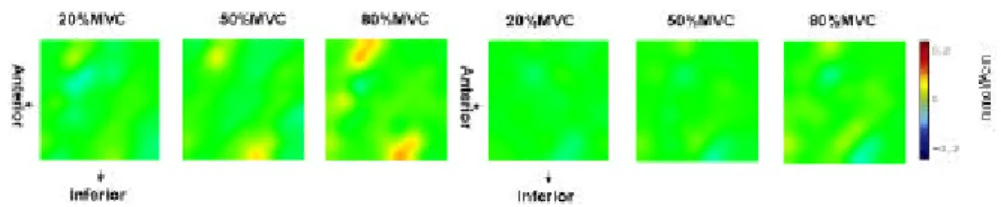
(4) 3. tensity of clenching was shown on a display and feed backed. Intensity was set at 20%, 50% and 80% of maximum voluntary clenching force (20%, 50% and 80%MVC) [8]. Each subject was required to perform clenching twice, at each of these three intensities. All tasks were performed using a block design (20-sec rest, 20-sec clenching, 20-sec rest). And the subjects were trained to clench without activating this muscle. An fNIRS system (NIRStation OMM-2001, Shimazu Co. Ltd., Japan) was used. The optodes were then positioned so as to cover the areas anterior and posterior to the central sulcus. The spectroscope's 15 source and 15 detector optodes were arranged alternately in a lattice pattern, with a distance of 30 mm between them, to form 49 source-detector pairs on an adjustable surface holder for positioning on the subject's head. Two measurements were conduced in each clenching force. OxyHb has been proposed to be the best indicator of change in rCBF in cognitive studies with NIRS [9]. So representative example of the OxyHb changes were shown in color map with deoxyHb map first. And the OxyHb data on a channel which seemed to be corresponding to the premotor area [8] for the 10-sec period commencing 5 sec after commencement of clenching at each intensity were analyzed in each subject and then averaged between all subjects as a group analysis. DeoxyHb were also analyzed to confirm general changes in blood flow. Statistical comparisons were made using a one-way analysis of variance (ANOVA) test followed by a Tukey multiple comparison tests for further comparisons among three different clenching forces (p<0.05) using SPSS(r) (SPSS Japan Inc. Tokyo, Japan).. 3 Results. Each color map of OxyHb and DeoxyHb represents the activated spatial distribution. Activation of the premotor area with bite force control was noted and the activations seemed to be increased as the clenching strengthened (Fig.2). A correlation between clenching force and OxyHb was found in the premotor cortex. In other words, OxyHb in the premotor cortex was increased as the clenching strengthened, at 20%MVC, 50%MVC and 80%MVC. ANOVA confirmed a significant difference between the three clenching forces in OxyHb but DeoxyHb (p<0.05). A significant difference was also found between 20%MVC and 80%MVC in OxyHb (Tukey test) (Fig.3)..
(5) 4. 4 Discussion The findings of this study are: activation of the premotor area with bite force control occurred in all subjects, and in the group analysis OxyHb in the premotor cortex was significantly increased as the clenching strengthened, at 20%MVC, 50%MVC and 80%MVC. These findings about OxyHb are similar to other studies showing that finger force is correlated with brain activity [1-3]. And DeoxyHb did not fall in group analysis, it is explained by the results of a study which imply the possibility of the deoxyHb is not necessarily shown decrease in brain activity [10]. Also the slight influence of systemic artifacts related to clenching is necessarily incontrovertible. The dorsal prefrontal cortex lies at the top of the motor hierarchy [11]. It can influence manual action indirectly by projections to the premotor area and from this area it has projections to the primary motor cortex, with other intricate connections among the prefrontal cortex, the pre-SMA and the rostal cingulated motor area, etc. [11]. The pre-SMA, the SMA, the premotor area and the prefrontal cortex are able to integrate information concerning the context and the response that is appropriate given the subject's goals [12, 13]. Motor preparation is associated with activations in the premotor cortex [14-16]. Thus, the premotor cortex does not seem to be concerned with control of the posture itself or of its adjustment, but with cognitive motion. Specifically, it is assumed that this area controls the process before the performance of an actual operation. The premotor cortex might convert sense information (mainly the visual information) into a target and body region information. In addition, the premotor cortex is assumed to participate in a process of integrating this converted information and generating operative information. It is thought that this processing starts from the ventral area in which the visual information is abundant according to the projection between the cortexes from the parietal lobe, and afterwards advances to the dorsal area [17] which has abundant input/output between this area, the prefrontal area and the first motor area. It is possible that the premotor cortex is involved in mastication, biting preparation and control. Changes in biting strength influence not only the primary motor cortex, but also the premotor cortex (being one of the higher motor-related cortexes). These results suggest that improvement of the function of the stomatognathic system improves the wide area of brain function. Thus, dental therapy, such as reconstruction of appropriate occlusal condition for edentulous patients [18] and treatment for eating disorder patients, might improve higher cognitive functions such as memory and learning, as well as mastication quality thorough improvement of brain activation..
(6) 5. 5. Conclusion This study aimed to elucidate the influence of bite force control on oxygenated hemoglobin levels in regional cerebral blood flow as an indicator of brain activity in the premotor area, by means of fNIRS. The results show that: activation of the premotor area with bite force control occurred in all subjects, and in the group analysis OxyHb in the premotor cortex was significantly increased as the clenching strengthened, at 20%MVC, 50%MVC and 80%MVC. These results suggest that there is a possibility that the premotor area is involved in bite force control.. References 1. 2. 3. 4. 5. 6. 7.. 8.. 9.. 10. 11. 12. 13.. 14. 15.. Dettmers, C., et al., Relation between cerebral activity and force in the motor areas of the human brain. J Neurophysiol, 1995. 74(2): p. 802-15. Nambu, I., Motor cortical activity measured with NIRS and its possible application for brain machine interface NAIST Academic Repository, 2006: p. 202-204. Thickbroom, G.W., et al., Isometric force-related activity in sensorimotor cortex measured with functional MRI. Exp Brain Res, 1998. 121(1): p. 59-64. Kaku, M., et al., Influences of reduced masticatory function on learning and memory ability. Dentistry in Japan, 2006. 42: p. 82-85. Kondo, K., M. Niino, and K. Shido, A case-control study of Alzheimer's disease in Japan-significance of life-styles. Dementia, 1994. 5(6): p. 314-26. Ishikawa, T., Brain regions activated during the mandibular movement task in functional magnetic resonance imege. J Stomatol Soc. Japan 2002. 69(1): p. 39-48. Narita N, Kamiya K, Yamaura K, Kawasaki S, Matsumoto T, et al. Chewing-related prefrontal cortex activation while wearing partial denture prosthesis: Pilot Study. J Prothodont Res. 2009; 53: doi:10.1016/j.jpor.2009.02.005 Mami Shibusawa, T.T., Kazunori and K.I. Nakajima, Kaoru Sakatani, Functional nearinfrared spectroscopy study on cerebral activity response to two different levels of hardness during food mastication. Neuroscience Letters, 2009. 449: p. 98-102. Hoshi, Y., N. Kobayashi, and M. Tamura, Interpretation of near-infrared spectroscopy signals: a study with a newly developed perfused rat brain model. J Appl Physiol, 2001. 90(5): p. 1657-62. Watanabe, E., Functional test manual of brain :Near infrared spectroscopy. MEDICAL REHABILITATION, (in Japanese), 2004. 40: p.44-50. Rushworth, M., Anatomical and functional subdivision within the primate latelal frontal cortex. Psysichobiolgy, 2000. 28: p. 187-196. Miller, E.K. and J.D. Cohen, An integrative theory of prefrontal cortex function. Annu Rev Neurosci, 2001. 24: p. 167-202. Passingham, R.E., I. Toni, and M.F. Rushworth, Specialisation within the prefrontal cortex: the ventral prefrontal cortex and associative learning. Exp Brain Res, 2000. 133(1): p. 10313. Richter, W., et al., Sequential activity in human motor areas during a delayed cued finger movement task studied by time-resolved fMRI. Neuroreport, 1997. 8(5): p. 1257-61. Rushworth, M.F., M. Krams, and R.E. Passingham, The attentional role of the left parietal cortex: the distinct lateralization and localization of motor attention in the human brain. J Cogn Neurosci, 2001. 13(5): p. 698-710..
(7) 6 16. Toni, I., M.F. Rushworth, and R.E. Passingham, Neural correlates of visuomotor associations. Spatial rules compared with arbitrary rules. Exp Brain Res, 2001. 141(3): p. 359-69. 17. Tanji, J., Recent findings on the use of premotor cortex of primates Igakunoayumi (in Japanese), 2005. 212(10 ): p. 975-982. 18. Miyamoto, I., et al., Rehabilitation with dental prosthesis can increase cerebral regional blood volume. Clin Oral Implants Res, 2005. 16(6): p. 723-7.. Figures & Legends. Fig. 1. Regulation and adjustment of clenching strength. A bite block equipped with an occlusal force sensor was prepared to measure clenching at the position where the right upper and lower canine cusps came into contact. Intensity of clenching was shown on a display and feedback was provided to the subjects.. Fig. 2. Representative examples of a subject’s relevant color maps of OxyHb (left) and DeoxyHb (light) at 10 seconds after each clenching task are shown. OxyHb increases and DeoxyHb decreases or the activations seemed to be increased as the clenching strengthened.. Fig. 3. Group analysis; a correlation between clenching force and OxyHb and DeoxyHb were found in the premotor cortex. One-way analysis of variance confirmed a significant difference between the three clenching forces (p<0. 05) not in DeoxyHb but in OxyHb. A significant difference was also found between 20%MVC and 80%MVC (Tukey test) in OxyHb..
(8)
図
関連したドキュメント
Furthermore, the upper semicontinuity of the global attractor for a singularly perturbed phase-field model is proved in [12] (see also [11] for a logarithmic nonlinearity) for two
This paper develops a recursion formula for the conditional moments of the area under the absolute value of Brownian bridge given the local time at 0.. The method of power series
We present sufficient conditions for the existence of solutions to Neu- mann and periodic boundary-value problems for some class of quasilinear ordinary differential equations.. We
In Section 13, we discuss flagged Schur polynomials, vexillary and dominant permutations, and give a simple formula for the polynomials D w , for 312-avoiding permutations.. In
Analogs of this theorem were proved by Roitberg for nonregular elliptic boundary- value problems and for general elliptic systems of differential equations, the mod- ified scale of
Then it follows immediately from a suitable version of “Hensel’s Lemma” [cf., e.g., the argument of [4], Lemma 2.1] that S may be obtained, as the notation suggests, as the m A
Definition An embeddable tiled surface is a tiled surface which is actually achieved as the graph of singular leaves of some embedded orientable surface with closed braid
Correspondingly, the limiting sequence of metric spaces has a surpris- ingly simple description as a collection of random real trees (given below) in which certain pairs of
