表 題 オキサシリン感性 mecA 陽性黄色ブドウ球菌におけるβラクタ ム薬高度耐性化機構の解明
(Mechanisms of acquisition of β-lactam resistance in
oxacillin-susceptible mecA-positive Staphylococcus aureus) 論 文 の 区 分 博士課程 著 者 名 ブーンシリ タニト Boonsiri Tanit 担当指導教員氏名 教授 崔 龍洙 所 属 自治医科大学大学院医学研究科 人間生物学 専攻 生体防御医学 専攻分野 微生物・免疫学 専攻科 令和 2 年 1 月 10 日申請の学位論文
CONTENTS
1. Abstract………. 1
2. Introduction……….. 5 3. Materials and Methods……… 7
3-1. Bacterial strains and growth conditions 3-2. Antibiotic susceptibility test
3-3. mecA detection via PCR
3-4. Isolation of mutants with reduced susceptibility to oxacillin from parental OS-MRSA strains
3-5. Whole-genome sequencing
3-6. Construction of the phylogenetic tree 3-7. Growth curve analysis
3-8. Determination of cellular ATP level
3-9. RNA extraction
3-10. Determination of mecA expression level by qRT-PCR 3-11. RNA-sequencing analysis
3-12. Construction of mecA and blaI knockout mutants 3-13. Complementation of mecA
3-14. Statistical analysis
4. Results... 21 4-1. Characterization of clinical OS-MRSA isolates
4-2. Genome analysis of the clinical OS-MRSA isolates
4-3. Identification of mutations associated with reduced susceptibility to oxacillin in OS-MRSA
4-4. Increased mecA expression was responsible for reduced oxacillin susceptibility in OS-MRSA
4-5. Correlation between mecA expression levels and oxacillin MICs in mutants with reduced oxacillin susceptibility
4-6. The bla operon is involved in reduced susceptibility to oxacillin
4-7. Transcriptome analysis revealed stringent response-like gene regulation in the oxacillin-resistant mutants
4-8. The mutants with reduced oxacillin susceptibility did not display a slow- growth phenotype
4-9. Intracellular ATP accumulation in the resistant mutants
5. Discussion……….... 52 6. Conclusion………... 62
7. References………... 63 8. Funding………... 71
1. Abstract
Staphylococcus aureus strains that are susceptible to the β-lactam antibiotic
oxacillin despite carrying mecA (oxacillin-susceptible methicillin-resistant S. aureus [OS-MRSA]) cause serious clinical problems globally because of their ability to acquire β-lactam resistance. Understanding the genetic mechanism(s) responsible for the acquisition of β-lactam resistance in OS-MRSA is therefore crucial for its clinical management. For this purpose, whole-genome sequencing-based analysis was performed using clinical OS-MRSA isolates and their oxacillin-resistant mutants. Genomic analysis revealed extensive genomic diversity among 43 OS-MRSA strains (11 MLST types and 4 SCCmec types) and identified 118 variants of 141 mutations in 46 genes and 8 intergenic regions among the 100 oxacillin-resistant mutants generated from 26 OS-MRSA strains. Among them, mutations were most frequently found in genes related to RNA polymerase (rpoBC), purine biosynthesis (guaA, prs,
hprT, rsh), glycolysis (pykA, fbaA, fruB), protein quality control (clpXP, ftsH), and
tRNA synthase (lysS, gltX), whereas no mutations existed in the mec and bla operons. The whole-genome transcriptional profile of the resistant mutants demonstrated that downregulation of genes associated with purine biosynthesis, protein quality control, and tRNA synthesis, mimicking the classic stringent response, decreased intercellular
GTP levels and targeted metabolic pathways toward the induction of mecA expression by upregulating blaR1 and altering the expression of genes involved in autolysis and peptidoglycan synthesis. Moreover, the resistance mutations exhibited a relatively low fitness cost, explaining the easy selection of β-lactam–resistant OS-MRSA mutants during the course of antimicrobial treatment.
Keywords: oxacillin-susceptible mecA-positive Staphylococcus aureus; MRSA; OS-MRSA; Staphylococcus aureus; β-lactam resistance; transcriptome; stringent response; (p)ppGpp; drug resistant mutant; whole genome sequence
Abbreviations
ATP: Adenosine triphosphate
CLSI: Clinical and Laboratory Standards Institute DNA: deoxyribonucleic acid
dNTP: Deoxyribonucleotide triphosphate GDP: Guanosine diphosphate
GTP: Guanosine triphosphate HP: Hypothetical protein
IR: Intergenic region LB: Luria-Bertani
MHA: Mueller–Hinton agar MHB: Mueller–Hinton broth
MIC: Minimum inhibitory concentration MLST: Multilocus sequence typing
MRSA: Methicillin-resistant Staphylococcus aureus MSSA: Methicillin-susceptible Staphylococcus aureus OD600: Optical density at a wavelength 600 nm
OS-MRSA: Oxacillin-susceptible MRSA PBP2a: Penicillin-binding protein 2a
(p)ppGpp: Guanosine tetra- and penta-phosphate qRT-PCR: Quantitative Reverse Transcription PCR RNA-seq: RNA sequencing
RNA: Ribonucleic acid RNAP: RNA polymerase rRNA: ribosomal RNAs
SNPs: single nucleotide polymorphisms ST: Sequence type of MLST
TSA: Tryptic soy agar TSB: Tryptic soy broth
2. Introduction
Staphylococcus aureus is an important bacterial pathogen that can cause
life-threatening infections in both humans and animals (1, 2). A known feature of S.
aureus is its evolutionary potential to develop antibiotic resistance under selection
pressure via antibiotic treatment. Methicillin-resistant S. aureus (MRSA) is resistant to the entire class of β-lactam antibiotics, including penicillin, methicillin, and cefazolin (3). It was first recognized as a problematic pathogen in hospital settings, but it has subsequently emerged in community settings and livestock (3-5). MRSA infections remain a major concern in the clinical setting because they are more difficult to treat than infections involving other ordinary susceptible strains of S.
aureus. β-lactam resistance in MRSA is primarily mediated by a non-native mecA
gene encoding modified penicillin-binding protein 2a (PBP2a), which has an extremely low affinity for β-lactams, whereas PBP2a expression in MRSA is dependent on the presence of functional MecI/MecR1/MecR2 and BlaI/BlaR1 regulators in the mec and bla operons, respectively (6, 7). However, the level of β-lactam resistance does not always correlate with that of PBP2a expression (6-8). Recently, oxacillin-susceptible mecA-positive S. aureus (OS-MRSA) strains have been increasingly reported worldwide in clinical isolates as well as in animals and
food (9-15). In clinical microbiology laboratories, an oxacillin minimum inhibitory concentration (MIC) ≥ 4 µg/mL or cefoxitin MIC ≥ 8 µg/mL is routinely used as a breakpoint for diagnosing MRSA, whereas the presence of mecA has been used as a marker for the genotypic identification of MRSA (16). Owing to its susceptibility to oxacillin, OS-MRSA might be misidentified as methicillin-susceptible S.
aureus (MSSA) in routine clinical laboratories in which mecA detection is unavailable
(17). In addition, despite being fundamentally susceptible to β-lactam treatment, OS-MRSA is prone to develop strong β-lactam resistance following antibiotic therapy due to its carriage of mecA (10-12, 14, 15). This ultimately leads to β-lactam treatment failure (18, 19). It is suggested that OS-MRSA possesses unknown regulatory mechanisms that contribute to the acquisition of β-lactam resistance. Therefore, this study aimed to unravel the genetic mechanism(s) responsible for the development of β-lactam resistance in OS-MRSA.
3. Materials and Methods
3-1. Bacterial strains and growth condition
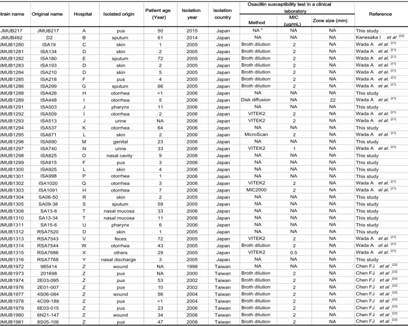
A total of 43 OS-MRSA isolates from various clinical samples were collected from routine clinical laboratories in hospitals across Japan and Taiwan between 1998 and 2015 (Table 1 (20-22)). Mueller–Hinton broth (MHB; Becton Dickinson Co., Ltd., Sparks, MD, USA) and tryptic soy broth (TSB; Becton Dickinson) were used to culture
S. aureus, whereas Escherichia coli was grown in Luria-Bertani (LB; Becton Dickinson)
medium. In some experiments, antibiotics were added to the medium at the following concentrations: ampicillin (Nacalai Tesque, Inc., Kyoto, Japan) at 100 µg/mL for E. coli, tetracycline (Nacalai Tesque) at 5 µg/mL for S. aureus, and chloramphenicol (Nacalai Tesque) at 10 µg/mL for S. aureus and E. coli. For preservation, bacterial cells were cultivated on tryptic soy agar (TSA; Becton Dickinson) and incubated at 37°C upon receipt. A single colony was then selected and grown overnight in TSB at 37°C. The overnight culture was aliquoted and stored at −80°C in 50% glycerol (Wako Pure Chemical Industries, Ltd., Tokyo, Japan) until use.
Table 1: List of clinical OS-MRSA isolates used in this study
aNA: Not available
Method (µg/mL) MIC Zone size (mm)
JMUB217 JMUB217 A pus 50 2015 Japan NAa NA NA This study
JMUB492 D2 B sputum 61 2014 Japan NA NA NA Kanesaka I et al.[20]
JMUB1280 ISA19 C skin 1 2005 Japan Broth dilution 2 NA Wada A et al.[21]
JMUB1281 ISA134 D skin 2 2005 Japan Broth dilution 2 NA Wada A et al.[21]
JMUB1282 ISA180 E sputum 72 2005 Japan Broth dilution 2 NA Wada A et al.[21]
JMUB1283 ISA193 D skin 2 2005 Japan Broth dilution 2 NA Wada A et al.[21]
JMUB1284 ISA210 D skin 5 2005 Japan Broth dilution 2 NA Wada A et al.[21]
JMUB1285 ISA218 F pus 4 2005 Japan Broth dilution 2 NA Wada A et al.[21]
JMUB1286 ISA299 G sputum 66 2005 Japan Broth dilution 2 NA Wada A et al.[21]
JMUB1288 ISA426 H otorrhea <1 2006 Japan NA NA NA This study
JMUB1289 ISA448 I otorrhea 5 2006 Japan Disk diffusion NA 22 Wada A et al.[21]
JMUB1291 ISA503 J pharynx 11 2006 Japan NA NA NA This study
JMUB1292 ISA509 I otorrhea 2 2006 Japan VITEK2 2 NA Wada A et al.[21]
JMUB1293 ISA513 J urine NA 2006 Japan VITEK2 2 NA Wada A et al.[21]
JMUB1294 ISA537 K otorrhea 64 2006 Japan NA NA NA This study
JMUB1295 ISA671 L skin 2 2006 Japan MicroScan 2 NA Wada A et al.[21]
JMUB1296 ISA690 M genital 23 2006 Japan NA NA NA This study
JMUB1297 ISA740 N urine 33 2006 Japan VITEK2 1 NA Wada A et al.[21]
JMUB1298 ISA825 O nasal cavity 9 2006 Japan NA NA NA This study
JMUB1299 ISA915 F pus 3 2006 Japan NA NA NA This study
JMUB1300 ISA925 L skin 4 2006 Japan NA NA NA This study
JMUB1301 ISA998 P otorrhea 1 2006 Japan NA NA NA This study
JMUB1302 ISA1020 Q otorrhea 3 2006 Japan VITEK2 2 NA Wada A et al.[21]
JMUB1303 ISA1091 H otorrhea 7 2006 Japan MIC2000 2 NA Wada A et al.[21]
JMUB1304 SA06-50 R skin 2 2005 Japan NA NA NA This study
JMUB1305 SA09-38 S sputum 59 2005 Japan NA NA NA This study
JMUB1308 SA13-6 T nasal mucosa 33 2006 Japan NA NA NA This study
JMUB1310 SA13-34 T nasal mucosa 11 2006 Japan NA NA NA This study
JMUB1311 SA15-6 U pharynx 6 2006 Japan NA NA NA This study
JMUB1312 RSA7520 D skin 1 2005 Japan NA NA NA This study
JMUB1313 RSA7543 V feces 72 2005 Japan VITEK2 2 NA Wada A et al.[21]
JMUB1314 RSA7544 W otorrhea 43 2005 Japan Broth dilution 2 NA Wada A et al.[21]
JMUB1315 RSA7686 X others 29 2005 Japan VITEK2 0.5 NA Wada A et al.[21]
JMUB1316 RSA7768 Y nasal discharge 3 2005 Japan NA NA NA This study
JMUB1972 985414 Z wound NA 1998 Taiwan NA NA NA Chen FJ et al.[22]
JMUB1973 201698 Z pus NA 2000 Taiwan Broth dilution 2 NA Chen FJ et al.[22]
JMUB1974 2E03-095 Z pus 53 2002 Taiwan Broth dilution 2 NA Chen FJ et al.[22]
JMUB1976 2E01-007 Z pus 10 2002 Taiwan Broth dilution 2 NA Chen FJ et al.[22]
JMUB1977 4S06-084 Z wound 56 2004 Taiwan Broth dilution 2 NA Chen FJ et al.[22]
JMUB1978 4C09-188 Z pus <1 2004 Taiwan Broth dilution 2 NA Chen FJ et al.[22]
JMUB1979 6E03-015 Z pus 23 2006 Taiwan Broth dilution 2 NA Chen FJ et al.[22]
JMUB1980 6N21-147 Z wound 34 2006 Taiwan Broth dilution 2 NA Chen FJ et al.[22]
JMUB1981 8S05-106 Z pus 47 2008 Taiwan Broth dilution 2 NA Chen FJ et al.[22]
Reference Oxacillin susceptibility test in a clinical
laboratory Strain name Original name Hospital Isolated origin Patient age
(Year)
Isolation year
Isolation country
3-2. Antibiotic susceptibility test
Oxacillin and cefoxitin MICs were determined using the E-test method according to Clinical and Laboratory Standards Institute (CLSI) guidelines (16). Briefly, overnight cultures of S. aureus strains grown in 4 mL of MHB at 37°C were adjusted to 0.5 McFarland turbidity (approximately 1 × 10⁸–2 × 10⁸ CFU/mL) and spread on Mueller–Hinton agar (MHA; Becton Dickinson) plates. The E-test gradient strip (bioMérieux SA, Marcy l’Étoile, France) was then placed on the bacterial lawn. The MIC was determined after incubation at 37°C for 24 h. The isolates with oxacillin MIC £ 2 µg/mL or cefoxitin MIC £ 4 µg/mL were considered oxacillin- and cefoxitin-susceptible, respectively.
3-3. mecA detection via PCR
DNA was extracted from OS-MRSA isolates grown overnight on TSA plates using MightyPrep reagent (Takara Bio Inc., Shiga, Japan) in accordance with the manufacturer’s instructions. PCR was then performed on the extracted DNA using Quick Taq® HS DyeMix (Toyobo Co., Ltd., Osaka, Japan). A primer pair (F and mecA-R, Table 2) was used to amplify a 519-bp region of mecA. The thermal cycling conditions included initial denaturation at 94°C for 2 min followed by 30 cycles of 94°C for 30 s,
55°C for 30 s, and 68°C for 1 min. Finally, the amplified products were electrophoresed on 1% agarose gel, stained with ethidium bromide, and visualized using AE-6933FXES Printgraph (Atto Co., Tokyo, Japan).
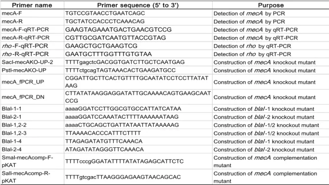
Table 2: List of primers used to construct mutants
Primer name Primer sequence (5' to 3') Purpose
mecA-F TGTCCGTAACCTGAATCAGC Detection of mecA by PCR mecA-R TGCTATCCACCCTCAAACAG Detection of mecA by PCR mecA-F-qRT-PCR GAAGTAGAAATGACTGAACGTCCG Detection of mecA by qRT-PCR mecA-R-qRT-PCR CGTTGCGATCAATGTTACCGTAG Detection of mecA by qRT-PCR
rho-F-qRT-PCR GAAGCTGCTGAAGTCG Detection of rho by qRT-PCR
rho-R-qRT-PCR GAATGCTTTGGTTTGTGTAA Detection of rho by qRT-PCR SacI-mecAKO-UP-2 TTTTgagctcGACGGTGATCTTGCTCAATGAG Construction of mecA knockout mutant PstI-mecAKO-UP TTTTctgcagTAGTAAACACTGAAGATGCC Construction of mecA knockout mutant mecA_fPCR_UP CGGATTGCTTCACTGTTTTGCAATATCCTCCTTATAT
AAG Construction of mecA knockout mutant
mecA_fPCR_DN CTTATATAAGGAGGATATTGCAAAACAGTGAAGCAATCCG Construction of mecA knockout mutant BlaI-1-1 aaaaGGATCCTTGGCGTGCCATTATCATAA Construction of blaI-1 knockout mutant BlaI-2-1 aaaaGGATCCAAATACTTTTAAAAAATAAG Construction of blaI-2 knockout mutant BlaI-1,2-2 aaaaCTGCAGCTGATTATAATTATAAAAAG Construction of blaI-1/2 knockout mutant BlaI-1,2-3 TTAAAACACCCATTTCTTTT Construction of blaI-1/2 knockout mutant BlaI-1-4 TTAGAGATATGTTTCAAACA Construction of blaI-1 knockout mutant BlaI-2-4 ATAGATATAGGGTTCAAACA Construction of blaI-2 knockout mutant
SmaI-mecAcomp-F-pKAT TTTTcccgGGATATTTTATATAGAGCATTCTC Construction of mutant mecA complementation
SalI-mecAcomp-R-pKAT TTTTgtcgacTTAAGGGAGAAGTAACAGCAC
Construction of mecA complementation mutant
3-4. Isolation of mutants with reduced susceptibility to oxacillin from parental OS-MRSA strains
To isolate mutants with reduced oxacillin susceptibility, all 43 OS-MRSA parental strains were exposed to oxacillin according to the E-test method as described for susceptibility testing. Briefly, the oxacillin E-test was performed using OS-MRSA strains inoculated onto MHA plates. A single colony growing inside the inhibition zone after 24– 48 h of incubation was randomly picked and sub-cultured in TSB for 24 h at 37°C. The overnight culture was then serially diluted 10-fold with 0.9% NaCl and spread onto a TSA plate. A single colony growing on the TSA plate was again randomly selected and inoculated into TSB for preservation in 50% glycerol at −80°C. The oxacillin susceptibility of the stocked cells was determined again using the E-test method to discriminate mutant colonies from persister colonies. The cells exhibiting higher oxacillin MICs were selected as oxacillin-reduced susceptibility mutants, which were then used for subsequent analysis.
3-5. Whole-genome sequencing
Genomic DNA was extracted from OS-MRSA and its mutants using the phenol-chloroform method and purified using a QIAamp DNA mini kit (Qiagen, Hilden,
Germany) following previously developed methods (23). The genomic sequences of parental strains were determined via mate-pair sequencing as previously described (23, 24). Briefly, a mate-pair library was prepared using a Nextera mate-pair library prep kit (Illumina, Inc., San Diego, CA, USA), and sequencing was performed using an Illumina MiSeq platform with the MiSeq reagent kit version 3 (Illumina). The mate-paired reads of OS-MRSA were trimmed using the FASTQ toolkit version 2.2.0 to generate high-quality reads and assembled using Velvet Assembler version 1.2.10 to construct genome scaffolds. The generated genomic sequences were finally annotated using Microbial Genome Annotation Pipeline (http://www.migap.org/). Meanwhile, the genomic sequences of in vitro-selected mutants with reduced oxacillin susceptibility were determined by sequencing paired-end reads as previously described (25). The paired-end library was prepared using a Nextera XT library prep kit and sequenced using the Illumina MiSeq platform with the MiSeq reagent kit version 3. The paired-end reads of each mutant were mapped against the genomic sequences of their corresponding parental OS-MRSA strains, and mutations were detected using CLC Genomics Workbench version 9 (CLCbio, Qiagen, Valencia, CA, USA). Mutations identified in each mutant were verified by Sanger sequencing using the Applied Biosystems 3130xl genetic analyzer (Thermo Fisher Scientific, MA, USA).
3-6. Construction of the phylogenetic tree
To construct the OS-MRSA phylogenetic tree, kSNP3 (26), available at https://sourceforge.net/projects/ksnp/, was first used to identify single nucleotide polymorphisms (SNPs) in the whole-genome sequencing data of OS-MRSA strains. The k-mer size was set to an optimum length of 13 as estimated by Kchooser for extracting SNPs from the sequence data. A maximum parsimony tree was then constructed using the majority of the SNPs present in at least 75% of the genomes. The generated phylogenetic tree was visualized using FigTree ver.1.4.3 (tree.bio.ed.ac.uk/software/figtree/).
3-7. Growth curve analysis
The bacterial doubling time was determined as described previously (27). Briefly, overnight cultures of parental OS-MRSA strains and the laboratory-selected mutants were adjusted to an OD600 of 0.2 in MHB. Then, aliquots of 100 µL were inoculated into 10 mL of MHB (final concentration of 1 × 10⁵ CFU/mL), and the cultures were grown at 37°C with agitation at 25 rpm in an automatic temperature gradient rocking incubator
measured every 5 min for 12 h, and the bacterial growth curve was generated by plotting the measured ODs against time. The doubling time was determined by fitting the growth curve to an exponential equation. Bacterial growth was measured using at least three independent experiments.
3-8. Determination of cellular ATP level
The parental OS-MRSA strains and the laboratory-selected mutants were cultured overnight in MHB at 37°C with agitation at 150 rpm. The overnight cultures were adjusted to an OD600 of 0.2 in MHB, and 100 µL of the OD-adjusted culture were inoculated into 10 mL of MHB. The cultures were grown at 37°C with agitation at 25 rpm in an automatic temperature gradient rocking incubator. One microliter of each mid-exponential phase culture (OD600 = 0.5) was then transferred to a fresh 1.5-mL tube and immediately centrifuged at 15,000 rpm for 1 min at 4°C to pellet cells. After centrifugation, the cell pellet was stored immediately at −80°C until analysis. To determine cellular ATP levels, a BacTiter-Glo™ Microbial Cell Viability Assay kit (Promega, WI, USA) was used according to the manufacturer’s instructions. Briefly, the cell pellet was resuspended in 1 mL of MHB, and 25 µL of the cell suspension were
Tokyo, Japan) and incubated at room temperature for 5 min. The luminescence was then read on an EnVision 2104 Multilabel Reader (Perkin Elmer, Waltham, MA, USA). The ATP concentration was determined with reference to an ATP standard curve prepared from ATP disodium salt hydrate (A2383, Merck KGaA, Darmstadt, Germany). ATP disodium salt was dissolved in distilled water, yielding 1 µM ATP standard solutions. Serial 10-fold dilutions of the ATP standard solution were created using MHB to prepare diluted standards that were then used to generate the standard curve. The cellular ATP concentration of each sample was presented as the mean of three independent experiments performed using three biological replicates.
3-9. RNA extraction
Overnight cultures of the parental OS-MRSA strains and the laboratory-selected mutants were adjusted to an OD600 of 0.4. The OD-adjusted cultures were then diluted 1:100 in 1 or 10 mL of MHB for qRT-PCR and RNA-seq, respectively. Each culture was grown to the early log-phase (OD600 = 0.3) before treatment with a final concentration of 0.1 µg/mL oxacillin or equal volume of distilled water (control) for 1 h (qRT-PCR) or until OD600 = 0.6 (RNA-seq). After oxacillin treatment, the bacterial cells were harvested
by centrifugation at 15,000 rpm for 1 min at 4°C (qRT-PCR) or at 8,000 rpm for 5 min at 4°C seq). Pelleted cells were resuspended in 600 µL (qRT-PCR) or 6 mL (RNA-seq) of TE buffer (10 mM Tris-HCl and 10 mM EDTA, pH 8.0) and lysed with 25 (qRT-PCR) or 30 µg (RNA-seq) of lysostaphin (Merck KGaA) by incubating the mixtures at 37°C for less than 5 min. Total RNA was then extracted using acidic-phenol saturated with 20 mM sodium acetate (pH 4.8) and chloroform and enriched via ethanol precipitation. Contaminating DNA was removed from the total RNA preparations by incubating the solutions with 2 (qRT-PCR) or 20 units (RNA-seq) of RNase-free DNase I (F. Hoffmann-La Roche Ltd, Basel, Switzerland) at 37°C for 30 min. Total RNA was finally purified using acidic-phenol/chloroform and eluted in RNase-free water.
3-10. Determination of mecA expression level by qRT-PCR
The extracted total RNA (100 ng per sample) was reverse-transcribed into complementary DNA (cDNA) using a PrimeScript™ 1st Strand cDNA Synthesis Kit (Takara Bio). qRT-PCR was performed using TB Green™ Premix Ex Taq™ (Tli RNaseH Plus, Takara Bio) on the Mx3005P™ Real-Time PCR instrument (Stratagene, La Jolla, CA, USA). A primer set (mecA-F-qRT-PCR and mecA-R-qRT-PCR, Table 2) was used
to amplify the 162-bp mecA sequence, whereas the 163-bp housekeeping gene rho was amplified using designated primers (rho-F-qRT-PCR and rho-R-qRT-PCR, Table 2) and used as the reference gene for normalization during gene expression analysis. The thermal cycling conditions included initial denaturation at 95°C for 30 s followed by 40 cycles of 95°C for 5 s and 60°C for 30 s.
3-11. RNA-sequencing analysis
To perform RNA-seq analysis, ribosomal RNAs (rRNAs) in total RNA preparations of the JMUB217 strain and its mutant derivatives were first depleted using a Ribo-Zero rRNA Removal Kit (Bacteria) from Illumina. Double-stranded cDNA was then synthesized using a PrimeScript Double Strand cDNA Synthesis Kit (Takara Bio). The generated cDNA served as the template for constructing the paired-end library using a Nextera XT library prep kit, and the library was subsequently sequenced using the Illumina MiSeq platform and the MiSeq reagent kit version 3. RNA-seq analysis was performed using CLC Genomics Workbench version 9, and the RNA-seq reads were aligned to the reference genomes of the parental strain JMUB217. Gene expression was normalized by calculating the reads per kilobase per million mapped reads, and
differentially expressed genes were identified using Baggerly’s test (β-binomial test) with false discovery rate correction. Genes with adjusted p < 0.05 were considered to be significantly differentially expressed.
3-12. Construction of mecA and blaI knockout mutants
To construct mecA and blaI-knockout mutants of the JMUB217 strain, the pKFT markerless gene deletion system was used as previously described (28). Briefly, to delete mecA, 1-kb upstream and downstream flanking sequences of the target gene were amplified by PCR using the primer sets SacI-mecAKO-UP-2/mecA_fPCR_UP and PstI-mecAKO-UP/mecA_fPCR_DN (Table 2), respectively, and KOD FX Neo (Toyobo). Then, second-round PCR was performed using the first-round PCR products as templates with the primer set SacI-mecAKO-UP-2/PstI-mecAKO-UP. The second-round PCR products and pKFT were digested with the restriction enzymes PstI and SacI (Takara Bio) and ligated using Ligation high ver. 2 (Toyobo), generating the plasmid pmecAKO. pmecAKO was transformed into E. coli DH5α, and the transformed cells were plated on LB agar with 100 µg/mL ampicillin. Concerning the generation of blaI-knockout mutants, DNA fragments containing blaI-1 (locus tag: JMUB217_1395) or
BlaI-1-1/BlaI-1,2-2 and BlaI-2-BlaI-1-1/BlaI-1,2-2 (Table 2), respectively. The PCR fragments and pKFT were then digested using the restriction enzymes BamHI and PstI (Takara Bio) and ligated using Ligation high ver. 2. The ligated DNA fragments were independently transformed into E. coli DH5α, and the transformed cells were plated on LB agar with 100 µg/mL ampicillin. The plasmids were extracted, and second-round PCR was conducted using the primer set 1,2-3/1-4 for blaI-1 knockout strains and BlaI-1,2-3/BlaI-2-4 for blaI-2 knockout strains. The self-ligated PCR fragments (pblaI-1KO and pblaI-2KO) were again individually transformed into E. coli DH5α, and transformed cells were plated on LB agar with 100 µg/mL ampicillin. Afterward, all three plasmids (pmecAKO, pblaI-1KO, and pblaI-2KO) were extracted from the E. coli DH5α transformants and transformed into E. coli BL21. The plasmids extracted from
E. coli BL21 were subsequently electroporated into S. aureus JMUB217 and mutants
with reduced oxacillin susceptibility as previously described (29), and the cells were cultured on TSA with 5 µg/mL tetracycline at 30°C. An isolated colony was then grown overnight in 4 mL of TSB containing 5 µg/mL tetracycline at 30°C. Single crossover was performed by growing the overnight culture on TSA with 5 µg/mL tetracycline at 43°C. Then, double crossover was performed by incubating the single crossover mutant
on TSA at 30°C. The double crossover event was confirmed by PCR and Sanger sequencing.
3-13. Complementation of mecA
To generate a mecA-complemented mutant, a DNA fragment containing wild-type mecA from strain JMUB217 was amplified using the primers SmaI-mecAcomp-F-pKAT and SalI-mecAcomp-R-SmaI-mecAcomp-F-pKAT (Table 2). The PCR fragment and SmaI-mecAcomp-F-pKAT were digested with SmaI and SalI (Takara Bio) and ligated using Ligation high ver. 2. The ligated DNA fragment was transformed into E. coli DH5α, and the transformed cells were plated on LB agar with 10 µg/mL chloramphenicol. Finally, the complementation plasmid was extracted and electroporated into the JMUB217 strain (29).
3-14. Statistical analysis
All statistical analyses were performed using Prism 8 (GraphPad Software, San Diego, CA, USA). The correlations between variables were calculated using the non-parametric Spearman’s correlation coefficient (rs), with p < 0.05 denoting statistical
4. Results
4-1. Characterization of clinical OS-MRSA isolates
A total of 43 OS-MRSA isolates recovered from various clinical specimens collected from Japan and Taiwan were included in this study (Table 1). The characteristics of OS-MRSA were re-confirmed via determination of their oxacillin susceptibility and the presence of mecA (Table 3). Our results revealed that all studied strains maintained the typical characteristics of OS-MRSA, including mecA positivity, but they were susceptible to oxacillin with MICs ranging from 0.125 to 2 µg/mL. According to CLSI, cefoxitin can also be used to detect MRSA. Thus, cefoxitin susceptibility testing was conducted to investigate whether there was discrepancy between oxacillin and cefoxitin susceptibility among the OS-MRSA isolates. The cefoxitin MICs for all MRSA isolates ranged 1.5–12 µg/mL, with 24 of the 43 OS-MRSA isolates (56%) exhibiting susceptibility to cefoxitin.
Table 3: Characteristics of the 43 clinical OS-MRSA isolates
aClade classified from a particular branch on the phylogenetic tree bSequence type
cNon-typeable dNot detected
eDetected by PCR and whole genome sequencing data
Clade a Strain Oxacillin Cefoxitin arcC aroE glpF gmk pta tpi yqiL STb SCC mec
type mecR1 mecI mecA e
JMUB1297 0.25 1.5 3 3 1 1 4 4 3 8 IVa + NDd + JMUB1282 0.125 2 3 3 1 1 4 4 3 8 IVa + ND + JMUB1315 0.25 3 3 3 1 42 4 4 3 1516 IVc + ND + 2 JMUB492 2 12 1 1 1 1 1 1 1 1 IVa + ND + 3 JMUB217 0.75 4 1 1 1 1 22 1 1 772 V + ND + 4 JMUB1293 0.125 1.5 1 4 1 4 12 1 10 5 II + + + JMUB1308 0.5 4 1 26 28 18 18 33 50 89 IVa + ND + JMUB1311 0.19 4 1 26 28 18 18 33 50 89 IVa + ND + JMUB1291 1.5 6 1 26 28 18 18 33 50 89 IVa + ND + JMUB1305 0.5 4 1 26 28 18 18 54 50 91 IVa + ND + JMUB1285 0.5 6 1 26 28 18 18 33 50 89 V + ND + JMUB1289 0.5 6 1 26 28 18 18 33 50 89 V + ND + JMUB1284 0.5 4 1 26 28 18 18 33 50 89 V + ND + JMUB1283 0.38 4 1 26 28 18 18 33 50 89 V + ND + JMUB1304 0.75 4 1 26 28 18 18 33 50 89 V + ND + JMUB1301 1 6 1 26 28 18 18 33 50 89 V + ND + JMUB1973 0.75 6 19 23 15 2 19 20 15 59 V + ND + JMUB1981 1 6 19 23 15 2 19 20 15 59 V + ND + JMUB1980 0.5 4 19 23 15 2 19 20 15 59 V + ND + JMUB1976 1.5 6 19 23 15 2 19 20 15 59 V + ND + JMUB1978 0.75 6 19 23 15 2 19 20 15 59 V + ND + JMUB1972 1 6 19 23 15 2 19 20 15 59 V + ND + JMUB1974 0.25 1.5 19 23 15 2 19 20 15 59 V + ND + JMUB1977 0.75 6 19 23 15 48 19 20 15 338 V + ND + JMUB1979 0.5 8 19 23 15 2 19 20 15 59 IVa + ND + JMUB1298 0.75 8 6 5 6 2 7 14 5 121 V + ND + JMUB1303 0.25 4 6 5 6 2 7 14 5 121 V + ND + JMUB1281 1 6 6 5 6 2 7 14 5 121 V + ND + JMUB1286 1 6 6 5 6 2 7 14 5 121 V + ND + JMUB1300 0.19 1.5 6 5 6 2 7 14 5 121 V + ND + JMUB1314 0.75 6 6 5 6 2 7 14 5 121 V + ND + JMUB1312 1 4 6 5 6 2 7 14 5 121 V + ND + JMUB1313 0.75 2 6 5 6 2 7 14 5 121 V + ND + JMUB1288 0.5 2 6 5 6 2 7 14 5 121 V + ND + JMUB1280 0.5 4 6 5 6 2 7 14 5 121 V + ND + JMUB1295 0.19 3 6 5 6 2 7 14 5 121 V + ND + JMUB1299 0.25 4 6 5 6 2 7 14 5 121 V + ND + JMUB1316 0.75 6 6 5 6 2 7 14 5 121 V + ND + JMUB1294 0.38 6 6 5 6 2 7 14 5 121 V + ND + JMUB1296 0.5 4 6 5 6 2 7 14 5 121 V + ND + JMUB1292 0.75 4 6 5 6 2 7 14 5 121 V + ND + JMUB1310 0.5 4 73 5 6 2 7 14 5 NTc V + ND + JMUB1302 1 6 73 5 6 2 7 14 5 NT V + ND + 7 6
MIC (μg/mL) MLST SCC mec typing
1
4-2. Genome analysis of the clinical OS-MRSA isolates
To determine the genetic background of the strains used in this study, the whole-genome sequences of the 43 clinical OS-MRSA isolates were determined, and their phylogenetic relationships were analyzed by constructing a phylogenetic tree using kSNP3 (Fig 1). The phylogenetic tree revealed extensive genomic diversity among the isolates, which could be classified into seven main phylogenetic clades. In addition, these isolates could also be grouped into 11 MLST types (ST1, ST5, ST8, ST59, ST89, ST91, ST121, ST338, ST772, ST1516, and ST-non-typeable [STNT]), and they carried four different types of SCCmec types (II, IVa, IVc, and V). The majority of OS-MRSA isolates were belonged to ST121-SCCmec type V (16 strains, 37%), followed by ST59-SCCmec type V (seven strains, 16%), ST89-SCCmec type V (six strains, 14%), ST89-SCCmec type IVa (three strains, 7.0%), ST8-SCCmec type IVa (two strains, 4.7%), STNT-SCCmec type V (two strains, 4.7%). In addition, seven singletons (ST1-STNT-SCCmec type IVa, ST5-SCCmec type II, ST59-SCCmec type IVa, ST91-SCCmec type IVa, ST338-SCCmec type V, ST772-ST338-SCCmec type V, and ST1516-ST338-SCCmec type IVc), each of which comprised 2.3% of all strains, were identified. SCCmec types V (33 strains, 77%) and IVa (eight strains, 19%) were predominant among the OS-MRSA isolates, whereas only one isolate harbored each of SCCmec type II and IVc, respectively. Moreover,
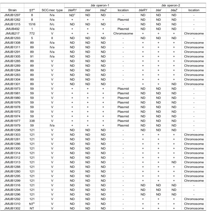
whole-genome sequencing demonstrated that 34 of the 43 (79%) OS-MRSA isolates carried a complete bla operon (Table 4), which could be classified into two genotypes, namely bla operon-1 and bla operon-2, based on the nucleotide sequences. These two operons shared nucleotide identities of 94% for blaZ, 92% for blaR1, and 94% for blaI. Twelve strains (28%) carried bla operon-1, and all but one (JMUB217) bla operon-1 was located on plasmids. Meanwhile, 23 isolates (53%) carried intact bla operon-2 in their chromosomes. JMUB217 carried both bla operons in its chromosome. An incomplete bla operon-2 lacking blaZ but having intact blaR1 and blaI was present in isolates JMUB1301 and JMUB1313. The absence of blaZ in the bla operons of these isolates was confirmed by PCR (data not shown). Lastly, seven isolates (16%) lacked a bla operon.
JMUB1297 (ST8, IVa) JMUB1282 (ST8, IVa) JMUB1315 (ST1516, IVc) JMUB492 (ST1, IVa) JMUB217 (ST772, V) JMUB1293 (ST5, II) JMUB1308 (ST89, IVa) JMUB1311 (ST89, IVa) JMUB1291 (ST89, IVa) JMUB1305 (ST91, IVa) JMUB1285 (ST89, V) JMUB1289 (ST89, V) JMUB1284 (ST89, V) JMUB1283 (ST89, V) JMUB1304 (ST89, V) JMUB1301 (ST89, V) JMUB1973 (ST59, V) JMUB1981 (ST59, V) JMUB1980 (ST59, V) JMUB1976 (ST59, V) JMUB1978 (ST59, V) JMUB1972 (ST59, V) JMUB1974 (ST59, V) JMUB1977 (ST338, V) JMUB1979 (ST59, IVa) JMUB1298 (ST121, V) JMUB1303 (ST121, V) JMUB1281 (ST121, V) JMUB1286 (ST121, V) JMUB1300 (ST121, V) JMUB1314 (ST121, V) JMUB1312 (ST121, V) JMUB1313 (ST121, V) JMUB1288 (ST121, V) JMUB1280 (ST121, V) JMUB1295 (ST121, V) JMUB1299 (ST121, V) JMUB1316 (ST121, V) JMUB1294 (ST121, V) JMUB1296 (ST121, V) JMUB1292 (ST121, V) JMUB1310 (STNT, V) JMUB1302 (STNT, V) Clade 1 Clade 2 Clade 3 Clade 4 Clade 5 Clade 6 Clade 7 0.07 ig 1: P h yl oge n et ic r el at ion sh ip s am on g cl in ic al oxac il li n -su sc ep ti b le m et h ic il lin -r es is tan t S . au re u s (O S -M R S A ) st rai n s. m axi m um pa rs im ony t re e of 43 O S -M R S A i sol at es w as ge ne ra te d fr om t he m aj or ity of s ingl e nu cl eot ide pol ym or phi sm s in t he c or e nom e and edi te d w it h F igT re e ve r. 1. 4. 3. T he s tr ai n na m e is f ol low ed by the s eque nc e type ( S T ) of M L S T a nd S C C me c type . OS -MR S A ol at es w er e cl as si fi ed int o se ve n m ai n cl ade s (c la de s 1– 7) .
Table 4: bla operon in OS-MRSA
aSequence type of MLST bNon-typable
cNot detected
Strain STa SCCmec type blaR1 blaI blaZ location blaR1 blaI blaZ location
JMUB1297 8 IVa NDc ND ND - ND ND ND
-JMUB1282 8 IVa + + + Plasmid ND ND ND
-JMUB1315 1516 IVc ND ND ND - ND ND ND
-JMUB492 1 IVa + + + Plasmid ND ND ND
-JMUB217 772 V + + + Chromosome + + + Chromosome
JMUB1293 5 II ND ND ND - ND ND ND
-JMUB1308 89 IVa ND ND ND - + + + Chromosome
JMUB1311 89 IVa ND ND ND - + + + Chromosome
JMUB1291 89 IVa ND ND ND - + + + Chromosome
JMUB1305 91 IVa ND ND ND - + + + Chromosome
JMUB1285 89 V ND ND ND - + + + Chromosome JMUB1289 89 V ND ND ND - + + + Chromosome JMUB1284 89 V ND ND ND - + + + Chromosome JMUB1283 89 V ND ND ND - + + + Chromosome JMUB1304 89 V ND ND ND - + + + Chromosome JMUB1301 89 V ND ND ND - + + ND Chromosome JMUB1973 59 V + + + Plasmid ND ND ND -JMUB1981 59 V + + + Plasmid ND ND ND -JMUB1980 59 V + + + Plasmid ND ND ND -JMUB1976 59 V + + + Plasmid ND ND ND -JMUB1978 59 V + + + Plasmid ND ND ND -JMUB1972 59 V + + + Plasmid ND ND ND -JMUB1974 59 V + + + Plasmid ND ND ND -JMUB1977 338 V + + + Plasmid ND ND ND
-JMUB1979 59 IVa + + + Plasmid ND ND ND
-JMUB1298 121 V ND ND ND - ND ND ND -JMUB1303 121 V ND ND ND - + + + Chromosome JMUB1281 121 V ND ND ND - + + + Chromosome JMUB1286 121 V ND ND ND - + + + Chromosome JMUB1300 121 V ND ND ND - + + + Chromosome JMUB1314 121 V ND ND ND - + + + Chromosome JMUB1312 121 V ND ND ND - + + + Chromosome JMUB1313 121 V ND ND ND - + + ND Chromosome JMUB1288 121 V ND ND ND - + + + Chromosome JMUB1280 121 V ND ND ND - + + + Chromosome JMUB1295 121 V ND ND ND - + + + Chromosome JMUB1299 121 V ND ND ND - + + + Chromosome JMUB1316 121 V ND ND ND - ND ND ND -JMUB1294 121 V ND ND ND - ND ND ND -JMUB1296 121 V ND ND ND - ND ND ND -JMUB1292 121 V ND ND ND - + + + Chromosome JMUB1310 NTb V ND ND ND - + + + Chromosome JMUB1302 NT V ND ND ND - + + + Chromosome
4-3. Identification of mutations associated with reduced susceptibility to oxacillin in OS-MRSA
To elucidate the pathway(s) leading to the acquisition of β-lactam resistance in OS-MRSA, laboratory-derived mutants with reduced susceptibility to oxacillin were obtained from the parental OS-MRSA strains via single-step exposure to oxacillin. Although resistant colonies growing inside the inhibition zone were generated from all 43 parental strains, not all selected colonies displayed increased oxacillin MICs after single-colony purification. Re-determination of the oxacillin susceptibility of the isolated colonies identified 100 in vitro-selected mutants with increased MICs to oxacillin (range, 1–256 µg/mL) from 26 parental OS-MRSA strains representing all seven phylogenetic clades (Fig 1), with 86 mutants exhibiting MICs exceeding 4 µg/mL (Table 5). The comparative genomic analysis of the 100 mutants and their corresponding parental OS-MRSA isolates identified a total of 141 mutations in the selected OS-OS-MRSA mutants (Table 5). The identified mutations were verified via Sanger sequencing (data not shown). Seventy mutants had only one mutation in a single gene, four mutants carried two mutations in a single gene, and 26 mutants had mutations in multiple loci including intergenic regions. Ninety-six mutants carried at least one nonsynonymous or frameshift mutation in their genomes, suggesting that the mutations may affect cellular metabolism.
silent mutations (HP7A450G, HP10G651A, guaAG1158A, and tilST1287G; Table 5). Among the 129 mutations identified in coding sequences, 98 (76%), 13 (10%), 10 (7.8%), and 8 (6.2%) were missense, nonsense, frameshift, and synonymous mutations, respectively. Of the 129 mutations, 121 nonsynonymous mutations were identified in 46 ORFs (Table 5 and Fig 2). However, no mutation was identified in mec or bla operons. As shown in Table 5, 129 mutations identified among 100 mutants were located in 46 genes. The mutated genes could be classified into 12 functional categories: (i) DNA/RNA polymerase, rpoC (22 mutations), rpoB (20 mutations), and dnaE (one mutation); (ii) purine biosynthesis, guaA (nine mutations), rsh (four mutations), hprT (three mutations), and relQ (one mutation); (iii) protein quality control, clpP (six mutations), clpX (three mutations), ftsH (one mutation), and yjbH (one mutation); (iv) membrane protein associated with glycopeptide resistance, mprF (four mutations), tcaA (one mutation), and
vraT (three mutations); (v) glycolysis, fruB (five mutations), fbaA (two mutations), ptsI
(one mutation), and pykA (one mutation); (vi) pentose phosphate pathway, rpiA (one mutation) and prs (five mutations); (vii) tRNA synthesis, thrS (one mutation), tilS (one mutation), gltX (one mutation), and lysS (one mutation); (viii) folate biosynthesis, folC (one mutation); (ix) peptidoglycan biosynthesis, sgtB (one mutation); (x) transcriptional regulation, mraZ (one mutation); (xi) extracellular matrix protein, ebhA (one mutation);
identified in eight intergenic regions were located between SA0499 and rpoB, between
sgtB and SA1692, between E8M03_00305 and hsdR, between SA2092 and ssaA2,
between SAS044 and SA1196, between norB and ebhA, between tnp and proP, and between SA1447 and alas, respectively. These results clearly demonstrated that the mutations associated with reduced oxacillin susceptibility in OS-MRSA occurred in a wide variety of types and locations.
Table 5: List of mutations in oxacillin-reduced susceptibility mutants Mutant strain Oxacillin MIC (µg/mL) Mutated gene Locus tag in N315 Nucleotide change Amino acid change
Product Functional category
JMUB1315-1 3 rpoB SA0500 G2780A G927D RNA polymerase beta
chain DNA/RNA polymerase
JMUB492-1 12 mprF SA1193 A2396G F799S Phosphatidylglycerol lysyltransferase
Membrane protein associated with glycopeptide resistance
JMUB492-2 12 mprF SA1193 A2396G F799S Phosphatidylglycerol lysyltransferase
Membrane protein associated with glycopeptide resistance
JMUB492-3 16 mprF SA1193 A2396G F799S Phosphatidylglycerol lysyltransferase
Membrane protein associated with glycopeptide resistance
JMUB492-4 16 mprF SA1193 A2396G F799S Phosphatidylglycerol lysyltransferase
Membrane protein associated with glycopeptide resistance
fruB SA0654 G106Del G39fs Fructose 1-phosphate
kinase Glycolysis HP1 SA0297 AAAG563-6Del K191fs Hypothetical protein, similar to ABC transporter (ATP-binding protein) Unknown function HP2 SA0705 C136T Q46* Hypothetical protein, similar to comF operon protein 1
Unknown function
tcaA SA2146 T643C N215D Membrane-associated protein TcaA Membrane protein associated with glycopeptide resistance intergenic-1 between SA0499 and SA500 (rpoB) T41A
-between genes encoding conserved hypothetical protein and RNA polymerase beta chain
Intergenic region
JMUB217-3 3 rpoB SA0500 T2255A M752K RNA polymerase beta
chain DNA/RNA polymerase
JMUB217-4 4 fruB SA0654 G106Del G39fs Fructose 1-phosphate
kinase Glycolysis
JMUB217-5 4 rpoB SA0500 C1441G H481D RNA polymerase beta
chain DNA/RNA polymerase
JMUB217-6 4 relQ SA0864 C536T A179V GTP pyrophosphokinase Purine biosynthesis pathway
JMUB217-7 4 rpiA SA2127 C191A A64E Ribose-5-phosphate isomerase A
Pentose phosphate pathway
JMUB217-8 4 rpoC SA0501 C2267A A756D RNA polymerase
beta-prime chain DNA/RNA polymerase
hprT SA0468 G466A G156S Hypoxanthine-guanine phosphoribosyltransferas e Purine biosynthesis pathway HP2 SA0705 C136T Q46* Hypothetical protein, similar to comF operon protein 1
Unknown function
JMUB217-10 4 fbaA SA1927 G62A G21D Fructose-bisphosphate
aldolase Glycolysis JMUB217-1 1.5
JMUB217-2 3
Table 5-continued Mutant strain Oxacillin MIC (µg/mL) Mutated gene Locus tag in N315 Nucleotide change Amino acid change
Product Functional category
JMUB217-11 4 rpoC SA0501 C1073T P358L RNA polymerase
beta-prime chain DNA/RNA polymerase JMUB217-12 6 fruB SA0654 A482G D161G Fructose 1-phosphate
kinase Glycolysis
JMUB217-13 6 fruB SA0654 C632A A211E Fructose 1-phosphate
kinase Glycolysis
JMUB217-14 8 rpoB SA0500 A1729T N577Y RNA polymerase beta
chain DNA/RNA polymerase
JMUB217-15 12 rpoC SA0501 C2215A R739C RNA polymerase
beta-prime chain DNA/RNA polymerase JMUB217-16 12 rpoC SA0501 CCA1856-7Ins F619_N620i
nsH
RNA polymerase
beta-prime chain DNA/RNA polymerase JMUB217-17 16 rpoC SA0501 G2317A E773K RNA polymerase
beta-prime chain DNA/RNA polymerase
JMUB217-18 24 rpoC SA0501 T1463G,
G2019T
V488G, K673N
RNA polymerase
beta-prime chain DNA/RNA polymerase JMUB217-19 24 rpoC SA0501 G2848C G950R RNA polymerase
beta-prime chain DNA/RNA polymerase
JMUB217-20 24 rpoB SA0500 T3047A,
C3126G
V1016E, H1042Q
RNA polymerase beta
chain DNA/RNA polymerase
JMUB217-21 24 guaA SA0376 TATC744-7Del I249fs GMP synthase Purine biosynthesis pathway
JMUB217-22 32 rpoB SA0500 A1935C Q645H RNA polymerase beta
chain DNA/RNA polymerase
JMUB217-23 64 rpoB SA0500 A2786C H929P RNA polymerase beta
chain DNA/RNA polymerase
JMUB217-24 256 rpoC SA0501 G1493A G498D RNA polymerase
beta-prime chain DNA/RNA polymerase
HP3 SA0224 G209T T70K Hypothetical protein, similar to 3-hydroxyacyl-CoA Unknown function HP4 B5M25_0443
5 G234A L78L Phage tail protein Unknown function
intergenic-2 between SA1691 (sgtB) and SA1692 A48G -between monofunctional glycosyltransferase (mgt) and hypothetical protein
Intergenic region
JMUB1293-2 8 folC SA1487 C574T P192S Folylpolyglutamate synthase
Folate biosynthesis pathway
vraT SA1702 G374A T125I Conserved hypothetical protein
Membrane protein associated with glycopeptide resistance
HP4 B5M25_0443
5 G234A L78L Phage tail protein Unknown function
JMUB1293-4 12 vraT SA1702 G521T P174Q Conserved hypothetical protein Membrane protein associated with glycopeptide resistance JMUB1293-1 8 JMUB1293-3 12
Table 5-continued Mutant strain Oxacillin MIC (µg/mL) Mutated gene Locus tag in N315 Nucleotide change Amino acid change
Product Functional category
HP3 SA0224 G209T T70K Hypothetical protein, similar to 3-hydroxyacyl-CoA Unknown function HP4 B5M25_0443
5 G234A L78L Phage tail protein Unknown function
intergenic-2 between SA1691 (sgtB) and SA1692 A48G -Monofunctional glycosyltransferase/Unch aracterized protein SA1692 Intergenic region HP3 SA0224 G209T T70K Hypothetical protein, similar to 3-hydroxyacyl-CoA Unknown function HP4 B5M25_0443
5 G234A L78L Phage tail protein Unknown function
intergenic-2 between SA1691 (sgtB) and SA1692 A48G -Monofunctional glycosyltransferase/Unch aracterized protein SA1692 Intergenic region
JMUB1293-7 24 rpoC SA0501 C2783T A928V RNA polymerase
beta-prime chain DNA/RNA polymerase JMUB1293-8 32 HP5 SA2220 A955G Y319H Conserved hypothetical
protein Unknown function JMUB1293-9 256 ftsH SA0469 G1627A Q543* Cell-division protein Protein quality control
guaA SA0376 C889T Q297* GMP synthase Purine biosynthesis pathway intergenic-3 between E8M03_0030 5 and E8M03_0031 0 (hsdR) A6G -Hypothetical protein/type I restriction endonuclease subunit R Intergenic region rsh SA1460 C1255Del, C1254Ins A419fs, Y418fs GTP pyrophosphokinase Purine biosynthesis pathway
HP6 SA0204 T449A E150V FMN-dependent
NADH-azoreductase Unknown function
HP7 SA2275 A450G G150G Uncharacterized
lipoprotein SA2275 Unknown function
intergenic-1 between SA0499 and SA500 (rpoB) T173A -Conserved hypothetical protein/RNA polymerase beta chain Intergenic region
JMUB1283-4 4 rpoB SA0500 G3374A P1125L RNA polymerase beta
chain DNA/RNA polymerase
JMUB1283-5 6 rpoB SA0500 T1874A I625N RNA polymerase beta
chain DNA/RNA polymerase
JMUB1283-6 12 guaA SA0376 C680A G227V GMP synthase Purine biosynthesis pathway
rpoB SA0500 C1930T R644C RNA polymerase beta
chain DNA/RNA polymerase
intergenic-4 between SA2092 and SA2093 (ssaA2) T159Ins -Hypothetical protein, similar to transcription regulator/Staphylococcal secretory antigen ssaA2
Intergenic region JMUB1293-6 16 JMUB1283-1 1 JMUB1283-2 1.5 JMUB1283-3 3 JMUB1283-7 24 JMUB1293-5 16
Table 5-continued Mutant strain Oxacillin MIC (µg/mL) Mutated gene Locus tag in N315 Nucleotide change Amino acid change
Product Functional category
JMUB1283-8 128 rpoB SA0500 G2780A G927D RNA polymerase beta
chain DNA/RNA polymerase
guaA SA0376 G944T S315* GMP synthase Purine biosynthesis pathway HP8 SA0507 A1089T K363N Hypothetical protein, similar to N-acyl-L-amino acid amidohydrolase Unknown function
JMUB1285-1 6 ptsI SA0935 C539A G180V
Phosphoenolpyruvate-protein
phosphotransferase
Glycolysis
prs SA0458 A602C D201A Ribose-phosphate pyrophosphokinase Pentose phosphate pathway intergenic-5 between SAS044 and SA1196 G114T -Probable tautomerase SA1195.1/Hypothetical protein, similar to DNA-damage repair protein
Intergenic region
JMUB1289-2 4 HP9 SA1389 G134T A45E Conserved hypothetical
protein Unknown function JMUB1301-1 6 guaA SA0376 C158T G53D GMP synthase Purine biosynthesis
pathway
JMUB1301-2 8 pykA SA1520 G41A S14L Pyruvate kinase Glycolysis
JMUB1301-3 8 guaA SA0376 T1498A Y500N GMP synthase Purine biosynthesis pathway
JMUB1301-4 16 rpoC SA0501 T1482A D494E RNA polymerase
beta-prime chain DNA/RNA polymerase
JMUB1311-1 3 vraT SA1702 G374A T125I Conserved hypothetical protein
Membrane protein associated with glycopeptide resistance JMUB1972-1 3 HP10 SA2091 G651A I217I Hypothetical protein Unknown function JMUB1972-2 12 rpoC SA0501 G3190A V1064I RNA polymerase
beta-prime chain DNA/RNA polymerase JMUB1972-3 12 rpoC SA0501 C1244T A415V RNA polymerase
beta-prime chain DNA/RNA polymerase JMUB1972-4 24 rpoC SA0501 A2295C,T2407
A
L765F, Y803N
RNA polymerase
beta-prime chain DNA/RNA polymerase JMUB1972-5 48 rpoB SA0500 G2783A R928Q RNA polymerase beta
chain DNA/RNA polymerase
JMUB1976-1 6 rpoC SA0501 G2351A T784I RNA polymerase
beta-prime chain DNA/RNA polymerase
clpX SA1498 A182C L61*
ATP-dependent Clp protease ATP-binding subunit ClpX
Protein quality control
HP11 E8M03_0794
0 T61C I21V AAA family ATPase Unknown function JMUB1284-1 256
JMUB1289-1 4
Table 5-continued Mutant strain Oxacillin MIC (µg/mL) Mutated gene Locus tag in N315 Nucleotide change Amino acid change
Product Functional category
HP12 SA2481 G952A A318T conserved hypotehtical
protein Unknown function
intergenic-6 between SA1269 (norB) and SA1267 (ebhA) C37T -Quinolone resistance protein NorB/Extracellular matrix-binding protein EbhA Intergenic region clpX SA1498 T207Ins H70fs ATP-dependent Clp protease ATP-binding subunit ClpX
Protein quality control
HP13 E8M03_0791
5 T178C M60V Hypothetical protein Unknown function
ebhA SA1267 G14762T G4921V Extracellular matrix-binding protein EbhA
Extracellular matrix protein
HP12 SA2481 G952A A318T Conserved hypotehtical
protein Unknown function
intergenic-6 between SA1269 (norB) and SA1267 (ebhA) C37T -Quinolone resistance protein NorB/Extracellular matrix-binding protein EbhA Intergenic region prs SA0458 C169T G57S Ribose-phosphate pyrophosphokinase Pentose phosphate pathway
HP14 SA0975 A420Ins V141fs Conserved hypothetical
protein Unknown function
mraZ SA1021 T133C L45L Transcriptional regulator
MraZ Transcriptional regulator JMUB1281-1 3 thrS SA1506 G1487A P496L Threonine--tRNA ligase tRNA systhesis
rpoB SA0500 C1898T S633L RNA polymerase beta
chain DNA/RNA polymerase
rpoC SA0501 C2186T P729L RNA polymerase
beta-prime chain DNA/RNA polymerase JMUB1281-3 4 gltX SA0486 AGA333-5Del E114L Glutamate--tRNA ligase tRNA systhesis
JMUB1281-4 8 clpP SA0723 G5A G2E
ATP-dependent Clp protease proteolytic subunit
Protein quality control
fruB SA0654 C811T Q271* Fructose 1-phosphate
kinase Glycolysis HP15 SA1815 T265G T89P Hypothetical protein, similar to Na+-transporting ATP synthase Unknown function sgtB SA1691 C556T Q186* Monofunctional glycosyltransferase Peptidoglycan biosynthesis JMUB1281-6 24 rpoB SA0500 C1898T S633L RNA polymerase beta
chain DNA/RNA polymerase
JMUB1281-7 32 guaA SA0376 G1158A V386V GMP synthase Purine biosynthesis pathway
JMUB1281-8 256 guaA SA0376 C973T Q325* GMP synthase Purine biosynthesis pathway JMUB1979-1 3 JMUB1979-2 64 JMUB1280-1 48 JMUB1281-2 3 JMUB1281-5 16
Table 5-continued Mutant strain Oxacillin MIC (µg/mL) Mutated gene Locus tag in N315 Nucleotide change Amino acid change
Product Functional category
JMUB1286-1 4 guaA SA0376 C806T G269D GMP synthase Purine biosynthesis pathway
JMUB1286-2 24 hprT SA0468 G308A T103I
Hypoxanthine-guanine phosphoribosyltransferas e
Purine biosynthesis pathway
JMUB1288-1 6 clpP SA0723 ATAA184-7Del L62fs
ATP-dependent Clp protease proteolytic subunit
Protein quality control
rpoC SA0501 A3191C V1064G RNA polymerase
beta-prime chain DNA/RNA polymerase
dnaE SA1525 T337A F113I DNA polymerase III
subunit alpha DNA/RNA polymerase
intergenic-7 between SA0369 (tnp) and SA0531 (proP) A121T -Transposase for IS1181/Putative proline/betaine transporter Intergenic region clpP SA0723 G101C S34* ATP-dependent Clp protease proteolytic subunit
Protein quality control
HP16 SA2095 G20C G7A Hypothetical protein, similar to D-octopine dehydrogenase Unknown function clpP SA0723 G453T N151K ATP-dependent Clp protease proteolytic subunit
Protein quality control
HP17 SA0330 A509G I170T
Hypothetical protein, similar to ribosomal-protein-serine N-acetyltransferase Unknown function clpP SA0723 G453T N151K ATP-dependent Clp protease proteolytic subunit
Protein quality control
HP17 SA0330 A509G I170T
Hypothetical protein, similar to ribosomal-protein-serine N-acetyltransferase
Unknown function
JMUB1296-1 12 prs SA0458 G951T S317R Ribose-phosphate pyrophosphokinase
Pentose phosphate pathway
rpoB SA0500 A1874T I625N RNA polymerase beta
chain DNA/RNA polymerase
HP18 SA2318 C338T A113V
Hypothetical protein, similar to L-serine dehydratase
Unknown function
rpoB SA0500 A1874T I625N RNA polymerase beta
chain DNA/RNA polymerase
HP18 SA2318 C338T A113V
Hypothetical protein, similar to L-serine dehydratase
Unknown function
JMUB1302-1 24 rsh SA1460 C781T Q261* GTP pyrophosphokinase Purine biosynthesis pathway JMUB1292-1 6 JMUB1292-2 6 JMUB1292-3 6 JMUB1292-4 6 JMUB1296-2 32 JMUB1296-3 48
Table 5-continued Mutant strain Oxacillin MIC (µg/mL) Mutated gene Locus tag in N315 Nucleotide change Amino acid change
Product Functional category
rpoB SA0500 C812T G271D RNA polymerase beta
chain DNA/RNA polymerase
intergenic-8 between SA1447and SA1446 (alaS) G151T -between hypothetical protein, similar to deoxyribonuclease and hypothetical protein, similar to alanine--tRNA ligase Intergenic region
JMUB1303-2 8 prs SA0458 T583G T195P Ribose-phosphate pyrophosphokinase
Pentose phosphate pathway
JMUB1310-1 3 hprT SA0468 G3A M1I
Hypoxanthine-guanine phosphoribosyltransferas e
Purine biosynthesis pathway
JMUB1310-2 6 rpoC SA0501 G2821A R941C RNA polymerase
beta-prime chain DNA/RNA polymerase
JMUB1310-3 8 clpP SA0723 C341A A114E
ATP-dependent Clp protease proteolytic subunit
Protein quality control
JMUB1310-4 8 rpoC SA0501 C1480T D494N RNA polymerase
beta-prime chain DNA/RNA polymerase JMUB1310-5 12 rsh SA1460 982T R328* GTP pyrophosphokinase Purine biosynthesis
pathway JMUB1310-6 16 tilS SA0467 T1287G G429G tRNA(Ile)-lysidine
synthase tRNA systhesis JMUB1310-7 24 fbaA SA1927 C22A E8* Fructose-bisphosphate
aldolase Glycolysis
JMUB1310-8 24 yjbH SA0860 T740Ins I247fs UPF0413 protein SA0860 Protein quality control JMUB1310-9 256 rpoB SA0500 C2780T G927D RNA polymerase beta
chain DNA/RNA polymerase
JMUB1312-1 24 rpoC SA0501 C1004T R335H RNA polymerase
beta-prime chain DNA/RNA polymerase
JMUB1314-1 4 clpX SA1498 C977T A326V
ATP-dependent Clp protease ATP-binding subunit ClpX
Protein quality control
JMUB1314-2 48 lysS SA0475 A1403C I468S Lysine--tRNA ligase tRNA systhesis JMUB1316-1 12 prs SA0458 T674C I225T Ribose-phosphate
pyrophosphokinase
Pentose phosphate pathway
JMUB1316-2 48 rpoB SA0500 C2908A Q970K RNA polymerase beta
chain DNA/RNA polymerase
Fig 2: Relationship between mutated genes and fold changes of oxacillin minimum inhibitory concentrations (MICs) in mutants with reduced oxacillin susceptibility. Each mutant with reduced oxacillin susceptibility is represented by a filled, color-coded circle indicating different phylogenetic clades. (A) Fold changes of oxacillin MICs in mutants carrying single or double mutations in same gene. (B) Fold changes of oxacillin MICs in mutants carrying single or double mutations in different genes.
Fold c ha nge of ox ac illi n M IC ( µg/m L) Functional category
A. Single gene mutation
B. Multiple gene mutation
rpoC rpoB guaA rsh hprT relQ clpP clpX ftsH yjbH mprF vraT fruB fba ptsI pykA prs rpiA thrS gltX tilS lysS folC HP5 HP9 HP10
1 2 4 8 16 32 64 128 256 512 1024 2048 rpoB/rpoCrpoB /HP1 8 rpoB/IR4 rpoB/IR8guaA /HP8 guaA/IR3 rsh /HP6 hprT /HP2 clpP/ HP1 6 clpP/ HP1 7 clpX/ HP1 1 prs/IR5 tcaA/ IR1 vraT /HP4 HP7 /IR1 HP1 2/IR 6 rpoC /dna E/IR 7 prs/ mra Z/HP1 4 fruB/ sgtB /HP1 5 fruB/ HP1 /HP2 HP3 /HP4 /IR2 clpX/ ebhA /HP1 2/HP1 3/IR 6 1 2 4 8 16 32 64 128 256 512 1024 2048 Clade 1 Clade 2 Clade 3 Clade 4 Clade 5 Clade 6 Clade 7 Mutated gene Fold c ha nge of ox ac illi n M IC ( µg/m L) Mutated gene
4-4. Increased mecA expression was responsible for reduced oxacillin susceptibility in OS-MRSA
To understand the role of the identified mutations in reduced oxacillin susceptibility, the JMUB217 strain (ST772-SCCmec type V) and its mutants were selected as representative strains for further study because we could generate 24 mutants carrying 26 variants in 11 genes and an intergenic region could be derived from the parental strain. In addition, the oxacillin MICs for these 24 mutants ranged 1.5–256 µg/mL, versus 0.75 µg/mL for JMUB217, and the JMUB217 strain appeared to be the only strain harboring two types of bla operons in its genome. First, we created a mecA-overexpressing mutant to investigate whether changes in its expression affect oxacillin susceptibility in OS-MRSA. pKAT containing mecA and its native promoter was introduced into JMUB217 to generate the mecA-overexpressing mutant JMUB217 (pmecA), and the generated mutant exhibited an increase in its oxacillin MIC from 0.75 to 48 µg/mL (Table 6). Next, we created mecA-knockout mutants of JMUB217 and measured the oxacillin MIC. The oxacillin MIC slightly decreased from 0.75 to 0.38 µg/mL following mecA deletion (Table 6), indicating that the presence of mecA itself confers a low level of oxacillin tolerance in JMUB217. We also overexpressed mecA in the mecA-deleted mutant JMUB217 (∆mecA), resulting in an increment of the oxacillin
Moreover, we also generated mecA-knockout strains from three oxacillin-resistant mutants (JMUB217-11, JMUB217-22, and JMUB217-24) carrying RpoCP358L, RpoBG645H, and RpoCG498D, respectively, and found that their oxacillin MICs decreased to 0.38 µg/mL from 4, 32, and 256 µg/mL, respectively, similar to that of the mecA-knockout mutants of JMUB217 (Table 6). These results indicated that mecA expression is a key factor promoting reduced oxacillin susceptibility in OS-MRSA.
Table 6: Oxacillin MIC of mecA overexpression and deletion derivatives in OS-MRSA JMUB217 and its oxacillin-reduced susceptibility mutants
Strain Description Oxacillin MIC (µg/mL)
parent 0.75 pmecA 48 ∆mecA 0.38 ∆mecA-pmecA 48 RpoC P358L 4 RpoC P358L -∆mecA 0.38 RpoB Q645H 32 RpoB Q645H -∆mecA 0.38 RpoC G498D 256 RpoC G498D -∆mecA 0.38 JMUB217 JMUB217-11 JMUB217-22 JMUB217-24
4-5. Correlation between mecA expression levels and oxacillin MICs in mutants with reduced oxacillin susceptibility
To examine the correlation between mecA expression and oxacillin susceptibility,
mecA expression was analyzed in 23 JMUB217-derived mutants with reduced
susceptibility to oxacillin in the presence and absence of oxacillin (0.1 µg/mL). In this analysis, one mutant carrying GuaAI249fs (JMUB217-21) was omitted because of its extremely slow-growing phenotype. The results indicated that the natural expression of
mecA in all mutants was extremely low in the absence of oxacillin, whereas its expression
in the presence of oxacillin was significantly increased in the mutant and wild-type strains in line with the level of oxacillin resistance. The correlation coefficient between mecA expression and the oxacillin MIC was 0.78 (p < 0.0001) (Fig 3). Although the correlation coefficient was relatively high, the resistant mutant with the highest oxacillin MIC did not display the strongest mecA expression. Specifically, JMUB217-24 had the highest oxacillin MIC of 256 µg/mL, but it had lower mecA levels than 22, JMUB217-19, JMUB217-20, and JMUB217-17, which had oxacillin MICs of 32, 24, 24, and 16 µg/mL, respectively (Table 5, Fig 3), indicating that mecA expression was not the only cause of oxacillin resistance in the mutants.
Fig 3: Relationship between oxacillin minimum inhibitory concentrations (MICs) and mecA expression levels in the oxacillin-susceptible methicillin-resistant S.
aureus (OS-MRSA) strain JMUB217 and its mutants with reduced oxacillin
susceptibility.
Expression of mecA in the OS-MRSA strain JMUB217 (circle) and its oxacillin-reduced susceptibility mutants (diamond) with (black color) and without oxacillin induction (white color) were measured using qRT-PCR and shown as fold changes normalized to
rho expression. The correlation between oxacillin MICs and mecA expression was
evaluated using Spearman’s correlation coefficient (rs) analysis. A two-sided p-value <
0.05 was considered significant.
4-6. The bla operon is involved in the reduced susceptibility to oxacillin
A previous study suggested that blaI expression levels were associated with reduced oxacillin susceptibility in OS-MRSA isolates (30). To understand the mechanism by which the bla operons affect oxacillin susceptibility in the tested OS-MRSA strains,
0.5 1 2 4 8 16 32 64 128 256 512 0 1 2 3 Oxacillin MIC (µg/mL) Relative value of mecA expression ( mecA / rho ) rs = 0.78 p < 0.0001 rs = 0.54 p = 0.0068 Mutant with oxacillin induction Mutant without oxacillin induction Parent without oxacillin induction Parent with oxacillin induction
revealed that bla regulatory genes (blaI and blaR1) were carried by 36 of the 43 (84%) OS-MRSA isolates, and the bla operons could be classified into two genotypes, both of which were carried by JMUB217 (Table 4). Therefore, we deleted one or both of these operons in JMUB217 to generate single and double blaI-knockout mutants, respectively, and determined their penicillin G and oxacillin MICs. Knockout of blaI-1 or blaI-2 alone did not significantly affect the penicillin G and oxacillin MICs of the mutants, whereas mutants in both genes were deleted exhibited an increase in the penicillin G MIC from 1.5 to 8 µg/mL (Table 7). Although that of oxacillin was also increased from 0.75 to 2 µg/mL, the double-gene-knockout mutants remained susceptible to oxacillin according to the CLSI criteria. These results indicated that the influence of blaI on oxacillin susceptibility is limited, and other genetic factors may more strongly affect oxacillin susceptibility than blaI in OS-MRSA.
Table 7: MIC of blaI deletion derivatives in OS-MRSA JMUB217
Strain Penicillin G Oxacillin
JMUB217 1.5 0.75
JMUB217∆blaI-1 1.5 1.5
JMUB217∆blaI-2 2 1
JMUB217∆blaI-1/-2 8 2
4-7. Transcriptome analysis revealed stringent response-like gene regulation in the oxacillin resistant mutants
To understand the mechanism by which mutations in the oxacillin-resistant mutants affect transcriptional profiles and ultimately bias gene regulation toward the expression of oxacillin resistance in the OS-MRSA mutants, the whole-genome expression profiles of five representative mutants of JMUB217 (JMUB217-7 carrying RpiAA64E [oxacillin MIC = 4 µg/mL], JMUB217-11 carrying RpoCP358L [oxacillin MIC = 4 µg/mL], JMUB217-18 carrying RpoCV488G,K673N [oxacillin MIC = 24 µg/mL], JMUB217-20 carrying RpoBV1016E,H1042Q [oxacillin MIC = 24 µg/mL], and JMUB217-22 carrying RpoBQ645H [oxacillin MIC = 32 µg/mL]) were analyzed and compared with that of the parental strain JMUB217. These transcriptome analyses were performed under both oxacillin-induced and drug-free growth conditions. As shown in Fig 4, in concordance with the qRT-PCR data, mecA expression was significantly increased in the mutants grown under oxacillin induction (Fig 4A). BlaR1, an activator of blaZ and mecA transcription that regulates the increased expression of the resistance genes in the mutants, was also highly expressed in the mutants (Fig 4A). In addition to mecA gene, other antibiotic resistance genes, including bla operons, the bacitracin resistance gene bcrAB, and the aminoglycoside resistance gene aac(6′)-aph(2″), were also upregulated not only
in the mutants but also in the parent strain JMUB217 under oxacillin induction. Only the expression of dfrG gene was increased specifically in the mutants.
In addition to the aforementioned alterations potentially associated with oxacillin resistance in the generated mutants, classic stringent response-like gene regulation was observed via transcriptomic analysis. As shown in Fig 4, significant downregulation of genes associated with purine biosynthesis, glycolysis, the pentose phosphate pathway, folate biosynthesis, protein quality control, tRNA synthesis, and peptidoglycan synthesis was found in a series of resistance mutants carrying mutations in rpoC, rpoB, and rpiA. The downregulation of genes involved in glycolysis, the pentose phosphate pathway, and folate and purine biosynthesis was clearly linked with lower intracellular GTP levels (Fig 4G–J). Meanwhile, rsh gene was downregulated in our studied strains (Fig 4I). The downregulation of rsh may resulted in the decreased expression of (p)ppGpp-mediated stringent response of S. aureus. The downregulation of rsh resembled the partial loss of function caused by mutation of rsh, as demonstrated by our whole-genome sequencing analysis (frameshift mutation [RshY418fs,A419fs] or nonsense mutation [RshQ261* and RshR328*]).
Some additional classic stringent-like responses gene expression profiles were found. The mutants with reduced oxacillin susceptibility also exhibited downregulation