Abstract [Objectives] The association between tuberculosis and lung cancer had drawn attention in terms of epidemiological and etiological connection, but the relationship between latent tuberculosis infection (LTBI) and lung cancer has rarely been discussed. To review the incidence and clinical characteristics of LTBI in lung cancer, we retrospectively studied lung cancer cases from a single center. [Methods] Clinical data of lung cancer diagnosed and treated at our hospital between 2004 and 2013, including interferon-gamma release assay (IGRA) results were analyzed. Patient groups included those with active tuberculosis (ATB), previous tuberculosis (PTB), LTBI, and non-LTBI. [Results] Of 1,518 lung cancer patients, 12 had ATB, and 61 had PTB. IGRA results were available for 511 of the remaining 1,445 patients, with 120 (23%) having LTBI and 341 non-LTBI. Multivariate analysis revealed that clinical features of LTBI patients were different from those of non-LTBI patients, including older age (p<0.0001; odds ratio [OR], 1.057; 95% confi dence interval [CI], 1.030_1.086), a higher proportion of smokers (p<0.0001; OR, 3.429; 95% CI, 1.883_6.245), and a higher resection rate (p=0.0024; OR, 2.190; 95% CI, 1.320_3.634), whereas they were not signifi cantly different from PTB patients, with the exception of a higher resection rate (p=0.0032; OR 2.948, 95% CI, 1.438_6.046). The overall survival of LTBI patients was similar to that of non-LTBI patients, but was better than that of PTB patients (p=0.0146; hazard ratio [HR], 1.608; 95% CI, 1.095_2.361). Three patients with PTB and none of the LTBI and/or non-LTBI patients developed active tuberculosis during a 5-year follow-up period, and the cumulative incidences ATB development among PTB patients were 1.74% (1 year), 4.31% (3 years), and 8.20% (5 years). [Conclusions] The rate of LTBI among patients with lung cancer in this study was 23%. The clinical status of LTBI was similar to that of PTB, but the prognosis was better. Risk of ATB development may be a consideration for PTB patients, but not for LTBI or non-LTBI patients.
Key words: Lung cancer, Interferon-gamma release assay, Latent tuberculosis infection, Previous tuberculosis
1Department of Respiratory Diseases, 2Department of Pathology, 3Department of Chest Surgery, National Hospital Organization Tokyo
National Hospital;4Clinical Research, Innovation and Education
Center, Tohoku University Hospital
Correspondence to : Atsuhisa Tamura, Department of Respiratory Diseases, National Hospital Organization Tokyo National Hospital, 3_1_1, Takeoka, Kiyose-shi, Tokyo 204_8585 Japan.
(E-mail: tamura-in@tokyo-hosp.jp)
(Received 30 Aug. 2017/Accepted 10 Nov. 2017)
−−−−−−−−Original Article−−−−−−−−
LATENT TUBERCULOSIS INFECTION IN LUNG CANCER:
A RETROSPECTIVE STUDY FROM A SINGLE CENTER
1
Atsuhisa TAMURA,
1Kei KUSAKA,
1Masahiro SHIMADA,
1Masahiro KAWASHIMA,
1Akira YAMANE,
1Hideaki NAGAI,
2Akira HEBISAWA,
3Takeshi FUKAMI,
1
Ken OHTA, and
4Fumiaki TAKAHASHI
INTRODUCTION
The interferon-gamma release assay (IGRA) is used for the diagnosis of tuberculosis (TB) infection by measuring levels of interferon gamma (IFN-γγ) released by effector T cells in response to stimulation by Mycobacterium tuberculosis-specifi c antigens, such as early secretory antigenic target-6 (ESAT-6), culture fi ltrate protein-10 (CFP-10), and Rv2654 (TB7.7). There are three IGRA methods for detection of TB infection: ESAT-6- and CFP-10-stimulation of IFN-γγ, measured with the QuantiFERON®-TB Gold enzyme-linked immunosorbent assay (QFT-G; Cellestis, a QIAGEN Com-pany, Chadstone, Victoria, Australia); an improved version of the QFT-TB Gold In Tube assay (QFT-GIT; QIAGEN,
Inc., Valencia, CA, USA), which stimulates peripheral lymphocytes with ESA6, CFP-10 and TB7.7 ; and the T-SPOT.TB test (T-SPOT; Oxford Immunotec Ltd., Abing-don, UK), an enzyme-linked immunospot assay to count the number of effector T cells producing IFN-γγ following stimulation with ESAT-6 and CFP-10. Essentially, IGRA is considered useful for the supplemental diagnosis of active tuberculosis (ATB) and for the differential diagnosis with other diseases. In addition, IGRA is important in the diagnosis of latent tuberculosis infection (LTBI) and is widely used for contact examinations. Furthermore, IGRA is used to manage risks for the development of ATB in patients who are at a high risk of the disease or are immunocompromised as a result of human immunodefi ciency virus/acquired immune defi ciency
whether or not patients had developed ATB during their clinical course, and prognoses. Staging was based on pathological staging of resected specimens for patients who underwent resection or clinical staging for those who did not. Lung cancer patients with HIV/AIDS, with a history of organ transplantation (and hence at risk of being severely immunocompromised), or those who had received treatment for LTBI at any time were excluded from the study.
In general, patients who underwent surgery at our hospital were followed up as outpatients for 5 years after surgery, while non-surgical patients and those with other lung diseases or those who developed recurrence after surgery continued to attend our hospital beyond this 5-year period. Nonetheless, this investigation of prognosis was based on data from 5 years after a diagnosis of lung cancer.
The protocol for this retrospective study was approved by the ethical committees of the National Hospital Organization Tokyo National Hospital (obtained September 30, 2015; approval number 289) and conducted in accordance with the principles of the Declaration of Helsinki. Informed consent was considered unnecessary, given the retrospective design of this study.
Evaluation
IGRA testing of patients with lung cancer was performed using the QFT-G assay until August 2009, the QFT-GIT assay between September 2009 and September 2013, and the T-SPOT assay from October 2013 onwards. The interpretation criteria of these IGRA tests depended on the guideline from Prevention Committee of the Japanese Society for Tubercu-losis17)18). Where multiple IGRA tests were performed during the clinical course of lung cancer, the test results from the date closest to the lung cancer diagnosis were used as the study data. Furthermore, the IGRA test results used as the study data were obtained either before the diagnosis of lung cancer or within the 5-year follow-up period.
Based on clinical data and IGRA test results, we identifi ed (i) patients with ATB at the time of lung cancer diagnosis and (ii) patients with PTB. A diagnosis of ATB was based on culture-positive M.tuberculosis from respiratory and other clinical specimens. In order to clarify PTB status, PTB was defi ned as a history of TB treatment at the time of lung cancer diagnosis accompanied by compatible radiographic fi ndings. Given that apical scarring, shadowing and/or pleural thicken-ing, and adhesions are not necessarily specifi c to TB, patients with such abnormalities but without a history of TB treatment were not considered to have PTB. Patients without either ATB or PTB were divided into those who underwent IGRA testing and those who did not. For IGRA testing, patients with positive IGRA data were defi ned as having LTBI while those with negative data were defi ned as non-LTBI.
The LTBI rate among patients with lung cancer in this study was calculated as (IGRA-positive patients other than those with ATB and PTB)/(patients who underwent IGRA testing syndrome (HIV/AIDS), transplantation, or chronic renal
failure requiring hemodialysis1) 2).
Lung cancer is responsible for a high proportion of cancer-related deaths worldwide, and the most important cause of lung cancer is exposure to tobacco smoking3). It is well known that smoking has an important impact on many aspects of TB4). Previous studies of the association between lung cancer and TB have focused on the connection between etiological and epidemiological factors5) 6), whereas many recent cohort studies have demonstrated that previous tuberculosis (PTB), irrespective of smoking, is an independent risk factor for lung cancer occurrence7) 8) and that TB-induced chronic infl ammation may lead to cancer development9). Furthermore, Heuvers et al.10) recently reported that the prognosis of lung cancer patients with PTB is poorer than for those without PTB. Many studies have reported the association between lung cancer and ATB, as malignancies, including lung cancer, increase the risk for ATB11) and the prognosis of patients with lung cancer is considered generally poorer for those with ATB compared with those without12). The comorbidity of lung cancer and ATB varies historically and geographically13). In Japan, several case studies on the comorbidity of lung cancer and ATB have revealed that ATB is observed in 2%_5% of lung cancer cases whereas lung cancer is seen in 1%_2% of ATB cases, and most of the ATB development in lung cancer patients was observed during supportive therapy following cancer progression14).
Despite the association between PTB/ATB and lung cancer observed in these studies, only a few recent studies have examined LTBI rates in patients with infl ammatory pulmonary diseases and lung cancer or investigated the clinical features and prognosis of lung cancer with LTBI15)16). However, in these studies, LTBI was not clearly distinguishable from PTB because a gold standard for disease grading in LTBI has not been established.
In this retrospective survey, we evaluated LTBI in lung cancer patients treated at our hospital for whom information on TB (including IGRA data) was available, and subsequently investigated the associations between LTBI and lung cancer.
METHODS Sample collection
Data registered in our hospital’s lung cancer database was reviewed to identify patients who received a diagnosis of lung cancer and underwent initial treatment at our hospital between January 2004 and December 2013. Clinical data recorded in the hospital’s lung cancer database were reviewed, and included background factors at the time of lung cancer diagnosis such as previous history of TB, lung cancer status (for example, site, histologic type, TNM condition and stage), and clinical information such as treatment (surgery, chemotherapy, and radiotherapy) obtained from medical records, as well as chest X-ray and computed tomography images from individual patients, IGRA data, information on
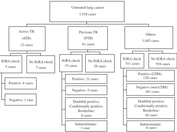
Fig. 1 Flow diagram of interferon gamma release assay in patients with lung cancer between 2004 and 2013.
Untreated lung cancer 1,518 cases Active TB (ATB) 12 cases IGRA check 5 cases Positive: 4 cases Negative: 1 case No IGRA check 7 cases (PTB) 61 cases IGRA check 33 cases Positive: 21 cases Negative: 5 cases Doubtful positive, Conditionally positive, Borderline: 6 cases Indeterminate: 1 case No IGRA check 28 cases Others 1,445 cases IGRA check 511 cases Positive (LTBI): 120 cases Negative (non-LTBI): 341 cases Doubtful positive, Conditionally positive, Borderline: 44 cases Indeterminate: 6 cases No IGRA check 934 cases Previous TB
other than those with ATB and PTB). To investigate the clinical characteristics of patients with LTBI, background factors, lung cancer status, prognosis, and other parameters were compared with those of patients with PTB and those with non-LTBI. Finally, medical records were used to identify patients who developed ATB within 5 years of the diagnosis of lung cancer and the cumulative incidence of ATB in each group was calculated.
Statistical analysis
All quantitative data, other than age and year of birth, were expressed as numbers and percentages. To identify the clinical profi le of patients with LTBI, their clinical characteristics on the date of lung cancer diagnosis were compared with those of patients with PTB and those of patients with non-LTBI. Lung cancer survival curves were obtained by the Kaplan‒Meier method and the risk of developing TB was estimated using Fisher’s exact test. Bonferroni’s correction method was used for analysis, with a level of signifi cance set at 0.05/2=0.025. The cumulative incidence of ATB was calculated as Fine and Gray’s cumulative incidence19), which treats death as a competing risk. All statistical analyses were conducted using the R statistical software Package cmprsk (R Project for Statistical Computing; https://www.r-project.org).
RESULTS
The 1,518 identifi ed subjects were classifi ed into three groups: 12 with ATB, 61 with PTB, and 1,445 others. The IGRA positive rate was 80% (4/5) for patients with ATB and
64% (21/33) for those with PTB. The remaining 1,445 patients were divided into two groups: 511 who underwent IGRA testing and 934 who did not. Of those who underwent IGRA testing, 120 had a positive result (patients with LTBI), 44 had a doubtful positive (QFT-G test), a conditionally positive (QFT-GIT test), or a borderline (T-spot test) result. Six had an indeterminate result. The remaining 341 patients had non-LTBI, indicating that the rate of LTBI among patients with lung cancer in this study was 23% (120/511) (Fig. 1). The timing of IGRA testing was left to the discretion of the attending physician and testing was performed for a variety of practical reasons, such as differential diagnosis before the diagnosis of lung cancer or to identify the onset of TB in the course of lung cancer treatment. IGRA testing was performed in 460 (84%) of the 549 cases before or simultaneously with a defi nitive diagnosis of lung cancer, and after diagnosis was made in 89 cases (16%) ; furthermore, 492 patients (90%) underwent testing within 1 year prior to or after diagnosis. A comparison of the clinical profi les of the 511 patients who underwent IGRA testing and the 934 patients who were not tested found no signifi cant differences in the clinical characteristics of the two groups (sex, p=0.315; age, p= 0.9656; year of birth, p=0.4345; performance status [PS], p= 0.247; histological type, p=0.1876; resection, p=0.4646; stage, p=0.9541; left or right lung, p=0.4033; and upper, middle, or lower lung, p=0.6211).
Table 1 shows the clinical characteristics of patients with ATB, PTB, LTBI, and non-LTBI. The background charac-teristics of the patients with ATB, PTB, and LTBI were
ATB (n=12) [%] PTB (n=61) [%] LTBI (n=120) [%] Non-LTBI (n=341) [%] Female Male
Median age (IQR) Median birth year (IQR) Smoking − (ref.) Smoking + Missing PS 0 _ 1 (ref.) PS 2 _ 4 Adenocarcinoma (ref.) Squamous cell carcinoma Other carcinomas Non-resection (ref.) Resection
Stages I _ II (ref.) Stages III _ IV Right lung (ref.) Left lung Upper lobe (ref.) Middle lobe Lower lobe 1 [ 8.3] 11 [ 91.7] 73 (68 _ 76) 1936 (1932 _ 1942) 0 [ 0.0] 12 [100.0] 0 9 [ 75.0] 3 [ 25.0] 2 [ 16.7] 5 [ 41.7] 5 [ 41.7] 8 [ 66.7] 4 [ 33.3] 5 [ 41.7] 7 [ 58.3] 5 [ 41.7] 7 [ 58.3] 6 [ 50.0] 2 [ 16.7] 4 [ 33.3] 6 [ 9.8] 55 [90.2] 74 (66 _ 77) 1935 (1930 _ 1943) 7 [12.3] 50 [87.7] 4 44 [72.1] 17 [27.9] 30 [49.2] 17 [27.9] 14 [23.0] 46 [75.4] 15 [24.6] 18 [29.5] 43 [70.5] 29 [47.5] 32 [52.5] 26 [42.6] 4 [ 6.6] 31 [50.8] 25 [20.8] 95 [79.2] 73 (67 _ 77) 1936 (1932 _ 1942) 16 [13.8] 100 [86.2] 4 92 [76.7] 28 [23.3] 69 [57.5] 30 [25.0] 21 [17.5] 64 [53.3] 56 [46.7] 49 [40.8] 71 [59.2] 68 [56.7] 52 [43.3] 54 [45.0] 10 [ 8.3] 56 [46.7] 115 [33.7] 226 [66.3] 69 (62 _ 75) 1941 (1935 _ 1947) 105 [31.6] 227 [68.4] 9 281 [82.4] 60 [17.6] 228 [66.9] 55 [16.1] 58 [17.0] 206 [60.4] 135 [39.6] 111 [32.6] 230 [67.4] 208 [61.0] 133 [39.0] 173 [50.7] 28 [ 8.2] 140 [41.1]
Table 1 Characteristics of lung cancer patients with active tuberculosis, previous tuberculosis, latent tuberculosis infection, and non-latent tuberculosis infection.
ATB, active tuberculosis; PTB, previous tuberculosis; LTBI, latent tuberculosis infection; IQR, interquartile range; ref.: reference
similar and fi tted the general profi le of patients with ATB, including a very high proportion of elderly male smokers with a median year of birth of 1935_1936. Other than for patients with ATB, adenocarcinoma was the most common type of cancer, accounting for the majority of patients with PTB, LTBI, and non-LTBI. The majority of patients in all groups had stage III_IV disease, and most did not undergo resection. To identify the clinical characteristics of patients with LTBI, we fi rst compared the characteristics of non-LTBI patients (Table 2) and then with those of patients with PTB (Table 3). Multivariate analysis revealed several major differences in background factors between LTBI and non-LTBI patients, with the former being older ( p<0.0001; odds ratio [OR], 1.057; 95% confi dence interval [CI], 1.030_1.086), having a higher proportion of smokers ( p<0.0001; OR, 3.429; 95% CI, 1.883_6.245) and a higher resection rate ( p=0.0024; OR, 2.190; 95% CI, 1.320_3.634), and tending to have poorer PS ( p=0.0445; OR, 1.856; 95% CI, 1.015_3.392). Multivariate analysis did not, however, reveal any differences in the background factors between patients with LTBI and those with PTB, and the only difference with respect to lung cancer was that the resection rate was higher for patients with LTBI ( p=0.0032; OR, 2.948; 95% CI, 1.438_6.046).
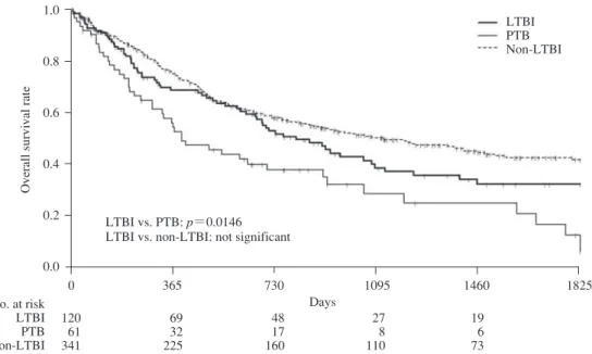
To investigate whether LTBI had any effect on lung cancer prognosis in addition to ATB or PTB, overall survival curves
after lung cancer diagnosis were compared between LTBI, PTB, and non-LTBI patients (Fig. 2). The median survival time for patients with LTBI was 810 days, which was signifi cantly better than that for those with PTB (397 days) ( p=0.0146; hazard ratio [HR], 1.608; 95% CI, 1.095_2.361). However, it was slightly worse than that for non-LTBI patients (1,123 days), although this difference was not signifi cant (p=0.1081; HR, 0.790; 95% CI, 0.593_1.054).
The most important clinical issue for lung cancer patients with LTBI is the possible increased risk of developing ATB during the clinical course of lung cancer. In this study, none of the 120 lung cancer patients with LTBI (median follow-up period, 1,394 days; interquartile range [IQR], 736_1,830 days) or the 341 lung cancer patients with non-LTBI (median follow-up, 1,423 days; IQR 860_1,830 days) developed TB within 5 years of the diagnosis of lung cancer. None of the 44 conditionally positive IGRA cases and 6 indeterminate IGRA cases developed ATB. However, 3 of the 61 PTB patients (mean follow-up, 991 days; IQR 688_1,830 days) developed ATB. Two of these three patients tested positive for IGRA, while the remaining patient did not undergo testing. The cumulative incidences of ATB development among patients with PTB were 1.74% (1 year), 4.31% (3 years), and 8.20% (5 years), which were signifi cantly higher than the incidence of TB among patients with LTBI (p=0.0102, based on the score
*Year of birth and stages were not included in the multivariate working model because of multicollinearity. Abbreviations: OR, odds ratio; CI, confi dence interval
Table 2 Logistic regression analysis (latent tuberculosis infection vs. non-latent tuberculosis infection)
Univariate analysis Multivariate analysis Backward elimination method
OR (95%CI) p-value OR (95%CI) p-value OR (95%CI) p-value
Female (ref.) Male Median age Birth year Smoking − (ref.) Smoking + PS 0 _ 1 (ref.) PS 2 _ 4 Adenocarcinoma (ref.) Squamous cell carcinoma Other carcinomas Non-resection (ref.) Resection
Stages I _ II (ref.) Stages III _ IV Right lung (ref.) Left lung Upper lobe (ref.) Middle lobe Lower lobe 1 1.934 (1.179 _ 3.170) 1.050 (1.025 _ 1.076) 0.954 (0.932 _ 0.977) 1 2.891 (1.625 _ 5.144) 1 1.425 (0.859 _ 2.366) 1 1.802 (1.072 _ 3.032) 1.196 (0.679 _ 2.110) 1 1.335 (0.878 _ 2.030) 1 0.699 (0.455 _ 1.074) 1 1.196 (0.785 _ 1.823) 1 1.144 (0.522 _ 2.506) 1.281 (0.829 _ 1.980) 0.0090 <0.0001 <0.0001 0.0003 0.1704 0.0264 0.5355 0.1765 0.1019 0.4056 0.7363 0.2639 1 1.228 (0.666 _ 2.266) 1.058 (1.030 _ 1.087) * 1 3.099 (1.531 _ 6.272) 1 1.874 (1.019 _ 3.446) 1 1.241 (0.699 _ 2.204) 0.936 (0.500 _ 1.754) 1 2.217 (1.325 _ 3.710) * * 1 1.184 (0.747 _ 1.876) 1 1.734 (0.730 _ 4.117) 1.134 (0.705 _ 1.824) 0.5104 <.0001 * 0.0017 0.0434 0.4610 0.8368 0.0024 * * 0.4714 0.2121 0.6051 Removed 1.057 (1.030 _ 1.086) * 1 3.429 (1.883 _ 6.245) 1 1.856 (1.015 _ 3.392) Removed 1 2.190 (1.320 _ 3.634) * * Removed Removed <0.0001 * <0.0001 0.0445 0.0024 * *
Table 3 Logistic regression analysis (latent tuberculosis infection vs. previous tuberculosis)
Univariate analysis Multivariate analysis Backward elimination method
OR (95%CI) p-value OR (95%CI) p-value OR (95%CI) p-value
Female (ref.) Male Age Birth year Smoking − (ref.) Smoking + PS 0 _ 1 (ref.) PS 2 _ 4 Adenocarcinoma (ref.) Squamous cell carcinoma Other carcinoma Non-resected (ref.) Resection Stages I _ II (ref.) Stages III _ IV Right lung (ref.) Left lung Upper lobe (ref.) Middle lobe Lower lobe 1 0.415 (0.160 _ 1.073) 1.002 (0.967 _ 1.039) 1.007 (0.972 _ 1.042) 1 0.875 (0.338 _ 2.264) 1 0.788 (0.391 _ 1.589) 1 0.767 (0.369 _ 1.597) 0.652 (0.293 _ 1.452) 1 2.683 (1.354 _ 5.319) 1 0.607 (0.314 _ 1.173) 1 0.693 (0.373 _ 1.287) 1 1.204 (0.345 _ 4.203) 0.870 (0.458 _ 1.652) 0.0695 0.8981 0.7065 0.7831 0.5051 0.4789 0.2954 0.0047 0.1375 0.2454 0.7713 0.6699 1 0.480 (0.147 _ 1.572) 1.017 (0.979 _ 1.057) * 1 1.777 (0.522 _ 6.052) 1 1.111 (0.492 _ 2.511) 1 0.778 (0.345 _ 1.750) 0.661 (0.268 _ 1.630) 1 2.949 (1.307 _ 6.655) * * 1 0.733 (0.372 _ 1.445) 1 1.450 (0.372 _ 1.445) 0.946 (0.461 _ 1.940) 0.2254 0.3899 * 0.3581 0.8000 0.5434 0.3685 0.0092 * * 0.3698 0.6082 0.8791 Removed Removed * Removed Removed Removed 1 2.948 (1.438‒6.046) * * Removed Removed * 0.0032 * *
Fig. 2 Overall survival rates of previous tuberculosis, latent tuberculosis infection, and non-latent tuberculosis infection in patients with lung cancer.
LTBI vs. PTB: p=0.0146 LTBI vs. non-LTBI: not significant
LTBI PTB Non-LTBI 1.0 0.8 0.6 0.4 0.2 0.0 0 365 730 1095 1460 1825
Overall survival rate
No. at risk LTBI PTB Non-LTBI 120 61 341 69 32 225 48 17 160 27 8 110 19 6 73 Days method).
With respect to the association between the type of IGRA test and results obtained, there was no signifi cant difference between the IGRA positive rates obtained with QFT-G (60/239, 25%), QFT-GIT (54/244, 22%), and T-SPOT (6/28, 21%) methods for patients without ATB or PTB (p=0.7222). Additionally, there was no difference between the ages of the patients tested with QFT-G, QFT-GIT, and T-SPOT (median age [IQR] 69 years [range 63.5_75 years], 70.5 years [range 74_77 years], and 72 years [range 65.5_77 years], respectively [p=0.2057]). With respect to year of birth, patients who underwent testing with QFT-G, the oldest type of test, belonged to a signifi cantly older generation (median birth year [IQR] 1938 [range 1932_1945], 1941 [range 1935_1947], and 1941 [1936_1942], respectively [p=0.0154]).
DISCUSSION
In this retrospective study, we determined that the rate of LTBI among patients with lung cancer was 23%. Previously, TB was highly prevalent in Japan; therefore, the rates of previous TB infection in 2010 were predicted based on older mathematical models to be 23.7%, 48.7%, and 73% for individuals aged 60_69, 70_79, and 80_89 years, respec-tively20). Another study also showed that the rates of previous TB infection in 2008 were 62.8% and 44.1% for individuals born in 1933 and 1943, respectively21). However, community investigations conducted in 2007 in Japan showed that the positive rate based on actual tests with QFT-G was 9.8% for individuals aged 60_69 years, far lower than the 53.1% predicted using the formula and caused by waning of the IFN-γ response after years of TB infection22). The rate of LTBI in lung cancer patients, based on our IGRA data, tended to be higher than that of the Japanese general population. In a
Taiwanese national population-based study, Su et al.23) re-ported that LTBI was a risk factor for the development of a number of cancers, including multiple myeloma, bladder, gastrointestinal, lung, and cancers of other organs. Rates of LTBI in patients with lung cancer have also been reported as 29.6% (8 positive for IGRA among 27 patients) in Italy15) and 28.2% (96 positive for IGRA among 340 patients) in Taiwan16). However, the results of these previous studies cannot be directly compared with those of the present study because of the IGRA examination selection bias, geographical differences in TB prevalence, and the incidence of LTBI in these studies was not clearly differentiated from that of PTB. However, the similar high rates of LTBI among lung cancer patients may be related to the well-known risk of lung cancer development in PTB and other previous lung diseases8). Compared with non-LTBI patients, patients with LTBI in this study included larger proportions of elderly people, who are more likely to develop ATB in Japan, and those who smoke, which is a risk factor for both TB and LTBI4)24). It is unclear, however, why the resection rate of LTBI patients was higher than that of non-LTBI patients. Nevertheless, background characteristics of LTBI patients were similar to those of PTB patients, with the exception of resection rates, while lung cancer status was also similar. These results suggest that patients with LTBI form a population closely related to patients with PTB. It is known that, in lung cancer patients with PTB, TB-induced anatomical lung damage may impose therapeutic limitations, particularly to surgical resection14). LTBI patients usually have no obvious signs of the lung parenchymal damage seen in PTB patients, and this differ-ence may be one reason for the higher rates of lung cancer resection and the better prognosis observed among patients with LTBI compared with those with PTB.
With regard to prognosis, Fan et al.16) reported that there was no difference between Taiwanese patients with lung cancer and LTBI (defi ned as IGRA positive, suggesting equivalence to LTBI together with PTB in the present study) and those classifi ed as non-LTBI (IGRA-negative), although the IGRA results of 30 patients with indeterminate status (indicating immunocompromised conditions) indicated a poorer prognosis than for LTBI and non-LTBI patients. In this study, very few patients (6 cases) had indeterminate IGRA results; therefore, we did not investigate this category of patients and observed no difference in prognosis among patients with LTBI (120 patients) and non-LTBI (341 patients). However, the prognosis of LTBI patients was signifi cantly better than that of patients with PTB, which may likely be the result of the aforementioned differences in lung cancer resection rates.
Based on the risk of ATB development2), the Japanese Society for Tuberculosis treatment guidelines for LTBI cite the following: LTBI treatment should be actively provided because of the high risk of developing ATB among patholo-gies involving immunodefi ciency, use of drugs with immu-nosuppressive effects, and other TB development risk fac-tors; however, lung cancer itself is not included in this list25). Of the 120 LTBI patients plus 50 IGRA-tested patients with conditionally positive results and/or indeterminate results, no patient developed ATB within 5 years of diagnosis of lung cancer. However, among the 61 PTB patients, 3 developed ATB. Since the incidence of ATB onset among patients with PTB (TB recurrence) is generally considered to be higher than that among those with previous TB infection2)26). the development of ATB during the treatment of lung cancer is of greater concern for patients with PTB than for those with LTBI. Furthermore, it is well known that liver damage as an adverse effect of the treatment of LTBI by isoniazid is not uncommon27). Thus, we found no evidence to extend the current Japanese LTBI treatment guidelines to include active treatment for LTBI in patients with lung cancer.
With respect to the results obtained based on different methods of IGRA testing, differences were observed between data from QFT-G and the subsequent QFT-GIT test28). Additionally, differences in the sensitivity and specifi city of QFT-GIT and T-SPOT were identifi ed in some studies29) but not others30). In this study, we found no difference in the positivity rate, although it was slightly higher when the older QFT-G test was used in a population with a higher rate of previous TB infection. However, there were no clear differ-ences between the different methods of conducting IGRA testing for the evaluation of LTBI.
This study had some limitations. This was a single-center retrospective study based on data from a limited number of subjects, and the data were obtained from a lung cancer database of patients over a limited 5-year period. There was no extensive analysis performed for other ATB risk factors, such as diabetes, gastrectomy, and contact history with an
infectious TB patient. Different methods for conducting IGRA testing were used and 16% of the patients underwent IGRA after lung cancer treatment. Furthermore, the study cohort was unbalanced because of the greater number of patients who did not undergo IGRA than those who did (934 vs. 511 patients, respectively). There was a bias in the decision to evaluate IGRA by the attending physicians; patients who underwent IGRA examination might have a greater tendency to have some fi ndings associated with TB infection than IGRA-unexamined patients. Despite these limitations and the absence of a LTBI gold standard, our results provide the fi rst published information on the rate of LTBI among lung cancer patients in Japan, clinical profi les of lung cancer patients with LTBI, development of ATB during the course of lung cancer treatment, its association with LTBI, and prognosis for lung cancer patients with LTBI.
In conclusion, the rate of LTBI among patients with lung cancer at our hospital was 23%. The clinical profi les of LTBI patients resembled those of PTB patients, but prog-nosis was better for the former than for the latter, while the clinical profi les of LTBI patients differed from those of nonLTBI patients although the prognoses were not signifi -cantly different. The risk of ATB occurrence in lung cancer patients with LTBI may be lower than in those with PTB. Further prospective, multi-center, long-term studies in larger numbers of patients are required to elucidate the association between lung cancer and LTBI, and it may be necessary to seek more conclusive evidence for the treatment of LTBI in lung cancer patients.
ACKNOWLEDGMENTS
No funding was received for this study. The authors have no confl icts of interest to declare.
REFERENCES
1 ) Pai M, Denkinger CM, Kik SV, et al.: Gamma interferon release assays for detection of Mycobacterium tuberculosis infection. Clin Microbiol Rev. 2014 ; 27 : 3 20.
2 ) Landry J, Menzies D: Preventive chemotherapy. Where has it got us? Where to go next? Int J Tuberc Lung Dis. 2008 ; 12 : 1352 1364.
3 ) Alberg AJ, Samet JM: Epidemiology of lung cancer. Chest. 2003 ; 123 : 21S 49S.
4 ) Slama K, Chiang CY, Enerson DA, et al.: Tobacco and tuberculosis: a qualitative systematic review and meta-analysis. Int J Tuberc Lung Dis. 2007 ; 11 : 1049 1061. 5 ) Friedrich G: Periphere Lungenkrebse auf dem Boden
pleu-ranaher. Virchows Arch (Pathol Anat). 1939 ; 304 : 230 247. 6 ) Haybittle JL: Study of cancer mortality in England and
Wales using birth-standardized populations. Br J Prev Soc Med. 1962 ; 16 : 93 104.
7 ) Engels EA, Shen M, Chapman RS, et al.: Tuberculosis and subsequent risk of lung cancer in Xuanwei, China. Int J Cancer. 2009 ; 124 : 1183 1187.
8 ) Brenner DR, Boffetta P, Duell EJ, et al.: Previous lung diseases and lung cancer risk: a pooled analysis from the International Lung Cancer Consortium. Am J Epidemiol. 2012 ; 176 : 573 585.
9 ) Nalbandian A, Yan BS, Pichugin A, et al.: Lung carcino-genesis induced by chronic tuberculosis infection : the experimental model and genetic control. Oncogene. 2009 ; 28 : 1928 1938.
10) Heuvers ME, Aerts JG, Hegmans JP, et al.: History of tuberculosis as an independent prognostic factor for lung cancer survival. Lung Cancer. 2012 ; 76 : 452 456. 11) Wu CY, Hu HY, Pu CY, et al.: Aerodigestive tract, lung
and haematological cancers are risk factors for tuberculo-sis: an 8-year population-based study. Int J Tuberc Lung Dis. 2011 ; 15 : 125 130.
12) Leung CC, Hui L, Lee RS, et al.: Tuberculosis is associ-ated with increased lung cancer mortality. Int J Tuberc Lung Dis. 2013 ; 17 : 687 692.
13) Christopoulos A, Saif MW, Sarris EG, et al.: Epidemiology of active tuberculosis in lung cancer patients: a systematic review. Clin Respir J. 2014 ; 8 : 375 381.
14) Tamura A: Tuberculosis and lung cancer. Kekkaku. 2016 ; 91 : 17 25.
15) Bordignon V, Bultrini S, Prignano G, et al.: High prevalence of latent tuberculosis infection in autoimmune disorders such as psoriasis and in chronic respiratory diseases, including lung cancer. J Biol Regul Homeost Agents. 2011 ; 25 : 213 220.
16) Fan WC, Ting WY, Lee MC, et al.: Latent TB infection in newly diagnosed lung cancer patients ― A multicenter prospective observational study. Lung Cancer. 2014 ; 85 : 472 478.
17) Prevention Committee of the Japanese Society for Tuber-culosis: Guideline for using QuantiFERON® TB-2G.
Kek-kaku. 2006 ; 81 : 393 397. (In Japanese)
18) Prevention Committee of the Japanese Society for Tubercu-losis: Guideline for using interferon-gamma release assay. Kekkaku. 2014 ; 89 : 717 725. (In Japanese)
19) Fine JP, Gray RJ: A proportional hazards model for
the subdistribution of a competing risk. J Am Stat Anal. 1999 ; 94 : 496 509.
20) Ohmori M: Estimating the year of eradication of tuber-culosis in Japan. Kekkaku. 1991 ; 66 : 819 828. (In Japanese with an English abstract)
21) Nagayama N: Tuberculosis in Japan at present and in near future. Kekkaku. 2001 ; 76 : 571 579. (In Japanese with an English abstract)
22) Mori T, Harada N: Higuchi K, et al.: Waning of the spe-cifi c interferon-gamma response after years of tuberculosis infection. Int J Tuberc Lung Dis. 2007 ; 11 : 1021 1025. 23) Su VY, Yen YF, Pan SW, et al.: Latent tuberculosis
infec-tion and the risk of subsequent cancer. Medicine. 2016 ; 95 : e2352.
24) Horne DJ, Campo M, Ortiz JR, et al.: Association between smoking and latent tuberculosis in the US population: an analysis of the National Health and Nutrition Examination Survey. PLoS One. 2012 ; 7 : e49050.
25) Prevention Committee and Treatment Committee of the Japanese Society for Tuberculosis: Treatment guidelines for latent tuberculosis infection. Kekkaku. 2014 ; 89 : 21 37. 26) Tuberculosis Research Committee (Ryoken): Relapse rate
of tuberculosis treated with standard regimen of chemo-therapy. Kekkaku. 2009 ; 84 : 617 625. (In Japanese with an English abstract)
27) Ito K, Hoshino H, Nakazono T, et al.: Liver damage in treatment of latent tuberculous infection by isoniazid. Kekkaku. 2006 ; 81 : 651 660. (In Japanese with an English abstract)
28) Matsumoto T, Yamazaki T: The evaluation of the utility of QuantiFERON® TB-Gold In-Tube; QFT-GIT. Kekkaku.
2014 ; 89 : 743 755. (In Japanese with an English abstract) 29) Diel R, Loddenkemper R, Nienhaus A: Evidence-based
comparison of commercial interferon-gamma release assays for detecting active TB: a metaanalysis. Chest. 2010 ; 137 : 952 968.
30) Higuchi K, Sekiya Y, Igari H, et al.: Comparison of specifi cities between two interferon-gamma release assays in Japan. Int J Tuberc Lung Dis. 2012 ; 16 : 1190 1192.
LATENT TUBERCULOSIS INFECTION IN LUNG CANCER:
A RETROSPECTIVE STUDY FROM A SINGLE CENTER
田村 厚久 日下 圭 島田 昌裕 川島 正裕 山根 章 永井 英明 蛇澤 晶 深見 武史 大田 健 高橋 史朗
要旨:〔目的〕当院の肺癌患者における潜在性結核感染症(latent tuberculosis infection : LTBI)の頻度
や特徴を後ろ向きに検討した。〔対象〕2004 年から 2013 年の間に当院で診断,治療を行った肺癌症例
のインターフェロンγ遊離検査(interferon-gamma release assay : IGRA)結果を含む臨床データを解析 した。この際,対象を活動性結核(active tuberculosis : ATB)合併例,結核既往(previous tuberculo-sis : PTB)例,LTBI 例(ATB,PTB を除く IGRA 陽性例),non-LTBI 例(ATB,PTB を除く IGRA 陰性例) に 分 け て 解 析 し た。〔 結 果 〕 全 肺 癌 1,518 例 か ら ATB 12 例,PTB 61 例 を 除 い た 1,445 例 中 511 例 で IGRA 結果が得られ,うち 120 例(23%)が LTBI 例,341 例が non-LTBI 例であった。臨床像の多変量 解析では LTBI 例は non-LTBI 例に比し,高齢(p<0.0001)で,喫煙者が多く( p<0.0001),肺癌切除 率が高かった(p=0.0024)が,PTB 例とは切除率が高い( p=0.0032)以外,差がなかった。生存解 析では,LTBI 例の予後は non-LTBI 例と差がなかったが,PTB 例より良かった( p=0.0146)。肺癌経 過 5 年間の結核発症は PTB 3 例にみられ,累積発症率は 1.74%( 1 年),4.31%( 3 年),8.20%( 5 年)
であったが,LTBI 例や non-LTBI 例からの結核発症はなかった。〔結論〕本研究の肺癌 LTBI 合併率は
23% であった。LTBI 例の臨床像は PTB 例に類似していたが,予後は PTB 例より良好であった。肺癌 経過中の結核発症は LTBI 例よりも PTB 例で注意すべきである。