1164
Original Article
A predictor of aerobic threshold for patients with heart failure with reduced ejection fraction
Sawako Yamamoto, RPT, MS1)*, YorimitSu Furukawa, RPT, PhD2), Seiji FukuShima, MD3), oSamu Nitta, RPT, PhD2)
1) Department of Rehabilitation, La Citta Del Sole of Geriatric Health Services Facility:
2-1-13 Ukima, Kita-ku, Tokyo 115-0051, Japan
2) Tokyo Metropolitan University, Japan
3) Department of Cardiology, Ukima Central Hospital, Japan
Abstract. [Purpose] The initial cardiopulmonary response to exercise is hypothesized to be a useful predictor of aerobic threshold in patients with heart failure. This study aimed to evaluate the correlation between aerobic thresh- old and cardiopulmonary responses to exercise onset by comparing patients with heart failure using preserved (≥50%) and reduced (<50%) left ventricular ejection fractions. [Participants and Methods] Twenty-eight males (age, 36–82 years; 12 with preserved and 16 with reduced left ventricular ejection fractions) underwent a progressive submaximal cardiopulmonary exercise test using a cycle ergometer. The aerobic threshold, time constant, and area under the oxygen uptake curve for the first 4 min (V̇O2AUC) were determined. [Results] A significant association was observed between aerobic threshold and V̇O2AUC in the reduced group but not in the preserved group. No significant correlations were found between time constant and V̇O2AUC or between aerobic threshold and time constant in either group. [Conclusion] The results suggest that V̇O2AUC measured from exercise onset to an initial 4-min period could provide an easily and safely obtained predictor to assess aerobic capacity in people with reduced left ventricular ejection fractions.
Key words: Reduced ejection fraction, Left ventricular ejection fraction, Aerobic threshold
(This article was submitted Apr. 27, 2018, and was accepted Jun. 22, 2018)
INTRODUCTION
Heart failure is highly prevalent and is increasing globally. Left ventricular diastolic dysfunction plays a considerable role in heart failure. Patients with heart failure can be divided into those with preserved left ventricular ejection fraction (LVEF) and reduced LVEF (HFpEF and HFrEF, respectively)1, 2). Cardiac dysfunction can lead to decreased aerobic capacity3); con- versely, higher aerobic capacity is associated with reduced mortality in heart failure4, 5). Oxygen uptake (V̇O2, defined as the product of cardiac output and arterial-mixed venous oxygen difference), increases during exercise. Maximal oxygen uptake (V̇O2 max), a measure of aerobic capacity, is an important consideration in people with heart failure. However, because of lack of muscle strength, fatigue, symptoms, or psychological factors, it can be difficult for patients with heart failure to attain the maximal exercise level needed to determine V̇O2 max6). Therefore, a safe and simple method is needed to determine the aerobic capacity of patients with heart failure.
We had earlier attempted to expect anaerobic threshold (AT) as an initial response to exercise. However, little is known about how this response changes with different aerobic capacities3, 7). The present study aimed to investigate the relationship between AT and the cardiopulmonary response to exercise onset in patients with heart failure, comparing this between HFpEF and HFrEF.
J. Phys. Ther. Sci. 30: 1164–1167, 2018
*Corresponding author. Sawako Yamamoto (E-mail: [email protected])
©2018 The Society of Physical Therapy Science. Published by IPEC Inc.
This is an open-access article distributed under the terms of the Creative Commons Attribution Non-Commercial No Derivatives (by-nc-nd) License. (CC-BY-NC-ND 4.0: https://creativecommons.org/licenses/by-nc-nd/4.0/)
The Journal of Physical Therapy Science
The Journal of Physical Therapy Science
1165
PARTICIPANTS AND METHODS
The study included 28 males with heart failure (age, 36–82 years). All participants had been admitted for cardiac rehabili- tation because of heart failure.
The participants were divided into two groups based on their LVEF: the HFpEF group with LVEF ≥50% (n=12; mean age, 68.7 years; range, 58–82 years) and the HFrEF group with LVEF<50% (n=16; mean age, 60.4 years; range 36–81 years).
Prior to the exercise test, the participants’ health history, risk factors for coronary disease, age, height, weight, and physi- cal characteristics were recorded. Informed consent was obtained from all patients before their participation in the study.
Inclusion criteria included diagnosis of heart failure, participation in cardiac rehabilitation programs, and aerobic capacity determined using cardiopulmonary exercise test (CPET). Exclusion criteria included detection of lethal arrhythmia on CPET, inability to decide AT, and oscillatory ventilation.
The participants underwent a submaximal CPET using a cycle ergometer, with expiratory gas analysis to determine their V̇O2max. There were rest and warm-up phases of 4 min each before initiation of Ramp protocol. The initial workload was set at 10 W and was subsequently increased by 10 W/min using Ramp protocol. The pedaling frequency was monitored to ensure it remained at ≥50 revolutions per min. The endpoint of CPET was determined by the appearance of a symptom associated with coronary disease or when the participant or the observer declared that the participant had reached his limit. Throughout the test, the participant was continuously monitored using electrocardiography.
Pulmonary ventilation and gas exchange parameters were determined breath-by-breath throughout the CPET using an Aeromonitor AE-300 (Minato Medical Science Co., Ltd., Osaka, Japan). The system was calibrated before each test using gas mixtures of known compositions.
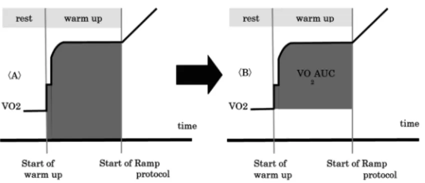
The participant’s AT, time constant (TC), and area under the oxygen uptake curve (V̇O2AUC) were evaluated. AT was determined using several methods based on conventional criteria: the point during the CPET at which there was the first departure from linearity in the plot of carbon dioxide output (V̇CO2) against V̇O2 (the V-slope method), the point when V̇O2 increased after being stable or decreased while V̇CO2 remained constant or was decreasing, and the point when the gas exchange ratio began to increase more steeply after being stable or slowly rising3). TC was determined from the initial increase in V̇O2 following exercise onset in the warm-up phase. V̇O2AUC was calculated as the area under the V̇O2 curve from the onset of exercise through the initial 4 min of the warm-up phase. This calculation excluded the influence of the resting state (Fig. 1).
Data are reported as means (SD). Independent t-tests were used to compare age, body mass index (BMI), LVEF, TC, V̇O2AUC, and AT between the HFpEF and HFrEF groups. The correlations between TC, V̇O2AUC, and AT were assessed using Pearson product-moment correlation coefficients. p<0.05 was considered statistically significant. All analyses were performed using IBM SPSS Statistical software (version 24).
This study was approved by the ethics committee of Tokyo Metropolitan University (approval number: 13099) and the ethics committee of Ukima Central Hospital (approval number: H25-1) and was conducted in accordance with the principles of the Declaration of Helsinki.
RESULTS
Table 1 showed the mean (SD) value for characteristics of the participants in the HFpEF (n=12) and HFrEF (n=16) groups in this study (Table 1). LVEF in the HFpEF and HFrEF groups were 65.7 (10.6%) and 36.1 (8.9%), respectively. There were no significant differences between any of the other measured parameters.
The prevalence of risk factors for coronary diseases and the participants’ indications for admission were determined Fig. 1. Method to calculate V̇O2 area of under curve (V̇O2AUC).
V̇O2AUC <B> was obtained by subtracting the integrated V̇O2 value during warm- up period from the total value <A>.
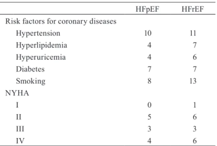
J. Phys. Ther. Sci. Vol. 30, No. 9, 2018 1166 according to the New York Heart Association (NYHA) classification in the HFpEF and HFrEF groups was shown (Table 2).
Pearson’s product-moment correlation coefficients for the HFpEF and HFrEF groups were shown (Table 3). Significant correlation showed between V̇O2AUC and AT in the HFrEF group.
DISCUSSION
A major finding of the current study was that V̇O2AUC and AT showed a strong, significant correlation in participants with HFrEF but not in those with HFpEF. Our study results suggest that V̇O2AUC with cardiac dysfunction such as HFrEF is associated with AT. Left ventricular diastolic dysfunction is a major factor of HFpEF1). In this study, we divided the participants into the HFpEF and HFrEF groups according to their LVEF. We expected these two groups would have different hemodynamic reactions owing to their left ventricular differences. Asanoi et al.8) reported that left ventricular end-diastolic volume increased below AT, whereas left ventricular end-systolic volume decreased above AT, suggesting that left ventricu- lar systolic function increases above the AT level. Thus, left ventricular diastolic dysfunction affected oxygen conveyance below AT in participants with HFpEF, whereas left ventricular systolic dysfunction affected oxygen conveyance above AT in participants with HFrEF.
We compared the differences in cardiac dysfunction between the two groups to test our hypothesis that AT could be predicted from parameters associated with the onset of exercise in participants with the absence of left ventricular systolic dysfunction such as HFrEF.
The AT has been shown to be related to oxygen conveyance9) as a component of exercise capacity. The oxygen uptake response, particularly of the TC of oxygen uptake, at the onset of exercise has been of remarkable value10) because of its correlation with aerobic capacity or cardiac output. AT is considered the level of exercise-related V̇O2 above which aerobic energy production is supplemented by anaerobic mechanisms; it is reflected by an increase in the lactate/pyruvate ratio in muscles or arterial blood11). Thus, consideration of both oxygen conveyance and uptake is needed to predict AT from factors related to exercise onset.
In the present study, the participants in both groups showed an increased TC and decreased AT. An increased TC suggests that the impaired cardiac function is due to poor left ventricular diastolic dysfunction in participants with HFpEF and poor left ventricular systolic dysfunction in participants with HFrEF. The decreased AT suggests moderate or severe cardiac dysfunction as described by the Weber-Janicki grade9); in the HFpEF group, the mean value was 8.4 ml/kg/min, whereas in the HFrEF group, it was 9.3 ml/kg/min.
Beside ventilation and muscle function, cardiac function plays a considerable role in human fitness11). In addition, cardiac function such as LVEF and stroke volume begun to decline at AT12). We believe that the reduced reserve of oxygen conveyance Table 1. The characteristics of participants
HFpEF
N=12 HFrEF
N=16 Age (years) 68.7 (7.7) 60.4 (12.3) Hight (cm) 164.7 (5.1) 166.3 (6.0) Weight (kg) 67.6 (7.8) 64.8 (16.2) BMI (kg/m2) 24.9 (2.7) 23.2 (4.7)
LVEF* (%) 65.7 (10.6) 36.1 (8.9)
TC (sec) 88.3 (52.2) 59.6 (23.0)
V̇O2AUC (ml/kg) 8.4 (1.7) 9.7 (1.4)
AT (ml/kg/min) 8.9 (1.5) 9.3 (1.4)
Mean (SD) values for the HFpEF and HFrEF groups.
HFpEF: heart failure with preserved left ventricular ejection fraction; HFrEF: heart failure with reduced left ventricular ejection fraction; BMI: body mass index; LVEF: left ventric- ular ejection fraction; TC: time constant; V̇O2AUC: area of under oxygen uptake curve; AT: aerobic threshold.
Independent t-test had been adopted to compare participants with HFpEF and HFrEF. No significant differential had been recognized between HFpEF and HFrEF eliminated LVEF.
*p<0.05.
Table 2. Prevalence of risk factors for coronary diseases and New York Heart Association grade (NYHA) on admis- sion of HFpEF and HFrEF
HFpEF HFrEF
Risk factors for coronary diseases
Hypertension 10 11
Hyperlipidemia 4 7
Hyperuricemia 4 6
Diabetes 7 7
Smoking 8 13
NYHA
I 0 1
II 5 6
III 3 3
IV 4 6
Table 3. Pearson’s product-moment correlation coefficient
HFpEF HFrEF
AT and V̇O2AUC 0.39 (p=0.21) 0.67 (p<0.05) TC and V̇O2AUC 0.14 (p=0.67) −0.22 (p=0.42) AT and TC −0.04 (p=0.91) −0.05 (p=0.86)
1167 above AT in participants with HFrEF. Oxygen conveyance and oxygen utilization is associated with aerobic capacity such as AT.
Reduced LVEF affected oxygen conveyance above AT, and led to appear the oxygen utilization. Thus, there were no correlations between V̇O2AUC and AT, TC and AT, or V̇O2AUC and TC in the HFpEF group. V̇O2AUC is an indicator of oxygen usage and is associated with AT in people with left ventricular systolic dysfunction, such as in participants belonging to the HFrEF group.
Other factors associated with AT include ventilation, lactic acid metabolism, muscle function, and mitochondrial function.
In this study, V̇O2AUC and TC were both determined soon after the onset of exercise; therefore, we expected to find a correlation between them. However, there was no significant correlation between the two parameters in either group. This suggests that V̇O2AUC, unlike TC, was influenced by oxygen use rather than oxygen conveyance. V̇O2AUC and TC were independent of oxygen use.
In addition, we found no significant correlation between AT and TC in either groups. The TC of oxygen uptake is im- portant, particularly given its significant association with cardiac output at the onset of exercise13). Cardiac output pursued increasing; however, it has been reported that stroke volume and LVEF decline after AT10). These previously described results suggest that there are several complex mechanisms underlying complementation in human physical fitness. Cardiac dysfunc- tion can be improved by cardiac rehabilitation because oxygen conveyance is associated with cardiac output, expansion of blood vessels14), and blood pressure; however, this does not improve LVEF. Furthermore, we expected that participants with HFpEF would show increased myocardial oxygen consumption because of retraction during exercise below the AT.
This study had several limitations. We could not find data on factors related to oxygen utilization such as muscle mass, fiber type, and mitochondrial function; thus, we could not determine correlations between V̇O2AUC and factors related to oxygen utilization. Moreover, we included only males, even though morbidity and mortality related to heart failure are higher in females. However, it discussed some knowledges with gender on human physical fitness, and we should include women in our future studies. We did not consider any mediating effects of insulin resistance or blood insulin concentration treatments, such as thiazolidinedione or sulfonylureas. Independent echocardiography data for cardiac diastolic dysfunction was not considered; however, oxygen pulse would be particularly suitable for determining stroke volume during exercise.
In conclusion, the findings of this study suggested that V̇O2AUC measured from exercise onset through the initial 4-min period of the warm-up phase is an easily and safely measured predictor of physical fitness, which could be useful for assess- ing the aerobic capacity in people with HFrEF.
Funding and Conflict of interest There are none.
REFERENCES
1) Alla F, Al-Hindi AY, Lee CR, et al.: Relation of sex to morbidity and mortality in patients with heart failure and reduced or preserved left ventricular ejection fraction. Am Heart J, 2007, 153: 1074–1080. [Medline] [CrossRef]
2) Ponikowski P, Voors AA, Anker SD, et al. ESC Scientific Document Group: 2016 ESC Guidelines for the diagnosis and treatment of acute and chronic heart failure: the task force for the diagnosis and treatment of acute and chronic heart failure of the European Society of Cardiology (ESC) developed with the special contribution of the Heart Failure Association (HFA) of the ESC. Eur Heart J, 2016, 37: 2129–2200. [Medline] [CrossRef]
3) Koike A, Hiroe M, Adachi H, et al.: Cardiac output-O2 uptake relation during incremental exercise in patients with previous myocardial infarction. Circula- tion, 1992, 85: 1713–1719. [Medline] [CrossRef]
4) Mancini DM, Eisen H, Kussmaul W, et al.: Value of peak exercise oxygen consumption for optimal timing of cardiac transplantation in ambulatory patients with heart failure. Circulation, 1991, 83: 778–786. [Medline] [CrossRef]
5) Myers J, Prakash M, Froelicher V, et al.: Exercise capacity and mortality among men referred for exercise testing. N Engl J Med, 2002, 346: 793–801. [Medline]
[CrossRef]
6) Sato S, Arakawa N, Kamata J, et al.: Relationship between exercise tolerance and respiratory pattern and muscular strength of legs in patients with chronic heart failure. J Phys Ther Sci, 2002, 14: 47–50. [CrossRef]
7) Koike A, Yajima T, Adachi H, et al.: Evaluation of exercise capacity using submaximal exercise at a constant work rate in patients with cardiovascular disease.
Circulation, 1995, 91: 1719–1724. [Medline] [CrossRef]
8) Asanoi H, Kameyama T, Ishizaka S, et al.: Ventriculoarterial coupling during exercise in normal human subjects. Int J Cardiol, 1992, 36: 177–186. [Medline]
[CrossRef]
9) McElroy PA, Janicki JS, Weber KT: Cardiopulmonary exercise testing in congestive heart failure. Am J Cardiol, 1988, 62: 35A–40A. [Medline] [CrossRef]
10) Koike A, Koyama Y, Itoh H, et al.: Prognostic significance of cardiopulmonary exercise testing for 10-year survival in patients with mild to moderate heart failure. Jpn Circ J, 2000, 64: 915–920. [Medline] [CrossRef]
11) Wasserman K, Hansen JE, Sue DY, et al.: Principles of exercise testing and interpretation. Philadelphia: Williams & Wilkins, 2012.
12) Koike A, Itoh H, Taniguchi K, et al.: Detecting abnormalities in left ventricular function during exercise by respiratory measurement. Circulation, 1989, 80:
1737–1746. [Medline] [CrossRef]
13) Matsumoto A, Itoh H, Yokoyama I, et al.: Kinetics of oxygen uptake at onset of exercise related to cardiac output, but not to arteriovenous oxygen difference in patients with chronic heart failure. Am J Cardiol, 1999, 83: 1573–1576. [Medline] [CrossRef]
14) Akashi YJ, Koike A, Osada N, et al.: Short-term physical training improves vasodilatory capacity in cardiac patients. Jpn Heart J, 2002, 43: 13–24. [Medline]
[CrossRef]