Acta Med. Nagasaki 48: 129-133
Circulating Sialyl Lewisa, Sialyl LewisX, and Sialyl Tn Antigens in Patients with Diffuse Type of Gastric Cancer
Tohru NAKAGOE1), Hiroshi ISHIKAWA 2), Terumitsu SAWAI 1), Takashi Tsuji 1), Shigekazu HIDAKA 1), Kenji TANAKA 1), Hiroaki TAKESHITA 1), Masayuki OHBATAKE 1), Atsushi NANASHIMA 1), Shinji AKAMINE 1), Hiroyuki YAMAGUCHI 1), Toru YASUTAKE 1), Takeshi NAGAYASU 1), Shimeru KAMIHIRA 3)
1) First Department of Surgery, Nagasaki University School of Medicine, Nagasaki, Japan 2) Department of Surgery, Sasebo Municipal Hospital, Sasebo, Nagasaki, Japan
3) Department of Laboratory Medicine, Nagasaki University Hospital, Nagasaki, Japan
The aim of this study was to clarify whether or not pre- operative serum levels of sialyl Lewisa (CA19-9), sialyl LewisX (SLX), and sialyl Tn (STN) antigens are predictors for diffuse type gastric cancer. Eighty-two patients with diffuse type and 96 patients with intestinal type cancers were studied. Univariate logistic regression analysis showed that the following factors were significantly associated with diffuse type cancer: high levels of serum STN, young
age (<62 years), female gender, tumor in the middle stom- ach, macroscopic type 3/type 4 cancer, presence of lym- phatic invasion, peritoneal dissemination, stage III/IV, and non-curative resection. Multivariate analysis revealed that diffuse type cancer was independently related to young age (<62 years), female gender, tumor in the middle stomach, and macroscopic type 3/type 4 cancer. In conclusion, none of the preoperative serum levels of CA19-9, SLX, and STN were predictors for diffuse type cancer.
ACTA MEDICA NAGASAKIENSIA 48: 129-133, 2003
Key Words: sialyl Lewisa; sialyl Lewisx; sialyl Tn; CEA; gas- tric cancer; diffuse type; intestinal type.
Introduction
Gastric cancer has been divided histologically into an intestinal type and a diffuse type' 2' . There has been a dramatic and unexplained decrease in the inci-
dence of gastric cancer, and this has occurred primar- ily in the intestinal type. However, unless gastric can- cer is identified early, the prognosis for cure remains poor''. The diffuse type gastric cancer has been associ ated with a poorer prognosis than the intestinal type'-".
Therefore, preoperative prediction of diffuse type gas- tric cancer is important to establish strategies for peri- operative adjuvant chemotherapy.
Sialyl Lewisa (CA 19-9), sialyl Lewisx (SLX), and sialyl Tn antigen (STN) antigens are among the mucin-associated carbohydrate moieties found in large quantities in gastric tumors'). CA 19-9 and SLX repre- sent examples of type 1 and type 2 terminal carbohy-
drate structures, respectively. STN is an example of a carbohydrate core structure". These three antigens may play an important role in cell adhesion. Their key role in local tumor invasiveness and metastasis has been previously discussed"'). These carbohydrate
antigens have also been detected in the sera of gastric cancer patients"'). Thus far, a correlation has been re- ported between serum levels of carbohydrate antigens and diffuse or intestinal type gastric cancer'-"'.
However, in these reports, clinical and pathological data were evaluated only by univariate analysis. We believe that the results of univariate analysis should be confirmed by multivariate analysis.
In this study, we examined the preoperative serum levels of CA 19-9, SLX, and STN in gastric cancer pa- tients who underwent gastrectomy. The aim of this study was to clarify whether or not the serum levels of these carbohydrate antigens are predictors for dif- fuse type gastric cancer. We also evaluated the serum CEA levels in the same patients with gastric cancer because CEA is commonly used to evaluate recurrence and prognosis of gastric cancer patients after surgery".
Patients and Methods Address Correspondence: Tohru Nakagoe, M.D.
First Department of Surgery, Nagasaki University School of Medicine, 1-7-1 Sakamoto, Nagasaki 852-8501, Japan
TEL: +81-95-849-7304, FAX: +81-95-849-7306 E-mail: [email protected]
Patients
A total of 178 gastric cancer patients (120 males and 58 females) were included in this study. The me- dian age of patients was 62.0 years (range, 29 to 86
Tohru Nakagoe et al : Circulating Antigens in Diffuse Type Gastric Cancer
years). All patients underwent resection for a gastric tumor at Nagasaki University Hospital and Sasebo Municipal Hospital between 1991 and 1993. Patients who had a synchronous or metachronous cancer of the stomach were excluded from this study. None of the patients had any evidence of other organ malig- nancies or history of preoperative treatment with anti- cancer drugs.
The following standardized procedures were per- formed: (i) gastric resection (the gastric resection line was 3 cm from the macroscopic edge for localized tu- mors and 5 cm for infiltrating tumors); (ii) prophylac- tic lymph node dissection greater than removal of group 1 lymph nodes"); and (iii) complete excision of invaded organs, irrespective of the number of sites on the organs, provided that there was no evidence of in- curable factors such as peritoneal dissemination, liver metastasis, and widespread nodal involvement"'. One hundred-twenty three patients underwent partial gastrectomy, and 55 patients had a total gastrectomy.
The number of patients who underwent curative re- section was 153. Twenty-five patients had noncurative resection. None of the patients died within 30 postop- erative days. Written informed consent was obtained from each patient. At the time these data were ana- lyzed, the median follow-up was 61.5 months (range:
42 days to 154.6 months).
Intestinal and diffuse types gastric cancer
Pathologic diagnoses and classification of the resected gastric cancer tissues were made according to the Japanese Classification of Gastric Carcinomal3' The histological classification was determined based on the predominant pattern of tumor as follows: papil- lary adenocarcinoma, tubular adenocarcinoma, poorly differentiated adenocarcinoma, signet-ring cell carci- noma, and mucinous carcinoma. Papillary or tubular adenocarcinoma can be interpreted as intestinal type, whereas poorly differentiated adenocarcinoma or sig- net-ring cell carcinoma can be regarded as diffuse type. Mucinous carcinoma can be interpreted as either intestinal or diffuse type, depending upon other pre- dominant elements (papillary adenocarcinoma, tubular adenocarcinoma, poorly differentiated adenocarcinoma, or signet-ring cell carcinoma)"'.
Serum levels of CA19-9, SLX, STN, and CEA were measured by the Otsuka Assay Laboratory (Tokushima, Japan) using the following commercially available radioimmunoassay kits: the Centocor CA 19-9 RIA kit (Centocor, Malvern, PA, USA)"), the FH-6 "Otsuka" kit (Otsuka Assay Lab., Tokushima, Japan)'6' , the STN
"Otsuka" kit (Otsuka Assay Lab
., Tokushima, Japan)"), and the CEA Roche 2 kit (Nippon Roche K.K., Tokyo, Japan)"), respectively. The data obtained were based on simultaneous assays for each antigen using the same set of sera. According to the manufacturers' in- structions, the diagnostic cut-off values for each marker were 37 U/ml for CA19-9, 38 U/ml for SLX, 45 U/ml for STN, and 2.5 ng/ml for CEA1-'$'. We classified patients into two groups based on the serum levels of each antigen: a high antigen group, serum antigen concentration greater than the recommended cut-off value; and a low antigen group, concentrations less than the cut-off value.
Statistical analysis
Logistic regression analysis was chosen to study the predictive value of risk factors because the outcome variable was binary (intestinal type cancer versus dif- fuse type cancer)'" O'. Potentially predictive variables were identified using a significance level of P<0.25 by univariate analysis, and these variables were then used in multivariate analysis. A 25% significance level was selected based on the recommendations of Hosmer and Lemeshow for building multivariate models20'.
Two variables with continuous data, such as age and maximal tumor diameter, were classified in two groups based on the median values (62.0 years and 4.0 cm, respectively). The disease-specific interval was cal- culated according to the Kaplan-Meier method 211 , and differences between disease-specific intervals were tested for significance using the log rank test22'
All tests were two-tailed and a P value of less than 0.05 was considered significant.
Results
Of the 178 gastric cancers, 96 (53.9%) were classified as intestinal type cancers and 82 (46.1%) were classi- fied as diffuse type cancers.
Measurement of the serum levels of antigens
To detect the presence of circulating cancer mark- ers, venous blood was obtained from patients after an overnight fast. Blood samples were immediately sepa- rated by centrifugation and sera were stored at -80'C.
Comparison of disease-specific survival after gastrectomy between patients with intestinal type cancer and those with diffuse type cancer
The survival time of patients with diffuse type
Tohru Nakagoe et al : Circulating Antigens in Diffuse Type Gastric Cancer
cancer was significantly shorter than the survival time of patients with intestinal type cancer (P=0.035) (Fig. 1). The cumulative 5-year survival rates were as follows: 59.8% among patients with diffuse type can- cer, and 74.2% among patients with intestinal type cancer.
Time after surgery (years)
Figure 1. Comparison of disease-specific survival between patients with intestinal type cancer and those with diffuse type cancers of the stomach.
Logistic regression analysis to determine the predictive factors for diffuse type cancer
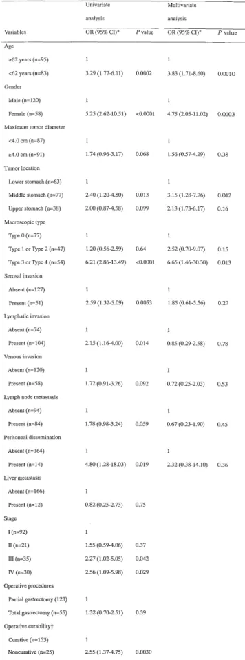
As shown in Table 1, 18 variables were included in this analysis. Univariate analysis showed that the fol- lowing ten variables were significantly associated with diffuse type cancer: age, gender, tumor location, mac- roscopic type, serosal invasion, lymphatic invasion, peritoneal dissemination, stage, operative curability, and serum STN status.
To avoid the problem of collinearity in multivariate analysis, the variables 'stage' and 'operative curability' were excluded. By means of univariate analysis, 11 variables (age, gender, maximum tumor diameter, tumor location, macroscopic type, serosal invasion, lymphatic invasion, venous invasion, lymph node me- tastasis, peritoneal dissemination, and serum STN status) were identified at a significance level of P<0.25 for diffuse type cancer. These variables were therefore included in the multivariate analysis. Consequently, the diffuse type cancer was found to be independently related to four variables: young age (<62 years), fe- male gender, tumor located in the middle stomach, and macroscopic type 3 or type 4 cancer. Of note, serum STN status was not related with diffuse type gastric cancer.
Table 1. Logistic regression analysis of predictive vari-
ables for diffuse type gastric cancer.
Univariate Multivariate
analysis analysis
Variables OR (95% CI)* P value OR (95% Cl)* P value
Age
a62 years (n=95) 1 1
<62 years (n=83) 3.29 (1.77-6.11) 0.0002 3.83 (1.71-8.60) 0.0010 Gender
Male (n=120) 1 1
Female (n=58) 5.25 (2.62-10.51) <0.0001 4.75 (2.05-11.02) 0.0003 Maximum tumor diameter
<4.0 cm (n=87) 1 1
z4.0 cm (n=91) 1.74 (0.96-3.17) 0.068 1.56 (0.57-4.29) 0.38 Tumor location
Lower stomach (n=63) 1 1
Middle stomach (n=77) 2.40 (1.20-4.80) 0.013 3.15 (1.28-7.76) 0.012 Upper stomach (n=38) 2.00 (0.87-4.58) 0.099 2.13 (1.73-6.17) 0.16 Macroscopic type
Type 0 (n=77) 1 1
Type 1 or Type 2 (n=47) 1.20 (0.56-2.59) 0.64 2.52 (0.70-9.07) 0.15 Type 3 or Type 4 (n=54) 6.21 (2.86-13.49) <0.0001 6.65 (1.46-30.30) 0.013 Serosal invasion
Absent (n=127) 1 1
Present (n=51) 2.59 (1.32-5.09) 0.0053 1.85 (0.61-5.56) 0.27 Lymphatic invasion
Absent (n=74) 1 1
Present (n=104) 2.15 (1.16-4.00) 0.014 0.85 (0.29-2.58) 0.78 Venous invasion
Absent (n=120) 1 1
Present (n=58) 1.72 (0.91-3.26) 0.092 0.72 (0.25-2.03) 0.53 Lymph node metastasis
Absent (n=94) 1 1
Present (n=84) 1.78 (0.98-3.24) 0.059 0.67 (0.23-1.90) 0.45 Peritoneal dissemination
Absent (n=164) 1 1
Present (n=14) 4.80 (1.28-18.03) 0.019 2.32 (0.38-14.10) 0.36 Liver metastasis
Absent (n=166) 1
Present (n=12) 0.82 (0.25-2.73) 0.75 Stage
I (n=92) 1
II (n=21) 1.55 (0.59-4.06) 0.37
III (n=35) 2.27 (1.02-5.05) 0.042
IV (n=30) 2.56 (1.09-5.98) 0.029
Operative procedures Partial gastrectomy (123) 1
Total gastrectomy (n=55) 1.32 (0.70-2.51) 0.39 Operative curabilityt
Curative (n=153) 1
Noncurative (n=25) 2.55 (1.37-4.75) 0.0030
Tohru Nakagoe et al : Circulating Antigens in Diffuse Type Gastric Cancer
Serum CA19-9 status
Low CA19-9 group (n=141) 1
High CA19-9 group (n=37) 0.75 (0.36-1.58) 0.45 Serum SLX status
Low SLX group (n=147) 1
High SLX group (n=31) 1.12 (0.51-2.44) 0.78 Serum STN status
Low STN group (n=150) I 1
High STN group (n=28) 2.42 (1.04-5.62) 0.039 1.25 (0.37-4.31) 0.72 Serum CEA status
Low CEA group (n=146) 1
High CEA group (n=32) 0.76 (0.35-1.67) 0.50
"` OR , odds ratio; CI, confidence interval.
( In accordance with the criteria of the Japanese research Society for gastric carcinoma ]';).
Discussion
In order to improve upon clinicopathological correla- tion, Lauren divided gastric cancer into intestinal and diffuse types"". The intestinal type has distinct large glands that are usually lined by well-polarized colum- nar cells with a well-developed brushed border. In the diffuse type of gastric cancer, the glandular structure is rarely present, and cells are scattered either as soli- tary cells or small clusters of cells. Typically, the in- testinal type is better circumscribed than the diffuse type 1, 2 , 13) The intestinal type gastric cancer appears to show a preference for liver metastasis, whereas the diffuse type is more likely to metastasize to the peri- toneum and lymph nodes'-". The diffuse type gastric cancer has been associated with a poorer prognosis than the intestinal type"'. The current study also re- vealed concordant results; i.e., the survival time of pa- tients with diffuse type cancer was significantly shorter than the survival time of patients with intesti- nal type cancer.
In order to establish the strategy of preoperative (neoadjuvant) and/or postoperative adjuvant chemo- therapy, a useful tumor marker that is able to predict diffuse type gastric cancer would be necessary. Thus far, no correlation between serum levels of CA 19-9 or
SLX and the Lauren's classification been reported'-"'.
Our current results were concordant with the results of their previous study. The current study revealed no correlation between serum CEA levels and Lauren's classification. However, Kodera et al.") reported that high serum CEA levels had a tendency to be associ- ated with the intestinal type of gastric cancer (P=0.085). Kodama et al."' also reported a significant association between high serum CEA levels and the intestinal type of cancer. However, this result was
based on a small number of patients with gastric can- cer (6 intestinal and 40 diffuse type cancers). Thus, al- though high serum levels of CEA appear to be associ- ated with the intestinal type of gastric cancer, these studies were evaluated by univariate analysis. The re- sults of univariate analysis should be confirmed by multivariate analysis in a large number of patients.
Takahashi et al.") reported no significant correlation between serum STN levels and the diffuse or intesti- nal type of gastric cancers. In the current study, the univariate analysis demonstrated that among the four carbohydrate antigens (CA 19-9, SLX, STN, and CEA), only high serum levels of STN was significantly asso- ciated with diffuse type cancer. However, the multi- variate analysis showed that the level of serum STN was not an independent predictor for diffuse type.
Some studies reported that immunohistochemical ex- pression of STN did not significantly correlate with Lauren's classification (the intestinal or diffuse type cancer)"-"'. However, some reports have revealed that the intestinal type of cancers express STN more often than diffuse type cancers.","). Thus, the correlation be- tween STN expression and Lauren's classification is controversial. Ikeda et al."' reported that the spread of STN into the surrounding stroma in diffuse type can- cer may be associated with peritoneal dissemination in Stage IV gastric cancer. We previously reported that preoperative serum levels of STN predict liver metas- tasis and poor prognosis in patients with gastric can- cer. However, the high serum levels of STN was asso- ciated with the diffuse type cancer"). Based on these results, we speculate that STN may not correlate with Lauren's classification (the intestinal or diffuse type c ancer), even though STN has an important role in metastasis of gastric cancer.
Body fluid (particularly blood and urine) testing for molecular markers (biomarkers) is easily accessible and useful in patients. The prognostic significance of circulating DNA in plasma or serum, and its genetic alterations in cancer are important trends"'. In the current study, among the four carbohydrate antigens (CA 19-9, SLX, STN, and CEA), we could not find an independent marker that predicted for the diffuse type gastric cancer. Further study using biomarkers is needed to clarify this issue.
References
1. Davis GR: Neoplasms of the stomach. Sleisenger MH and Fordtran JS, ed., Gastrointestinal disease. Pathophysiology/ Diagnosis/
Management. 5th ed., W.B. Sounders Co., Philadelphia, 1993.
2. Lauren P: The two histological main types of gastric carcinoma:
Diffuse and so-called intestinal-type carcinoma. An attempt at a histo-clinical classification. Acta Pathol Microbiol Scand 64: 31-49,
Tohru Nakagoe et al : Circulating Antigens in Diffuse Type Gastric Cancer 1965.
3. Esaki Y, Hirayama R, Hirokawa K: A comparison of patterns of metastasis in gastric cancer by histologic type and age. Cancer 65:
2086-2090, 1990.
4. Lee KH, Lee JH, Cho JK, et al.: A prospective correlation of Lauren's histological classification of stomach cancer with
clinicopathological findings including DNA flow cytometry. Pathol
Res Pract 197: 223-229, 2001.
5. Dabelsteen E: Cell surface carbohydrates as prognostic markers in human carcinomas. J Pathol 179: 358-369, 1996.
6. Berg EL, Magnani J, Warnock RA, Robinson MK, Butcher EC:
Comparison of L-selectin and E-selectin ligand specificities: the L-
selectin can bind the E-selectin ligands sialyl Le, and sialyl Lea.
Biochem Biophys Res Commun 184: 1048-1055, 1992.
7. Bresalier RS, Ho SB, Schoeppner HL, et al: Enhanced sialylation of mucin-associated carbohydrate structures in human colon cancer
metastasis. Gastroenterology 110: 1354-1367, 1996.
8. Imada T, Rino Y, Takahashi M, et al: Serum CA 19-9, SLX, STN and CEA levels of the peripheral and the draining venous blood
in gastric cancer. Hepatogastroenterology 46: 2086-2090, 1999.
9. Nakagoe T, Sawai T, Tsuji T, et al.: Predictive factors for preop- erative serum levels of sialy Lewis", sialyl Lewisa and sialyl Tn
antigens in gastric cancer patients. Anticancer Res 22: 451-458,
2002.
10. Kodera Y, Yamamura Y, Torii A, et al: The prognostic value of preoperative serum levels of CEA and CA19-9 in patients with gastric cancer. Am J Gastroenterol 91: 49-53, 1996.
11. Kodama I, Koufuji K, Kawabata S, et al.: The clinical efficacy of CA 72-4 as serum marker for gastric cancer in comparison with CA19-9 and CEA. Int Surg 80: 45-48, 1995.
12. Takahashi I, Maehara Y, Kusumoto T, et al.: Predictive value of preoperative serum sialyl Tn antigen levels in prognosis of pa- tients with gastric cancer. Cancer 72: 1836-1840, 1993.
13. Japanese Research Society for Gastric Cancer ed. Japanese classifi- cation of gastric caricinoma. Tokyo, Kanehara Co, Ltd, 1995.
14. Maehara Y, Kakeji Y, Kabashima A, et al.: Role of transforming
growth factor-beta 1 in invasion and metastasis in gastric carci- noma. J Clin Oncol 1999; 17: 607-14.
15. Del Villano BC, Brennan S, Brock P, et al: Radioimmunometric
assay for a monoclonal antibody-defined tumor marker, CA19-9.
Clin Chem 29: 549-552, 1983.
16. Imura H, Endo J, Ohkura H, et al: Initial basic and clinical evalua- tion of a solid-phase immunoradiometric assay for sialyl SSEA-1
antigen: (1) Evaluation of assay conditions and normal values. Jpn
J Cancer Chemother 14: 1315-1321, 1987.
17. Imura H, Mori T, Ohkura H, et al: Basic and clinical evaluation of an immunoradiometric competitive inhibition assay for sialyl Tn antigen: (1) Evaluation of assay conditions and normal values. Jpn
J Cancer Chemother 16: 3213-3219, 1989.
18. Nakamura RM, Plow EF, Edginton TS: Current status of carcinoembryonic antigen (CEA) and CEA-S assays in the evalua-
tion of neoplasm of the gastrointestinal tract. Ann Clin Lab -Sci 8:
4-10, 1978.
19. Infante-Rivard C, Esnaola S, Villeneuve JP: Clinical and statistical validity of conventional prognostic factors in predicting short-
term survival cirrhotics. Hepatology 7: 660-664, 1987
20. Hosmer DW, Lemeshow S: Applied logistic regression. New York, John Wiley & Sons, 1990.
21. Kaplan EL, Meier P: Nonparametric estimation from incomplete observation. J Am Stat Assoc 16: 95-101, 1977.
22. Peto R, Pike MC, Armitage P, et al: Design and analysis of ran- domized clinical trials requiring prolonged observation of each pa-
tient. Br J Cancer 35: 1-27, 1977.
23. Baldus S.E., Zirbes T.K., et al.: Histopathological subtypes and prognosis of gastric cancer are correlated with the expression of
mucin-associated sialylated antigens: Sialosyl-Lewisa, Sialosyl-
Lewis" and sialosyl-Tn. Tumor Biol 19: 445-453, 1998.
24. Terashima S, Takano Y, Ohori T, et al.: Sialyl-Tn antigen as a useful predictor of poor prognosis in patients with advanced
stomach cancer. Surg Today 28: 682-686, 1998.
25. Yamada T, Watanabe A, Yamada Y, et al.: Sialosyl Tn antigen ex- pression is associated with the prognosis of patients with ad-
vanced gastric cancer. Cancer 76: 1529-1536, 1995.
26. Victorzon M, Nordling S, Nilsson 0, Roberts PJ, Haglund C: Sialyl Tn antigen is an independent predictor of outcome in patients
with gastric cancer. Int J Cancer 65: 295-300, 1996.
27. Ikeda Y, Mori M, Kajiyama K, Haraguchi Y, Sasaki 0, Sugimachi K: Immunohistochemical expression of sialyl Tn, sialyl Lewisa,
sialyl Lewisa-b, and sialyl Lewis' in primary tumor and metastatic
lymph nodes in human gastric cancer. J Surg Oncol 62: 171-176,
1996.
28. Yoshida A, Sotozono M, Nakatou T, Okada Y, Tsuji T: Different expression of Tn and sialyl-Tn antigens between normal and dis-
eased human gastric epithelial cells. Acta Med Okayama 52: 197-
204, 1998.
29. Ikeda Y, Mori M, Kamakura T, Saku M, Sugimachi K:
Immunohistochemical expression of sialyl Tn and sialyl Lewisa an- tigens in stromal tissue correlates with peritoneal dissemination
in stage IV human gastric cancer. Eur J Surg Oncol 21: 168-175, 1995.
30. Nakagoe T, Sawai T, Tsuji T, et al.: Pre-operative serum levels of sialyl Tn antigen predict liver metastasis and poor prognosis in
patients with gastric cancer. Eur J Surg Oncol 27: 731-739, 2001.
31. Qin LX, Tang ZY: The prognostic molecular markers in hepatocellular carcinoma. World J Gastroenterol 8: 385-92, 2002.