Acta Med. Nagasaki 36: 275-280
Steroid Hormone Therapy in the Treatment of Vulvar Dystrophy
Naseem MAHMUD
Nagasaki University School of Medicine, Department of Obstetrics and Gynecology, Nagasaki, Japan.
Received for Publication, June 26, 1991
SUMMARY : Twenty-nine patient with vulvar dystrophy were classified histological- ly and were subjected to a proposed protocol of treatment by steroid hormone application to the local skin. Clinical response was evaluated in twenty-three of these patients.
Observation by colposcopy and toluidine blue test were the two techniques used to direct biopsies. The results showed that 4 or 5 patient with lichen sclerosus and 16 of 18 patients with Hyperplastic and mixed dystrophy had complete symptomatic response with testosterone and corticosteroid respectively. Moreover 22 (96%) had more than 50% decrease in total lesion morphologically. It is therefore concluded that for velvar dystrophy, a benign epithelial disorder of the vulva steroid hormone therapy is indeed a logical and an effective one.
INTRODUCTION
The vulvar dystrophy is an enigma and one of the most controversial topic as for the pathogenesis, clinical out-come and the malig- nant potentiality. Recently a more satisfactory terminology, more emphasis on histopathology, conservative approaches, more prospective stud- ies and better knowledge were attained due to the advancement in the techniques. And clinical investigative procedure have definitely con- tributed to the better understanding of the vulvar dystrophic diseases. Inspite of all this advancement, to what extent the agony and morbidity of the sufferers has decreased is still an issue of persuance.
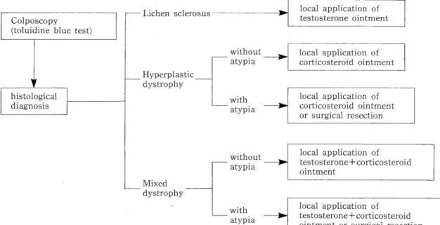
There has been very few systematic studies in Japan concerning vulvar dystrophy. In this institute a study was undertaken to evaluate the clinical outcome of vulvar dystrophies, where the patients were subjected to a proposed protocol of treatment (Fig. 1).
MATERIALS AND METHODS
The patients referred to the Gynecology Department of Nagasaki University Hospital with vulvar complaints with the subjective symptom of `itching' had undergone biopsy. In all the cases toluidine blue test and colposcopy directed biopsies were obtained. In addition to the toluidine positive areas, multiple biosies were obtained from several other sites because of the multicentric nature of the vulvar dy- strophic diseases.
Biopsy site and the size of the lesion were documented and photographed for references for the treatment assesment. Diagnosis being confirmed after histological examination, the patients were subjected to the proposed protocol of treatment. The steroid hormones were ap- plied to the local skin two to three times daily for at least four weeks and the frequency of application were decreased gradually to two times per week, with the alleviation of the symp- toms. The patients were examined bi-weekly
Fig. 1 Treatment protocol for vulvar dystrophy
at which time the gross appearance of vulva, symptomatic relief and side-effects were noted.
RESULTS
The patients who had undergone biopsy of epithelial lesions of the vulva were 75 in total from January 1986 till December 1990. Among them vulvar dystrophy was found in 29 cases (37.1%). The initial histological diagnosis of these 29 patients are listed in Table 1.
The age distribution according to histological diagnosis in lichen sclerosus was 56.4 (SD 11.4) years. The age of the patients with hyperplastic dystrophy without atypia was 54.08 (SD 12.4) years and for hyperplastic dystrophy with
atypia was 50.5 (SD 14) years. The age of the patients with mixed dystrophy was quite var- iable, with majority of these patients being over the age of 50 years. In contrary to other fre- quent statements the vulvar dystrophc lesions were obtained in some patients of reproductive
age group too (Fig. 2).
Itching was the most frequent complaint encountered in this study. Also mass, burning sensation, pain and disturbance in urination were. the other symptoms commonly found.
However, some of the patients were without any
symptom (Table 2).
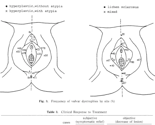
The frequency of vulvar dystrophy by site is presented in Fig. 3. Lichen sclerosus was found in the labia majora and minora and also rel- atively frequent in the prepuce of the clitoris and posterior labial commissure. The occur- ence was almost symmetrical in 4/5 (80%) of the cases. Hyperplastic dystrophy without atypia -was frequent in labia majora, minora and was
liable to spread to the prepuce of the clitoris and posterior labial commissure. In case of hyperplastic dystrophy with atypia was mostly localized, in the vestibule and frequently ob- served in the labia majora and minora.
The treatment advocated, after histological diagnosis paralleling the protocal, application of corticosteroid (Fluocinolone acetonide 0.25- mg/g in 10g) ointment in hyperplastic dystrophy without atypia and resulted in alleviation of itch.
The symptomatic relief was prompt in this group, followed by demonstrable gross change of the vulva, which was evident by reduction of the size of the lesion. The final result was histological changes towards regression. In patients with hyperplastic dystrophy with atypia, corticosteroid completely removed the disease without any recurrences in 6/9 (66.7%) patients. In two other patients it was ac-
Table 1. Epithelial Lesions of The Vulva [Nagasaki University (Jan 1986-May 1990)]
Epithelial Lesions Cases (%)
Lichen sclerous 5 ( 6.4)
Hyperplastic, without atypia 9(11.5)
Dystrophy H mild-mod 11 (14.1) 29(37.1)
yperplastic, with atypic severe I ( 1.3)
Mixed 3 ( 3.8)
Other benign Condyloma acuminatum 1 ( 1.3)
l Hydradenoma 1 ( 1.3) 32(43.6)
lesions V
ulvitis and others 32 (41.0)
In situ cancerous Carcinoma in situ 1 ( 1.3) 3 ( 3
.9)
lesion Paget's disease 2 ( 2.6)
Microinvasive carcinoma 1 ( 1.3)
Invasive Squamous cell carcinoma 9(11.5) 12 (15.4)
carcinoma I
nvasive Paget's disease 1 ( 1.3) Adenocarcinoma (Skene gland) 1 ( 1.3)
Table 2. Symptoms of Vulvar Dystrophy symptoms
lesion itching burning mass pain disturbance urinary symtoms no
Lichen sclerosus (5 case) 80 % 40% - 20 % 20 % -
Hyperplastic, (9 case) without atypia 77.8% - 11.1% 11.1% - 11.1%
Hyperplastic, (12 case) with atypia 58.3% - 16.7% 8.3% - 16.7%
Mixed (3 case) 66.7% - 33.3% - 33.3% 33.3%
Fig. 2. Age distribution in patients with vulvar dystrophies
complished by local excision of the diseased area. In these two patients the lesion was small, localized and well demarcated, which could eas- ily be approached for excision.
The treatment for lichen sclerosus with 2%
testosterone propionate in a vaseline base ap- plied locally has had dramatic response, noted by the symptomatic relief of itching. Subjective
Fig. 3. Frequency of vulvar dystrophies by site (%)
Table 3. Clinical Response to Treatment
subjective objective
cases (symptomatic relief) (decrease of lesion)
complete partial 100% 50%% 50%>
Lichen sclerosus 5 4 1 2 2 1
Hyperplastic, without atypia 8 7 1 4 4 -
mild 3 3 - 3 - -
Hyperplastic, with atypia moderate 5 4 1 3 2 -
severe 1 1 1 -
Mixed 1 1 1 -
Total 23 20 3 12 10 1
(87.0%) (13.0%) (52.2%) (43.5%) (4.3%)
symptoms disappeared or decreased in 4/5 (80%) of the cases. The improvement in the external morphology was more vivid in these patients.
The treatment offered for mixed dystrophy without atypia was corticosteroid and testoster- one ointment application was of great benifit.
The clinical response after treatment in 23 of the vulvar dystrophic patient are represented in Table 3.
DISCUSSION
In the previous times the treatment of choice for vulvar dystrophy was surgery by mutilat-
ing vulvectomy. Most clinicians at that time believed that this method relieves symptoms and provides an adequate prophylaxis against the inevitable development of carcinoma. In the
recent times there are several reports investi- gating the relationship of vulvar dystrophy to
the development of carcinoma'' 2. 3. "And these reports revealed that the risk of carcinoma of the vulva developing in a woman with vulvar dystrophy is approximately 2-5% at presentation with additional risk of developing malignancy from vulvar dystrophy is extremely small.
Previously, several kinds of topical agents has been tried for vulvar dystrophy. Vitamin A5' and Mercuric Sulphide' were used in the early times, but they did not result in the resolution of the disease, either morphologically or micro- scopically. Jasionowski) has tried a topical application of progesterone, which resulted in a good response but the treatment was advo- cated in a small group of patients only. As a medical approach, intradermal injection of triamnicilone' and absolute alcohol" have been used. The result of these agents were not sat- isfactory.
August") employed cryosurgery on 12 patients and reported satisfactory results, without fur- ther treatment after three years in only 25% of the cases.
Surgical therapy for vulvar dystrophies has been disappointing because most patients ex- perienced recurrences'. Rettenmaier" report- ed on skinning vulvectomy in vulvar dystrophic patients and observed recurrences in 50% of the patients at 45-96 months after initial surgery.
Di Paola121 treated vulvar dystrophy by grafting technique after vulvectomy. Two years later, the grafted skin has developed recurrence.
Recently, laser therapy has been used for the treatment of this disease"). But a high recur- rence rate has been found after this treatment.
Some authorities suggest that persisting lesion after laser therapy may respond more favorably to topical steroid, but this mode of treatment has been evaluated in only small number of patients. The therapeutic value of laser therapy in vulvar dystrophies is yet to be proved.
Effective treatment of vast majority of pa- tients can be achieved medically by steroid hormone. The local application of corticoster- oid has been proved to be of great benifit in most patients becoming free of symptoms. In a prospective study by Kaufman8' 24 out of 39 hyperplastic dystrophy without atypia had com- plete relief of symptoms with both morpho- logical and histological changes to regression.
Friedrich" similarly reported on prompt relief of symptoms in hyperplastic dystrophic patients by the treatment using topical application of a cream containing crotamine diluted with a hydrocortisone compound.
Cinberg''" was the first who noted the ef- ficiency of topical testosterone in lichen sclero- sus. His treatment was employed in a large series by several authorities""". Recently Friedrich" in a report summarized the results of testosterone ointment for the treatment of lichen sclerosus and mixed dystrophy. In ad- dition to his own results, he reported on the results of several authorities. Of 184 patients treated, 172 (93%) had shown improvement after testosterone therapy, both macroscopically and microscopically.
Vulvar dystrophy is a common gynecological problem causing sufferings and morbidity in large number of women. It is to be emphasiz-
ed that biopsy is mandatory before advocat- ing treatment in order to exclude malignancy during the first visit and that re-biopsy is also essential for the confirmation of regression of the disease. The present report has demon- strated the dramatic effects of steroid hormone therapy on vulvar dystrophy, irrespective of
dysplasia. In the study, 4 of the 5 patients of lichen sclerosus and 16 of the 18 patients of hyperplastic and mixed dystrophy had com-
plete symptomatic response to testosterone and corticosteroid respectively. Moreover, morpho- logically 22/23 (96%) of the patients had more than 50% decrease in total lesion. These data indicate that the protocol of treatment describ- ed here can eliminate the agonising symptoms without a need for mutilating surgery. It is likely that the sufferings and morbidity in vulvar dystrophic patients could be controlled
by this treatment.
REFERENCES
1) Jeffcoate TNA, Woodcock AS. Premalignant
Condition of The Vulva With Particular Re- ference To Chronic Dystrophies. Br MedJ2 : 127-
134, 1961.
2) Jeffcoate TNA. Chronic vulvar dystrophies. Am J Obstet Gynecol 95: 61-74, 1966.
3) Kaufman RH, Gardner HC, Brown D Jr, Beth Y.
Vulvar dystrophies ; an evaluation. Am J Obstet
Gvnccol 120: 363 367, 1974.
4) Hart WR, Norris HJ, Helwig EB. Relation of Lichen Sclerosus et Atrophicus of the Vuiva to
development of Carcinoma. Obslet Gynecol 45:
369 377, 1975.
5) Kaufman RH, Gardner HC. Vulvar dystrophies.
Clin Obstet Gynecol 21 : 1082 1106, 1978.
6) Friedman EG Jr. Vulvar dystrophy. Clin Obstet Gynne(,ol 28: 178-187, 1985.
7) Jasionowski EA, Jasionowski PA. Further obser- vations on the effect of topical progesterone on
vulvar disease. Am J Obstet Gynecol134 : 565 567,
1979.
8) Kaufman RH. Hyperplastic Dystrophy. J Reprod Mcd 17: 137 145, 1976.
9) Woodruff JI), Babaknia A. Local Alcohol Injection of the Vulva: Discussion of 35 Cases.
Obstet Gynccol 54: 512 514, 1974.
10) August PJ, Milward TM. Cryosurgery in the trearnent of Lichen sclerosus et atrophicus of
the vulva. Br J Dcrmatol 103: 667-670, 1980.
11) Rettenmaier MA, Braly PS, Roberts WS, Berman ML, Disaica PJ. Treatment of Cutaneous Vulvar
Lesios with Skinning Vulvectomy. J Reprod Med
30 : 478 480, 1985.
12) Di Paola GR, Rueda-Leverone NG, Belardi MG.
Lichen sclerosus of the Vulva Recurrent after
Myocutaneous Graft. A Case Report. J Reps-od
Med 27: 666-668, 1982.
13) Kaufman RH, Friedrich EG. The Carbon Dioxide Laser in the Treatment of Vulvar Disease. Cline
Obstet Gynecol 28: 220-229, 1985.
14) Cinberg BL. Post menopausal pruritus vulvae.
Am J Obstet Gynccol 49: 647 657, 1945.
15) Friedman EG Jr. Topical Testosterone for Benign Vulvar Dystrophy. Obstct Gynecol37 : 677
686, 1971.
16) Frierich EG Jr. Lichen Sclerosus. J Rcprod Med 17: 147-154, 1976.
![Table 1. Epithelial Lesions of The Vulva [Nagasaki University (Jan 1986-May 1990)]](https://thumb-ap.123doks.com/thumbv2/123deta/10139331.1973210/3.771.143.627.141.396/table-epithelial-lesions-vulva-nagasaki-university-jan.webp)