九州大学学術情報リポジトリ
Kyushu University Institutional Repository
経直腸経管腔的内視鏡手術のための新しい着脱型閉 塞バルーンユニットの開発
徐, 号
九州大学大学院医学研究院先端医療医学部門
https://doi.org/10.15017/26343
出版情報:Kyushu University, 2012, 博士(医学), 課程博士 バージョン:
権利関係:(C) Informa Healthcare USA, Inc.
1 Title page
New detachable occlusion balloon unit for transrectal natural orifice translumenal endoscopic surgery
Running title: New detachable occlusion balloon unit for NOTES
Hao Xu, MD1; Takeshi Ohdaira, MD2; Yoshihiro Nagao, MD3; Norifumi Tsutsumi, MD3; Megumu Mori, MD1; Munenori Uemura, PhD1; Kazutaka Toyoda, PhD1; Satoshi Ieiri, MD3; Makoto Hashizume, MD, PhD, FACS1,2,3
1Department of Advanced Medical Initiatives, Faculty of Medical Sciences, Kyushu University, Fukuoka, Japan
2Center for Advanced Medical Initiatives, Kyushu University, Fukuoka, Japan
3Department of Advanced Medicine and Innovative Technology, Kyushu University Hospital, Fukuoka, Japan
Correspondence to:
Hao Xu, MD
Department of Advanced Medical Initiatives Faculty of Medical Sciences, Kyushu University 3-1-1 Maidashi, Higashi-ku, Fukuoka 812-8582, Japan TEL: +81-92-642-5993
FAX: +81-92-642-5199
E-mail: jokyo@dem.med.kyushu-u.ac.jp
2
Table of contents
Abstract ……… 3
Introduction ………. 4
Materials and Methods ………... 5
Specifications of the balloon unit ……….. 5
In vivo study ……… 6
Assessment of balloon placement time ……….. 7
Assessment of water-tightness and bacterial clearance ……… 7
Assessment of air-tightness and the effectiveness of maintaining visibility in the working area ……… 8
Assessment of complications of balloon placement ……….. 10
Results ……….. 11
Assessment of effectiveness of balloon placement ……… 11
Assessment of water-tightness and effectiveness of bacterial clearance ………. 12
Assessment of air-tightness and effectiveness of visual field maintenance ………. 12
Assessment of complications due to balloon placement ………14
Discussion ………. 15
Conclusion ……… 18
Disclosure ……….. 19
References ………. 20
Table ………. 26
Table 1 ………. 26
Table 2 ………. 27
Figure and Figure Legends ……….28
Figure 1 ……… 28
Figure 2 ……… 29
Figure 3 ……… 30
Figure 4 ……… 31
3 Abstract
Introduction: Transrectal natural orifice translumenal endoscopic surgery (NOTES) requires a good endoluminal view and adequate intrarectal bacterial clearance in the working area. We developed a new occlusion balloon unit with an easily detachable inflation device, which allows the surgeon a clear working area distal to the balloon. Materials and methods: The effectiveness of the sealing balloon and the extent of macroscopic and histopathological injury to the bowel wall at the site of balloon placement were examined in 12 pigs. Results: The mean time to place and inflate the balloon unit was 12.0 ± 3.5 min, Effective air-tightness lasted for 21.0 ± 12.0 min. There was no leakage of dye (methylene blue) past the balloon when pressure was maintained above 6.70 ± 0.08 kPa (6.62–6.78 kPa). After gut irrigation, good visibility was maintained in the working area for 6 hours, and adequate bacterial clearance was maintained for 3 hours. There were no macroscopic signs of intestinal wall damage at the site of balloon placement. Histopathological examination showed only patchy mucosal damage and submucosal thrombus at the site of balloon placement.
Conclusion: This newly-developed occlusion balloon unit helps to establish good visibility and adequate bacterial clearance for endoluminal surgical procedures.
Keywords
occlusion balloon, transrectal, bacterial clearance, NOTES
4 Introduction
Natural orifice translumenal endoscopic surgery (NOTES) has been reported in humans since 2004 [1]. However, there are ongoing challenges to performing NOTES, as outlined in the Natural Orifice Surgery Consortium for Assessment and Research (NOSCAR) white paper [2-4].
Local bacterial clearance is of particular concern when using the transcolonic route. Several studies have reported the occurrence of intraperitoneal abscesses after transcolonic surgery, despite attempts to sterilize the working area in the bowel [5-9]. We considered that improvement of bacterial clearance in the colon would increase the safety of transrectal NOTES, and developed a method of transrectal bowel irrigation. This method uses an occlusion balloon unit that provides a water-tight occlusion of the intestinal lumen to achieve bacterial clearance with a high-speed irrigation device, and an air-tight seal to maintain visibility in the working area.
The present study assessed the balloon unit in terms of water-tightness, air-tightness, ability to maintain bacterial clearance, balloon inflation pressure, effectiveness in maintaining visibility in the working area, and intestinal wall damage.
5 Materials and Methods
Specifications of the balloon unit
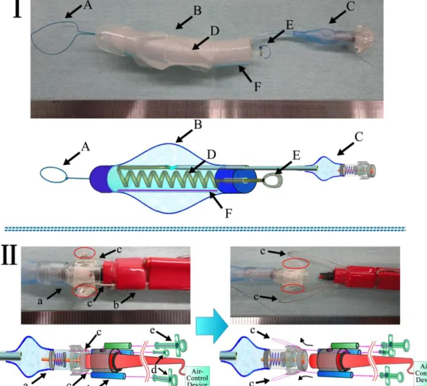
The design of the occlusion balloon unit is shown in Figure 1(I-A–I-F). The occlusion balloon is capsule-shaped to reduce intestinal injury. The balloon is oval in shape and is made of
biocompatible silicone. These features allow the balloon to change shape during intestinal peristalsis, thus minimizing the risk of injury to the bowel wall. The balloon measures 70 mm in length and 11 mm in width when deflated, and has a maximum diameter of 80 mm when inflated.
The most important design feature of this unit is the ability to detach the inflation device
(Figure1: I-C, II-a–II-e) from the balloon unit to allow the surgeon a clear working area distal to the balloon. The balloon unit also features radio-opaque line (Figure1: I-F).
The inflation device used in this study was developed by us. The unit includes a power supply, pressure gauge, and inflation device, and inflates the balloon at 0.18 L/min, requirements of an occlusion balloon unit for used in endoluminal surgery are shown in Table 1.
6 In vivo study
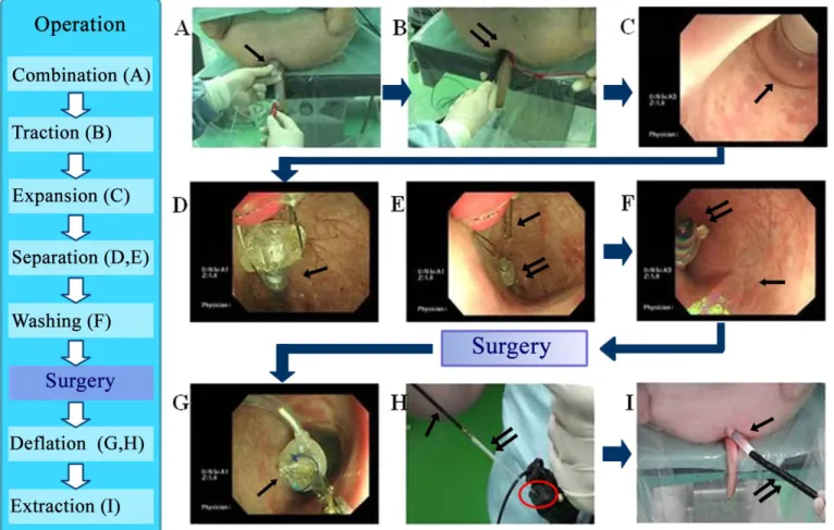
Twelve female pigs, weighing approximately 40 kg each, were used in this study. Two experimental models were used: a model to study visibility in the working area, and a model to study bacterial clearance in the working area. All animals were fed cereal preoperatively (Miniature Swine Diet M-16; CLEA Japan, Inc., Tokyo, Japan). Under general anesthesia, the balloon unit was inserted through the anus and advanced to the target position in the colon with endoscopic guidance. The balloon was then inflated to occlude the intestinal lumen (Figure 2-A, B, and C). After balloon inflation, the two wire anchoring loops connecting the inflation device to the balloon unit were loosened. The entire inflation device was pushed towards the check valve attached to the balloon, and the anchoring loops were extended, which pushed the wires along the 45 slope on the outside of the
anchoring hooks and released the loops (Figure 2-D and E). After bowel irrigation (Figure 2-F), the spiral deflation tube (1.5 m length, 0.7 mm diameter) of the balloon that was pulled through the endoscope channel to the outside of the body. The end was cut off the deflation tube with scissors, and a 10-mL syringe was connected to the deflation tube stump deflate the balloon (Figure 2-G and H). The balloon was then withdrawn through the anus (Figure 2-I).
7 The rectum contained a large amount of solid stool because no purgative had been used prior to the procedure. A high-speed irrigation device (HydroFlex Multi-application Irrigation Pump;
BARD, RI, USA) connected to the endoscope channel easily cleared the space between the anus and the peritoneal reflection of the rectum for balloon insertion.
Assessment of balloon placement time
The time taken to position and inflate the balloon was recorded. The displacement of the balloon over time and the time for balloon withdrawal were also measured.
Assessment of water-tightness and bacterial clearance
Assessment of occlusion
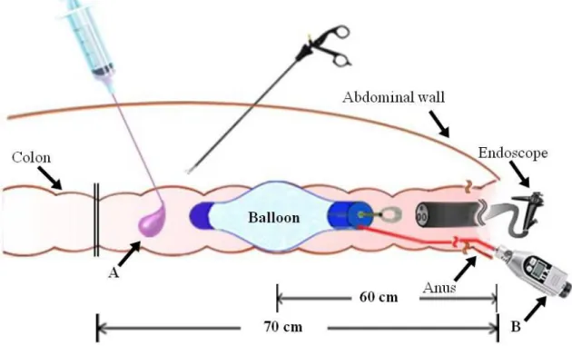
Ten measurements were performed in each of two pigs. The balloon unit was placed 60 cm to the anal verge with endoscopic guidance, and the balloon was inflated to enable laparoscopic
identification of the site. Two ligatures were placed around the colon at 70 cm proximal to the anal verge, using 1-0 silk/3-0 nylon. Methylene blue (Figure 3-A) (20 mL) was injected into the
8 bowel between the ligatures and the balloon. The balloon pressure was then gradually reduced, and a pressure sensor (AP-V80; Keyence, Tokyo, Japan) (Figure 3-B) was used to determine the pressure at which methylene blue leaked into the past balloon to the anal side (Figure 3).
Assessment of effectiveness of irrigation
The balloon was placed 20 cm proximal to the desired working area and inflated. The high-speed irrigation device (HydroFlex Multi-application Irrigation Pump; BARD, RI, USA) was used to irrigate the gut on the anal side of the balloon at a flow rate of 500 mL/min. The time required to fill the intestinal lumen was measured.
Residual stool was removed until the intestinal lumen had the same macroscopic appearance as would be expected after routine administration of polymer purgatives for oral
endoscopy .Bacterial were then measured. The irrigation solutions used were distilled water, isotonic saline, and 1% neomycin solution.
Assessment of air-tightness and the effectiveness of maintaining visibility in the working area
9 After balloon occlusion, the bowel was insufflated via an endoscope placed 15 cm from the anal verge .The time required to establish visibility in the working area and the inflation requirements were recorded. The length of time that visibility of the working area was maintained, was also measured. The time from maximum visibility in the working area until a 50% reduction in volume, when treatment was considered difficult to perform, was recorded.
To assess whether bacterial clearance was maintained in the working area during surgery,
cholecystectomy was performed in two pigs by transrectal hybrid NOTES using a 3-mm diameter forceps [10]. Injury to the bowel wall was assessed macroscopically and histopathologically.
The effectiveness of using the occlusion balloon during endoscopic submucosal dissection (ESD) was assessed was performed in two pigs. A hook (PCF-H180AL/I; Olympus, Tokyo, Japan) was used to simulate a rectal lesion 20 mm in diameter on the anal side of the balloon. Maintenance of visibility in the working area and the efficiency of clearance of smoke from electrocoagulation were assessed. The lesion was located on the left wall of the rectum at 20 cm from the anal verge, and the balloon was placed 10 cm proximal to the lesion. Electrocoagulation was performed for 30 s, with concurrent inflation and aspiration through the endoscope. The time from the start to completion of ESD was recorded, and the inflation status of the balloon was monitored.
10 Assessment of complications of balloon placement
Physiological adverse effects of balloon placement were assessed by monitoring heart rate and arterial oxygen saturation during balloon inflation in three pigs. Balloon inflation time (to a diameter of 80 mm was set to 3 seconds .Physiological effects were assessed at three different balloon locations20, 40, and 60 cm) from the anal verge.
Injury to the bowel wall caused by balloon inflation was assessed macroscopically and
histopathologically. The bowel wall was examined at the site of balloon inflation and on both the oral and anal sides of the occlusion site. The bowel was examined macroscopically for signs of mucosal rupture, bleeding, hematoma, and edema, and microscopically for signs of submucosal thrombus and inflammation.
The study protocol was registered with and approved by the Animal Ethics Committee and Animal Center at Medical Sciences Kyushu University (permission number A-21-165).
11 Results
Assessment of effectiveness of balloon placement
The relatively short length of the balloon and its excellent ability to mold to the intestinal tract resulted in good maintenance of its position. The balloon moved 1–2 cm towards the anus during intestinal peristalsis and then back towards the mouth during intestinal relaxation, maintaining its position overall.
The time required to place and inflate the balloon when there was no air leakage was 10.0 ± 2.5 min. We assessed the mechanism for detaching the inflation device from the balloon unit and the ease of inflating and withdrawing the balloon, which were the most important requirements of the unit, In two of the pigs , we verified that the inflation device could be detached from the balloon when the unit had bilateral anchoring wires. The original unit with a single anchoring wire was used in four of the pigs, and was also detached easily. Air leaked from the check valve after detachment of the inflation device in two of the pigs. The balloon therefore cannot be relied on to maintain its inflation in clinical settings.
12 Removal of the deflation tube from the balloon and subsequent deflation were performed
efficiently in all animals. Endoscopic withdrawal of the balloon assisted by gentle pulling on the deflation tube was very easy. The time required for balloon withdrawal was 2.0 ± 1.0 min.
Assessment of water-tightness and effectiveness of bacterial clearance
Gentian violet solution leaked past the balloon at a mean pressure of 6.70 ± 0.08 kPa (6.62–6.78 kPa) and a mean balloon diameter of 7.15 ± 0.95 cm (6.2–8.1 cm).
High-speed irrigation after balloon inflation reduced the total bacterial counts .The reduction in bacterial count differed depending on the irrigation solution used. Post-irrigation changes in bacterial counts over time are shown in Table 2.
Assessment of air-tightness and effectiveness of visual field maintenance
The distance of the balloon from the anal verge (20, 30, 40, and 60 cm), time required to establish visibility in the working area, inflation requirements, and time until 50% loss of volume in the working area were as follows. All inflations were at high speed, using the PCF-H180AL/I hook.
13 Deflation time was 2.05 ± 0.25 s (1.8–2.3 s) at 20 cm, 3.45 ± 0.65 s (2.8–4.1 s) at 30 cm, 3.55 ± 0.25 s (3.3–3.8 s) at 40 cm, and 5.0 ± 0.2 s (4.8–5.2 s) at 60 cm. The time until 50% loss of volume in the working area was 10.5 ± 1.5 min (12.0–9.0 min) at 20 cm, 12.5 ± 1.5 min (14.0–11.0 min) at 30 cm, 28.5 ± 6.5 min (35.0–22.0 min) at 40 cm, and 29.5 ± 1.5 min (31.0–28.0 min) at 60 cm.
Inflation in small volumes increments was found to rapidly establish visibility in the working area, and sustain visibility for a long time. However, the working area was lost more quickly when the balloon was placed closer to the anus.
The mean time to inflate the balloon was 9.0 ± 3.5 min , the mean time to deflate the balloon was 2.0 ± 1.0 min, and the mean time for bowel preparation was 11.0 ± 4.5 min; The time to completion of irrigation therefore was <10 minutes. Intestinal wall damage at the site of balloon placement in the two pigs that underwent cholecystectomy by NOTES was so minimal that it could not be recognized macroscopically.
Once visibility of the working area was established, it was maintained relatively well. A good visual field was maintained during ESD, and the procedure was easy to perform.
Effectiveness of balloon occlusion
14 We saw no leakage of intestinal contents (solid or liquid) from the proximal side of the balloon during the 6 hours after gut irrigation.
Assessment of complications due to balloon placement
The physiological effects of balloon placement were assessed by monitoring heart rate and arterial oxygen saturation in six pigs. No changes in these vital signs were observed, even when the balloon was inflated to its maximum diameter.
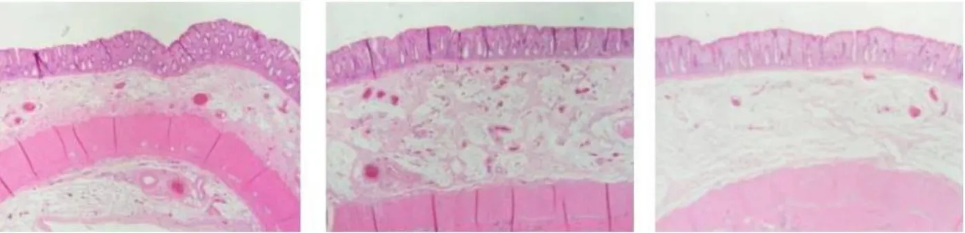
The macroscopic and histopathological examinations findings of the intestinal wall after balloon occlusionare shown in Figure 4. No macroscopic signs of mucosal rupture, bleeding, hematoma, or edema were observed at the balloon placement sites. Histopathological examination showed localized mucosal injury and submucosal thrombus. Submucosal edema was also observed on the anal side of the balloon placement site. There were no microscopic findings indicating rupture of the bowel wall.
15 Discussion
We developed a new occlusion balloon unit and demonstrated that this device may help to improve the endoluminal view and bacterial clearance during transrectal NOTES.
Previous animal and human studies of occlusion balloon devices have reported balloon placement times of 18 min or longer [11-13]. In this study, the maximum time required for balloon
placement was only 12 min. The balloon unit is placed using a conventional endoscope and conventional biopsy forceps, and is easy to operate. We therefore believe that surgeons will find it relatively easy to introduce it into clinical practice.
We showed that irrigation of the bowel in preparation for transrectal NOTES can be performed in the immediate preoperative period. Furthermore, the total bacterial count in the bowel did not increase over time-while the balloon was inflated, confirming that the balloon contributes to maintenance of adequate bacterial clearance. This reduces the risk of peritoneal infection by transrectal NOTES [14, 15]. The short time required for bowel preparation was verified when we performed cholecystectomy using transrectal NOTES. Currently, bowel preparation rarely
involves the use of antibiotic solutions. We are investigating establishment of a completely antiseptic environment by balloon occlusion and irrigation with low-concentration ozone water.
16 When performing colonic ESD, air insufflation to a relatively high intraluminal pressure may be necessary to visualize the surgical field. This may lead to postprocedural pain due to colonic distension. We consider that the present balloon unit, which was shown to markedly improve the ability to visualize the working area, could effectively be used for in ESD of extensive early colon cancer.
Previous studies which used miniature endoscopes to place inflatable balloons in the bowel demonstrated the safety of such procedures [11-13, 16-20].
Physiological assessment of three pigs in this study during the balloon placement procedure did not show any adverse effects. However, we did observe physiological effects when the entire colon was distended by a large volume of air. This indicates that our procedure may be useful for reducing physiological adverse effects, because it allows procedures to be performed with
delivery of a smaller volume of air to a restricted area of the colon.
Some studies have evaluated the effects of localized pressure on the wall of the small intestine in terms of blood flow, and subsequent ischemia or necrosis [21-26]. However, there are no reports of such studies of the large intestine. In this study, we evaluated the pathological changes
17 resulting from localized pressure on the bowel wall. Because of ethical considerations in animal experiments, it is impossible to fully evaluate the relationship between the localized pressure and ischemia or necrosis. However, some clinical reports have described ischemia or necrosis of the bowel wall that were considered to be caused by excessive localized pressure [27-29]. We are considering future studies to clarify the relationship between localized pressure on the bowel wall and ischemia or necrosis, if they are approved by our animal ethics committee.
The most important consideration when placing a balloon in the colon is avoiding injury to the bowel wall at the site of balloon placement. In this study, we observed only localized damage to the mucosa at the site of balloon placement. We speculate that this is because of the ability of the balloon to mold to the gut and move slightly with peristalsis. We judged it possible to safely perform balloon occlusion of the colon if the design of the balloon did not cause excessive friction. Movement of the balloon associated with intestinal peristalsis carries a risk of gradual leakage of stool past the balloon. This risk can be reduced by reducing balloon movements and increasing the ability of the balloon to mold to the intestinal mucosa. However, such features may cause damage to the bowel wall. We therefore used a silicone balloon to reduce friction. In future studies, we plan to place an antiseptic tampon that is easy to withdraw on the anal side of the balloon.
18 It is not possible to constantly monitor balloon pressure using our device, because the balloon unit is detached from the inflation device. We intend to address this in future studies. In addition, the immunological and bacteriological consequences of balloon occlusion should be studied further.
Although NOTES is still in the developmental stages, hybrid NOTES has already been used in clinical practice. As NOTES is the ultimate minimally invasive surgery that causes minimal stress to the patient’s body, it will hopefully become established as a routine modality in clinical practice in the near future [30, 31].
In conclusion, our newly-developed occlusion balloon unit with an easily detachable inflation device was found to improve endoluminal views and maintain a useful working area for endoluminal surgery. Its ability to prevent fluid leakage makes it a promising device for
providing an irrigated endoluminal surgical field. Furthermore, use of the unit may improve the intraperitoneal visual field by reducing the need for bowel distension during NOTES.
19 Disclosure
Drs. Hao Xu, Takeshi Ohdaira, Yoshihiro Nagao, Norifumi Tsutsumi, Megumu Mori, Munenori Uemura, Kazutaka Toyoda, Satoshi Ieiri, and Makoto Hashizume have no conflicts of interest or financial ties to disclose.
20 References
1. Kalloo AN, Singh VK, Jagannath SB, Niiyama H, Hill SL, Vaughn CA, et al. Flexible transgastric peritoneoscopy: a novel approach to diagnostic and therapeutic interventions in the peritoneal cavity. Gastrointest Endosc 2004; 60: 114-7.
2. Hawes RH. ASGE/SAGES working group on natural orifice translumenal endoscopic surgery.
White paper October 2005. Gastrointest Endosc 2006; 63: 199-203.
3. Rattner D, Kalloo A. ASGE/SAGES working group on natural orifice translumenal endoscopic surgery. October 2005. Surg Endosc 2006; 20: 329-33.
4. Benhidjeb T, Witzel K, Bärlehner E, Stark M. The New European Surgical Academy (NESA): the natural orifice surgery concept. Vision and rationale for a paradigm shift.
(Article in German). 2007; 78: 537-42.
5. McGee MF, Marks JM, Onders RP, Chak A, Rosen MJ, Williams CP, et al. Infectious implications in the porcine model of natural orifice transluminal endoscopic surgery (NOTES) with PEG-tube closure: a quantitative bacteriologic study. Gastrointest Endosc 2008; 68: 310-8.
21 6. Gonzalez-Pezzat I, Soto-Perez-de-Celis E, Pantoja-Millan JP. Late perforation and
abscess formation at the site of the jejunojejunal anastomosis following laparoscopic Roux-en-Y gastric bypass surgery. Obes Surg 2009; 19: 661-3.
7. Allemann P, Probst H, Demartines N, Schäfer M. Prevention of infectious
complications after laparoscopic appendectomy for complicated acute appendicitisthe role of routine abdominal drainage. Langenbecks Arch Surg 2011; 396: 63-8.
8. Senagore AJ, Delaney CP. A critical analysis of laparoscopic colectomy at a single institution: lessons learned after 1000 cases. Am J Surg 2006; 191: 377-80.
9. Kehagias I, Karamanakos SN, Panagiotopoulos S, Panagopoulos K, Kalfarentzos F.
Laparoscopic versus open appendectomy: which way to go? World J Gastroenterol 2008;
14: 4909-14.
10. Ohdaira T, Tsutsumi N, Xu H, Mori M, Uemura M, Ieiri S, et al. Ultra-minimally invasive local immune cell therapy and regenerative therapy by multi-piercing surgery for abdominal solid tumor: therapeutic simulation by natural orifice translumenal endoscopic
surgery-assisted needlescopic surgery using 3-mm diameter robots. J Hepatobiliary Pancreat Sci 2011; 18(4): 499-505.
22 11. Delvaux M, Gay G. International Conference on Capsule and Double-Balloon
Endoscopy (ICCD). Endoscopy 2011; 43(6): 533-9.
12. Beppu K, Osada T, Inoue K, Matsumoto K, Shibuya T, Sakamoto N, et al. Intestinal involvement in Wegener’s granulomatosis diagnosed and followed up by double balloon enteroscopy. Intern Med 2011; 50: 219-22.
13. Takeda T, Shibuya T, Osada T, Izumi H, Mitomi H, Nomura O, et al. Metastatic renal cell carcinoma diagnosed by capsule endoscopy and double balloon endoscopy. Med Sci Monit 2011; 17: CS15-7.
14. Ohdaira T, Ikeda K, Tajiri H, Yasuda Y, Hashizume M. Usefulness of a flexible port
for natural orifice transluminal endoscopic surgery by the transrectal and transvaginal routes.
Diagn Ther Endosc 2010; 2010: 473080.
15. Ohdaira T, Hashizume M. The possibility of NOTES using trans-colorectal route.
Endoscopia Digestiva 2011; 22: 1617-30.
16. Chan YC, Tsai YC, Fang SY. Subcutaneous emphysema, pneumothorax,
pneumomediastinum, and pneumoperitoneum during colonoscopic balloon dilation: a case report. Kaohsiung J Med Sci 2010; 26: 669-72.
23 17. Gay G, Delvaux M. Double-balloon colonoscopy after failed conventional
colonoscopy: a pilot series with a new instrument. Endoscopy 2007; 39: 788-92.
18. Rösch T, Adler A, Wiedenman BH, Hoepffner N. A prospective pilot study to assess technical performance of a new single use colonoscope with inverted sleeve technology.
Gastrointest Endosc 2007; 65: AB340.
19. Kita H, Yamamoto H, Yano T, Miyata T, Iwamoto M, Sunada K, et al. Double balloon endoscopy in two hundred fifty cases for the diagnosis and treatment of small intestinal disorders. Inflammopharmacology 2007; 15(2): 74-7.
20. Rex DK, Goodwine BW. Method of colonoscopy in 42 consecutive patients presenting after prior incomplete colonoscopy. Am J Gastroenterol 2002; 97: 1148-51.
21. Yamamoto H, Sekine Y, Sato Y, Higashizawa T, Miyata T, Iino S.et al. Total enteroscopy with a nonsurgical steerable double-balloon method. Gastrointest Endosc 2001; 53: 216-220.
22. Bretthauer M, Hoff GS, Thiis-Evensen E, Huppertz-Hauss G, Skovlund E.How much gas do we insufflate during colonoscopy? Air and carbon dioxide volumes insufflated during colonoscopy. Gastrointest Endosc 2003; 58: 203-206.
24 23. Bretthauer M, Lynge AB, Thiis-Evensen E, Hoff G, Fausa O, Aabakken L. Carbon dioxide
insufflation in colonoscopy: safe and effectiveness in sedated patients. Endoscopy 2005;
37: 706-709.
24. Xin L, Liao Z, Jiang YP, Li ZS. Indications, detectability, positive findings, total enteroscopy, and complications of diagnostic double-balloon endoscopy: a systematic review of data over the first decade of use. Gastrointest Endosc 2011; 74: 563-570.
25. Ikeya K, Osawa S, Kuriyama S, Hamaya Y, Yamada T, Sugimoto M, et al. Decompression side tube-equipped double-balloon enteroscopy extends intubation depth and reduces patient discomfort. Endoscopy2012; 2:E256-7.
26. Fukumoto A, Tanaka S, Shishido T, Takemura Y, Oka S, Chayama K. Comparison of detectability of small-bowel lesions between capsule endoscopy and double-balloon endoscopy for patients with suspected small-bowel disease. Gastrointest Endosc 2009 69(4):857-65.
27. Cappell MS, Inglis B, Levy A. Two case reports of gastric ulcer from pressure necrosis related to a rigid and taut percutaneous endoscopic gastrostomy bumper. Gastroenterol Nurs2009; 32(4):259-63.
25 28. Nib Soehendra, Hans Seifert. Therapeutic Endoscopy: Color Atlas of Operative Techniques
for the Gastrointestinal Tract.2004; P46.47.
29. Barker DE, Kaufman HJ, Smith LA, Ciraulo DL, Richart CL, Burns RP. Vacuum pack technique of temporary abdominal closure: a 7-year experience with 112 patients. J Trauma 2000; 48(2):201-6.
30. Xu H, Tomikawa M, Konishi K, Ieiri S, Tanoue K, Hashizume M. Future prospect on
natural orifice translumenal endoscopic surgery (NOTES). Fukuoka Acta Medica 2009;
100(2): 43-50.
31. Tomikawa M, Xu H, Hashizume M. Current status and prerequisites for natural orifice translumenal endoscopic surgery (NOTES). Surg Today 2010; 40: 909-16.
26 Table
Table 1. Requirements of the balloon unit 1. Molding during intestinal peristalsis
2. A balloon deflation mechanism for emergencies
3. An easy method of detaching the inflation device from the balloon unit during procedures 4. A mechanism for manual withdrawal of the balloon
5. Radio-opaque marking
27 Table 2. Total bacterial counts (colony forming units /field)
Irrigation solutions, n = 6
Time after irrigation
Immediate 1hour 2hours 3hours
Unirrigated part of the colon 4886 4882 4872 4881
Distilled water 326 335 330 342
Isotonic saline 162 173 178 182
1% neomycin solution 6 11 10 12
28 Figure and Figure Legends
Figure 1. Design of the balloon unit and the procedure for detaching the inflation device from the balloon
(I) Design of the balloon unit:(A) ring used for traction by the balloon unit during placement. (B) occlusion balloon.
(C) check valve. (D) deflation tube. (E) ring for grasping the of deflation tube (F) radio-opaque line
(II) Procedure for detaching the inflation device from the balloon unit. Left: the inflation device is attached to the check valve of the balloon unit. Right: the anchoring wire rings are unhooked and the inflation device is detached from the balloon unit. (a) check valve. (b) proximal end of the inflation device. (c) anchoring wire. (d) controls to extend the proximal end of the inflation device. (e) control to extend the anchoring ring wires and the check valve.
Red circles: hooks for the anchoring wires, with a 45 slope on the outer edge.
29
Figure 2. Use of the occlusion balloon during in vivo operations
(A) The balloon unit is connected to the inflation device and inserted into the anal canal, followed by the endoscope.
Single arrow: occlusion balloon. (B) The inflation tube is pushed in proportion to the insertion speed of the
endoscope. Double arrow: endoscope. (C) The ring on the proximal end of the balloon unit is grasped with forceps to position the unit. The balloon is then inflated to occlude the intestinal lumen. Single arrow: inflated balloon. (D) The balloon is completely inflated, and the anchoring wires are unhooked to detach the inflation device from the balloon unit. Single arrow: check valve. (E) The balloon unit and inflation device are completely detached. Single arrow:
anchoring ring wire. Double arrow: check valve. (F) Irrigation is initiated immediately after balloon inflation. Single arrow: water jet from the tip of the endoscope. Double arrow: check valve. (G) The forceps are passed through the endoscope channel and the ring of the spiral deflation tube is grasped. Single arrow: ring of the spiral deflation tube.
(H) The spiral deflation tube of the balloon is withdrawn through the endoscope channel and guided to the outside of the body. Single arrow: endoscopic forceps. Double arrow: spiral deflation tube. Red circle: endoscope channel. (I) The spiral tube is used to deflate the balloon and guide it through the anal canal to the outside of the body. Single arrow: occlusion balloon. Double arrow: endoscope.
30
Figure 3. Balloon fluid leak test using methylene blue solution
(A) methylene blue. (B) pressure gauge.
31
Figure 4. Microscopic submucosal thrombus was observed in a very limited area at the site of balloon placement
Edema was observed in the bowel wall on the oral side of the balloon placement site. No significant change was seen on the anal side of the balloon placement site. No mucosal changes were observed in the bowel wall
(hematoxylin-eosin staining, 40, 100).