• 検索結果がありません。

Bronchial Epithelial Cells BEAS-2B

N/A
N/A
Protected

Academic year: 2021

シェア "Bronchial Epithelial Cells BEAS-2B"

Copied!
11
0
0

読み込み中.... (全文を見る)

全文

(1)

Tumor Necrosis Factor-alpha and Transforming Growth Factor-beta Synergistically Upregulate Endothelin-1 Expression in Human

Bronchial Epithelial Cells BEAS-2B

Yutaka T SUCHIYA , Aya W AKABAYASHI , Satoshi M ATSUKURA

, Yuki O SAKABE , Ayaka S EKIGUCHI , Daisuke I NOUE , Yusuke K AKIUCHI , Toshitaka F UNAKI , Yohei Y AMAZAKI ,

Hiromi T AKAYASU , Hidetsugu T ATENO , Eisuke K ATO , Makoto H AYASHI , Gen I SHII , Fumihiro Y AMAGUCHI

and Fumio K OKUBU

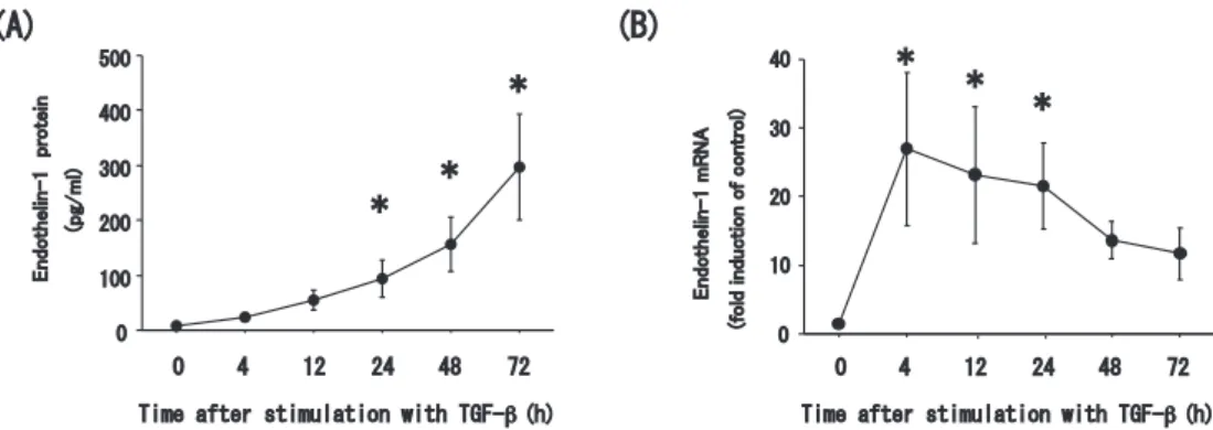
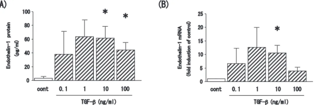
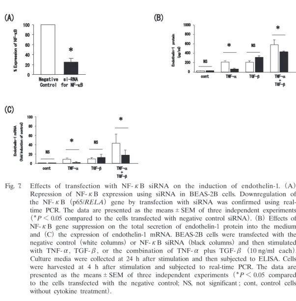
Abstract : Endothelin-1 is a peptide with many functions including bronchoconstric- tion and the stimulation of fibroblasts, and myofibroblasts, and airway smooth muscle cell proliferation. These functions are related to airway remodeling and endothelin-1 is known to be upregulated in the epithelium of patients with severe asthma. We thus sought to elucidate the mechanisms underlying endothelin-1 expression in bronchial epithelial cells in vitro. The human bronchial epithelial cell line BEAS-2B was grown in culture and then treated with tumor necrosis factor- alpha (TNF-α) , interleukin-4 (IL-4) , interleukin-13 (IL-13) , and transforming growth factor-beta (TGF-β) . Expression of endothelin-1 mRNA and protein was quantified by real-time polymerase chain reaction and enzyme-linked immuno- sorbent assay, respectively. We also repressed expression of the key transcription factor in the pathogenesis of severe asthma, nuclear factor-kappa B (NF-κB) , using small interfering RNA (siRNA) . TNF-α and TGF-β significantly increased the release of endothelin-1 protein into the culture medium of BEAS-2B cells at 24 h after treatment compared to untreated cells ; however, the Th2 cytokines, IL-4 and IL-13, had no effect. Endothelin-1 mRNA expression was also upregulated by TNF-α and TGF-β with a peak time point at 4 h after stimulation. Finally, the combination of TNF-α and TGF-β synergistically increased both endothelin-1 protein secretion and mRNA expression, and this upregulation was significantly sup- pressed in cells transfected with siRNA to repress NF-κB expression. TNF-α and TGF-β synergistically upregulate the expression of endothelin-1 in human bronchial epithelial cells, possibly via the activity of NF-κB. Our findings thus suggest NF- κBa as a potential therapeutic target for the regulation of airway remodeling.

Key words : bronchial epithelial cells, endothelin-1, NF-κB, TGF-β, TNF-α Original

Department of Respirology, Showa University Fujigaoka Hospital, 1—30 Fujigaoka, Aoba-ku, Yokohama 227-8501, Japan.

To whom corresponding should be addressed.

(2)

Introduction

 The pathophysiology of asthma is characterized by chronic airway inflammation and remodel- ing such as subepithelial fibrosis, deposition of extracellular matrix protein, hypertrophy and hyperplasia of airway smooth muscle, and goblet cell hyperplasia. Increased bronchial wall thick- ness caused by such airway remodeling contributes to airflow obstruction and the subsequent treatment resistance in severe asthma

1)

.

 Endothelin-1 is a 21-amino acid peptide with vasoconstrictive and bronchoconstrictive activi- ties

2)

. It also functions to stimulate the proliferation of fibroblasts, myofibroblasts, and airway smooth muscle cells, as well as the deposition of extracellular matrix and the differentiation of fibroblasts into myofibroblasts

3, 4)

. These functions of endothelin 1 might therefore play a role in the pathogenesis of airway remodeling. Indeed, recent studies indicate that the expression of endothelin-1 is increased in severe asthma and contributes to airway remodeling therein

5, 6)

, while data from murine models of asthma corroborated that endothelin-1 contributes to airway remod- eling

7)

. All of these studies identified bronchial epithelial cells as an important source for endo- thelin-1 production, supported by in vitro experiments showing that cultured bronchial epithelial cells also produce endothelin-1

8)

. In the present study, we used this in vitro system to further investigate the mechanisms underlying endothelin-1 production in human bronchial epithelial cells.

Methods

Cell culture and reagents

 The human bronchial epithelial cell line BEAS-2B was purchased from the American Type Culture Collection (Manassas, VA, USA) . This cell line was derived from non-tumor bronchial epithelial cells transformed with the SV40 virus

9)

. The cells were cultured in Dulbeccoʼs modi- fied Eagleʼs medium (DMEM)/Hamʼs F12 (F12) medium supplemented with 10% fetal bovine serum (FBS) , 100 U/ml penicillin, and 100 ng/ml streptomycin (Invitrogen, Tokyo, Japan) and maintained at 37 C in a humidified 5% CO

2

atmosphere. We purchased recombinant human cytokines, tumor necrosis factor-alpha (TNF-α) , transforming growth factor-beta1 (TGF-β1) , interleukin-4 (IL-4) , and interleukin-13 (IL-13) , from R&D Systems (Tokyo, Japan) . Cells were treated with each cytokine as described below and then the supernatants and RNA from the cells were collected and stored at ­80 C.

Enzyme-linked immunosorbent assay(ELISA)assay for endothelin-1 protein production

 The concentrations of secreted endothelin-1 in the cell culture supernatants were measured using enzyme-linked immunosorbent assay (ELISA) kits (R&D Systems) . Standards and samples were added to 96-well microtiter plates coated with the anti-endothelin-1 antibody.

After incubation at room temperature for 2 h, the wells were washed 5 times with washing

buffer. A horseradish peroxidase-conjugated secondary antibody against endothelin-1 was then

added to the wells and incubated at room temperature for 2 h. The wells were washed 5 times

with washing buffer, before adding a substrate solution (stabilized hydrogen peroxide and tetra-

(3)

methylbenzidine) and incubating the plate at room temperature for 30 minutes to visualize the antibody binding. Sulfuric acid was added to arrest the reaction, and the absorbance at 450 nm was measured using an ELISA plate reader (Glomax-Multi Detection System, Promega, Tokyo, Japan) .

RNA purification and cDNA synthesis

 Total RNA was extracted from cells after incubation with and without the indicated reagents using ISOGEN (Nippon-Gene, Tokyo, Japan) . cDNAs were synthesized from isolated RNA templates using a High-Capacity cDNA Archive Kit (Applied Biosystems, Tokyo, Japan) .

Real-time polymerase chain reaction (PCR)

 A pre-designed TaqMan probe for endothelin-1 was purchased from Applied Biosystems, and then labeled with a fluorescent reporter dye (FAM) at the 5ʼ end and a downstream quencher dye (TAMRA) at the 3ʼ end. The TaqMan Ribosomal RNA probe, labeled with a fluorescent reporter dye (VIC) , was used as an internal control. Each reaction consisted of 2X Universal Master Mix (Applied Biosystems) , primers, labeled probes, and 100 ng cDNA in a total volume of 50 µl. Amplification conditions consisted of an initial incubation at 95 C for 10 min followed by 40 cycles of 95 C for 15 s and 60 C for 1 min. Amplification and fluorescence measurements were carried out during the elongation step using a Real-Time PCR system 7,500 (Applied Biosystems) . Data are shown as fold induction of mRNA compared with non-stimulated control cells.

Repression of NF-κB expression with siRNA

 To analyze the transcriptional mechanisms of endothelin-1 expression in bronchial epithelial cells, we focused our study on the role of nuclear factor-kappa B (NF-κB) which is a key transcription factor in the pathogenesis of severe asthma. We utilized small interfering RNA

(siRNA) -mediated repression of NF-κB expression, as previously reported

10)

. BEAS-2B cells were seeded into 6-well plates and allowed to grow to 50% confluence, before being transfected with 50 nm siRNA for NF-κB (p65 subunit ; RELA) or scrambled RNA (negative control)

(Applied Biosystems) plus 5 µl Lipofectamine 2,000 (Promega) and grown in DMEM/F12 with 10% FBS without antibiotics for 24 h. Then, the cell media were changed to DMEM/F12 with 10% FBS, 100 U/ml penicillin, and 100 ng/ml streptomycin. After another 24 h, the cells were incubated with or without the indicated cytokines for 4 h (for PCR) or for 24 h (for ELISA) . Then cells were harvested and the supernatant was collected and stored at ­80 C.

Statistical analysis

 Data were expressed as the means standard error of the mean (SEM) . Statistical differ-

ences were determined by analysis of variance with a Fisher protected least significant differences

test. Data were analyzed with Stat-View 5.0 (Abacus Concepts, Inc., Berkeley, CA, USA) .

(4)

Results

Increase of endothelin-1 protein production by TNF-α and TGF-β

 The cytokines used in this study were chosen because of their reported pathogenic associa- tion with airway remodeling in asthma

1)

. We first examined the effect of each cytokine on the secretion of endothelin-1 into the culture medium of BEAS-2B cells. TNF-α and TGF- β each increased endothelin-1 secretion at 24 h after treatment (P < 0.05 compared with non- treated control cells) (Fig. 1) . In contrast, the Th2 cytokines IL-4 and IL-13 did not increase the secretion of endothelin-1. We therefore focused our study on the regulation of endothelin-1 expression by TNF-α and TGF-β and the underlying mechanisms thereof.

Time-dependent expression of endothelin-1 protein and mRNA after stimulation with TNF-α

 As an initial experiment, we observed the time course of endothelin-1 protein release into the cell culture medium and that of its cellular mRNA expression in response to TNF-α treat- ment. The peak secretion of endothelin-1 occurred at 24 h after stimulation with 10 ng/ml TNF- α (P < 0.05 compared with non-treated control cells) (Fig. 2A) . On the other hand, the peak expression of endothelin-1 mRNA was observed at 4 h after the same stimulation, preceding protein production (P < 0.05 compared with non-treated control cells) (Fig. 2B) .

Time-dependent expression of endothelin-1 protein and mRNA after stimulation with TGF-β  We also analyzed the time course of endothelin-1 protein secretion and mRNA expression after stimulation with TGF-β (10 ng/ml) . Peak secretion of endothelin-1 protein into the medium of BEAS-2B cells was observed at 24 to 72 h after stimulation with TGF-β (P < 0.05 compared with non-treated control cells) (Fig. 3A) . However, for TNF-α, the peak expression of endothelin-1 mRNA was detected at 4 h after TGF-β stimulation (P < 0.05 compared with non-treated control cells) (Fig. 3B) . Therefore, we focused subsequent analyses of endothelin-1 secretion and mRNA production at 24 h and 4 h, respectively, after stimulation with TNF-α and/or TGF-β.

Secretion of endothelin-1 protein and mRNA expression after stimulation with different doses of TNF-α

 Endothelin-1 protein secretion appeared to increase dose-dependently at 24 h after TNF-α stimulation of the BEAS-2B cells (P < 0.05 compared with non-treated control cells) , with maxi- mum expression observed with 10-100 ng/ml TNF-α (Fig. 4A) . Similarly, the mRNA expression of endothelin-1 at 4 h after stimulation was also upregulated by TNF-α in a dose-dependent manner (P < 0.05 compared with non-treated control cells) (Fig. 4B) . Therefore, we chose the dose of 10 ng/ml TNF-α for subsequent endothelin-1 analyses.

Secretion of endothelin-1 protein and mRNA expression after stimulation with different doses of

TGF-β

(5)

 Endothelin-1 secretion from the BEAS-2B cells also seemed to increase does-dependently at 24 h after TGF-β stimulation (P < 0.05 compared with non-treated control cells) (Fig. 5A) . The difference in secretion was statistically significant between treated and control cells at 1-10 ng/ml TGF-β, while the expression of endothelin-1 mRNA was upregulated by 10 ng/ml TGF-β at 4 h after stimulation (P < 0.05 compared with non-treated control cells) (Fig. 5B) . Accord- ingly and as for TNF-α, we chose the dose of 10 ng/ml TGF-β for subsequent analyses of endothelin-1.

Fig. 1.   Increased endothelin-1 protein secretion into the medium of cultured bronchial epithelial cells, BEAS-2B, treated with TNF-α and TGF- β. BEAS-2B cells were incubated with 10 ng/

ml IL-4, IL-13, TNF-α, or TGF-β cytokines for 24 h and the supernatants were subjected to ELISA for endothelin-1. The data are presented as the means SEM of three independent experiments [

P < 0.05 compared with non- treated control cells (cont)] .

Fig. 2.   Time course of endothelin-1 induction with TNF-α. (A) Total amount of

endothelin-1 protein secreted into the medium, and (B) expression of endothelin-1

mRNA for BEAS-2B cells stimulated with 10 ng/ml TNF-α. The culture medium

and cells were collected and subjected to ELISA or real-time PCR at the indicated

time points after stimulation. The data are presented as the means SEM of three

independent experiments (

P < 0.05 compared with non-treated control cells) .

(6)

Synergistic upregulation of endothelin-1 expression by TNF-α and TGF-β

 We next observed the effects of stimulation with a combined treatment of TNF-α and TGF- β. Combination treatment significantly increased the total level of endothelin-1 protein in the medium of BEAS-2B cells at 24 h after treatment (P < 0.05 compared with stimulation by TNF-α or TGF-β alone) (Fig. 6A) . This synergistic stimulation of endothelin-1 was also observed in the mRNA expression levels (P < 0.05 compared with stimulation by TNF-α or TGF-β alone) (Fig. 6B) .

Induction of endothelin-1 expression by TNF-α is dependent upon the transcription factor NF-κB  Finally, we explored the role of transcription factor NF-κB in the mechanisms of endothelin-1 regulation in bronchial epithelial cells. First, real-time PCR was used to confirm that the NF-

Fig. 3.   Time course of endothelin-1 induction with TGF-β. (A) Total amount of endothelin-1 protein secreted into the medium, and (B) expression of endothelin-1 mRNA for BEAS-2B cells stimulated with 10 ng/ml TGF-β. Culture medium and cells were collected and subjected to ELISA or real-time PCR at the indicated time points after stimulation. The data are presented as the means SEM of three independent experiments (

P < 0.05 compared with non-treated control cells) .

Fig. 4.   Endothelin-1 induction with different doses of TNF-α. (A) Total amount of

endothelin-1 protein secreted into the medium, and (B) expression of endothelin-1

mRNA for BEAS-2B cells stimulated with the indicated doses of TNF-α. Culture

media were collected at 24 h after stimulation and then subjected to ELISA. Cells

were harvested at 4 h after stimulation and subjected to real-time PCR. The data

are presented as the means SEM of three independent experiments (

P < 0.05

compared with non-treated control cells) .

(7)

κB (p65/RELA) gene was significantly downregulated in cells transfected with the siRNA for NF-κB, compared with cells transfected with a negative control siRNA (scrambled) (P < 0.05 compared with cells transfected with the negative control) (Fig. 7A) . Both the increased secretion of endothelin-1 protein and the mRNA upregulation following treatment with TNF-α or the combination of TNF-α and TGF-β were significantly suppressed in the NF-κB si-RNA- transfected cells (P < 0.05 compared with cells transfected negative control) (Fig. 7B, C) .

Discussion

 In this study we demonstrated that pro-inflammatory cytokine, TNF-α, and growth fac- tor, TGF-β, could synergistically stimulate the mRNA expression and protein secretion of

Fig. 5.   Endothelin-1 induction with different doses of TGF-β. (A) Total amount of endothelin-1 protein secreted into the medium, and (B) expression of endothelin-1 mRNA for BEAS-2B cells stimulated with the indicated doses of TGF-β. Culture media were collected at 24 h after stimulation and then subjected to ELISA. Cells were harvested at 4 h after stimulation and subjected to real-time PCR. The data are presented as the means SEM of three independent experiments [

P < 0.05 compared with non-treated control cells (cont)] .

Fig. 6.   Synergistic stimulation of endothelin-1 induction with TNF- α and TGF-β. (A)

Total amount of endothelin-1 protein secreted into the medium, and (B) expression

of endothelin-1 mRNA for BEAS-2B cells stimulated with TNF-α, TGF-β, or the

combination or TNF-α plus TGF-β (10 ng/ml each) . Culture media were collected

at 24 h after stimulation and then subjected to ELISA. Cells were harvested at 4

h after stimulation and subjected to real-time PCR. The data are presented as the

means SEM of three independent experiments [

P < 0.05 compared with non-

treated control cells (cont) ,

**

P < 0.05 compared to the cells stimulated with TNF-

α or TGF-β alone].

(8)

endothelin-1 protein from bronchial epithelial cells as measured by real-time PCR and ELISA, respectively. In contrast, Th2 cytokines did not change endothelin-1 expression or secretion. We further showed that these mechanisms partially depended on the transcription factor, NF-κB, by siRNA-mediated NF-κB knockdown experiments.

 The cytokines IL-4 and IL-13 are reportedly important in the pathogenesis of asthma, and are known to cause airway remodeling and severe asthma. However, asthma is a disease encom- passing multiple different phenotypes

11, 12)

, and cytokines, such as TNF-α and TGF-β, might also play an important role in certain phenotypes of severe asthma. TNF-α is a pleiotropic cytokine that upregulates the expression of adhesion molecules, cytokines, and chemokines in the bronchial mucosa of patients with asthma, implicating an important role for TNF-α in the pathogenesis of severe asthma

13, 14)

. Recent data also showed that TNF-α inhibits eosinophil apoptosis

15)

, and that the TNF-α pathway could be associated with airway hyper-responsiveness in obesity, one of the risk factors for the severity of asthma

16)

. The exact mechanistic relationship between airway remodeling and TNF-α has not been clarified ; however, it is possible that endothelin-1, induced

Fig. 7.   Effects of transfection with NF-κB siRNA on the induction of endothelin-1. (A)

Repression of NF-κB expression using siRNA in BEAS-2B cells. Downregulation of the NF-κB (p65/RELA) gene by transfection with siRNA was confirmed using real- time PCR. The data are presented as the means SEM of three independent experiments

P < 0.05 compared to the cells transfected with negative control siRNA) . (B) Effects of

NF-κB gene suppression on the total secretion of endothelin-1 protein into the medium

and (C) the expression of endothelin-1 mRNA. BEAS-2B cells were transfected with the

negative control (white columns) or NF-κB siRNA (black columns) and then stimulated

with TNF-α, TGF-β, or the combination of TNF-α plus TGF-β (10 ng/ml each) .

Culture media were collected at 24 h after stimulation and then subjected to ELISA. Cells

were harvested at 4 h after stimulation and subjected to real-time PCR. The data are

presented as the means SEM of three independent experiments (

P < 0.05 compared

to the cells transfected with the negative control; NS, not significant ; cont, control cells

without cytokine treatment) .

(9)

by TNF-α, might contribute to the pathogenesis of airway remodeling.

 In turn, TGF-β is a multifunctional cytokine that exerts many biological effects including immunosuppressive and inflammation-regulatory effects. On the other hand, TGF-β also seems to be pivotal in the pathogenesis of airway remodeling and severe asthma

17)

, inducing fibrocyte differentiation and stimulating both fibroblast proliferation and the synthesis of extracellular matrix proteins

18)

. TGF-β can also initiate the epithelial-mesenchymal transition, a mechanism characterized by the acquisition of a mesenchymal phenotype in bronchial epithelial cells and observed in the epithelial cells of patients with severe asthma

19)

. TGF-β is also expressed in eosinophils, one of the key inflammatory cells in asthma, and its expression therein is upregulat- ed in severe asthma

20, 21)

. Recent reports also indicated that TGF-β expression correlates with the thickening of the basement membrane in children with asthma, even though these patients were using inhaled corticosteroids

22)

. In concordance with this and as we demonstrated in this study, TGF-β might contribute to the pathogenesis of airway remodeling through the upregula- tion of endothelin-1 expression in the bronchial epithelium.

 In this study, TNF-α and TGF-β synergistically upregulated both the expression of endothe- lin-1 in human bronchial epithelial cells and the subsequent protein secretion from these cells, supporting an important role for both cytokines in the pathogenesis of airway remodeling and severe asthma. These effects seemed to depend on transcription, because TNF-α and TGF-β stimulation also upregulated endothelin-1 mRNA expression in a synergistic manner. To examine the potential transcriptional regulation further, we also tested the role of NF-κB in endothelin-1 expression and secretion. NF-κB is a key transcription factor for initiating inflammation in asthma. Many kinds of cytokines, chemokines, adhesion molecules, and growth factors can be activated by NF-κB ; indeed, our studies demonstrated that NF-κB activates inflammatory chemokines such as CCL-11, CCL-5, CXCL-8, and CXCL-10 in bronchial epithelial cells

10, 23, 24)

. Several reports also indicated that NF-κB is activated in the epithelium of patients with asthma and is related to disease severity

25, 26)

. NF-κB is also an important regulator of growth factor expression and cell viability. In addition, the contribution of NF-κB to airway remodeling has been reported in a murine asthma model

27)

. These data are concordant with those obtained herein, and together implicate NF-κB as a pivotal player in the regulation of endothelin-1 in bronchial epithelial cells.

 Such accumulating data supports the potential for endothelin-1 as a possible therapeutic target of asthma. Several antagonists, including bosentan, a dual antagonist of the endothelin receptors ETA and ETB, have been shown to repress airway inflammation, hyper-reactivity, and remodel- ing in a murine asthma model

28, 29)

. However, to our knowledge, only a few clinical studies have targeted endothelin-1 in patients with asthma. In a pilot study to determine the effect of bosentan administration over four weeks in seven patients with poorly controlled asthma, Coyle and Metersky

30)

were unable to demonstrate improvement in either lung function or asthma symptoms. Thus, a longer course of therapy and larger numbers of subjects might be necessary for a meaningful evaluation of the efficacy of endothelin-1 antagonists in asthma.

 Taken together, our results demonstrated that TNF-α and TGF-β synergistically upregulate

(10)

endothelin-1 in bronchial epithelial cells, and that the transcription factor NF-κB might play an important role in this mechanism. These findings suggest that NF-κB might represent a thera- peutic target against not only inflammation, but also airway remodeling.

Acknowledgments

 The authors would like to thank the Showa University Medical Foundation for grant support, and Drs. Kazuaki Inoue, Hironori Sagara, and Keiko Takeuchi for their kind assistance.

Conflict of interest disclosure

 There are no conflicts of interest to declare.

References

1) Manuyakorn W, Howarth PH, Holgate ST. Airway remodelling in asthma and novel therapy. Asian Pac J Allergy Immunol. 2013;31:3-10.

2) Yanagisawa M, Kurihara H, Kimura S, et al. A novel potent vasoconstrictor peptide produced by vascular endo- thelial cells. Nature. 1988;332:411-415.

3) Dawes KE, Cambrey AD, Campa JS, et al. Changes in collagen metabolism in response to endothelin-1: evidence for fibroblast heterogeneity. Int J Biochem Cell Biol. 1996;28:229-238.

4) Glassberg MK, Ergul A, Wanner A, et al. Endothelin-1 promotes mitogenesis in airway smooth muscle cells. Am J Respir Cell Mol Biol. 1994;10:316-321.

5) Pegorier S, Arouche N, Dombret MC, et al. Augmented epithelial endothelin-1 expression in refractory asthma. J Allergy Clin Immunol. 2007;120:1301-1307.

6) Weng CM, Chen BC, Wang CH, et al. The endothelin A receptor mediates fibrocyte differentiation in chronic obstructive asthma. The involvement of connective tissue growth factor. Am J Respir Crit Care Med. 2013;188:298- 308.

7) Yao X, Wang W, Li Y, et al. IL-25 induces airways angiogenesis and expression of multiple angiogenic factors in a murine asthma model. Respir Res (Internet) . 2015;16:39. (accessed 2016 Jul 13) Available from : http://www.ncbi.

nlm.nih.gov/pmc/articles/PMC4390095/

8) Takizawa H, Desaki M, Ohtoshi T, et al. Erythromycin and clarithromycin attenuate cytokine-induced endothelin-1 expression in human bronchial epithelial cells. Eur Respir J. 1998;12:57-63.

9) Reddel RR, Ke Y, Gerwin BI, et al. Transformation of human bronchial epithelial cells by infection with SV40 or adenovirus-12 SV40 hybrid virus, or transfection via strontium phosphate coprecipitation with a plasmid containing SV40 early region genes. Cancer Res. 1988;48:1904-1909.

10) Matsukura S, Kokubu F, Kurokawa M, et al. Synthetic double-stranded RNA induces multiple genes related to inflammation through Toll-like receptor 3 depending on NF-kappaB and/or IRF-3 in airway epithelial cells. Clin Exp Allergy. 2006;36:1049-1062.

11) Fajt ML, Wenzel SE. Asthma phenotypes and the use of biologic medications in asthma and allergic disease: the next steps toward personalized care. J Allergy Clin Immunol. 2015;135:299-310;quiz 311.

12) Brown SD, Brown LA, Stephenson S, et al. Characterization of a high TNF-alpha phenotype in children with moderate-to-severe asthma. J Allergy Clin Immunol. 2015;135:1651-1654.

13) Howarth PH, Babu KS, Arshad HS, et al. Tumour necrosis factor (TNFalpha) as a novel therapeutic target in symptomatic corticosteroid dependent asthma. Thorax. 2005;60:1012-1018.

14) Menzella F, Lusuardi M, Galeone C, et al. Tailored therapy for severe asthma. Multidiscip Respir Med(Internet) .

(11)

2015;10:1. (accessed 2016 Jul 13)Available from : http://www.ncbi.nlm.nih.gov/pmc/articles/PMC4323120/

15) Kankaanranta H, Ilmarinen P, Zhang X, et al. Tumour necrosis factor-alpha regulates human eosinophil apoptosis via ligation of TNF-receptor 1 and balance between NF-kappaB and AP-1. PLoS One(Internet) . 2014;9:e90298.

(accessed 2016 Jul 13) Available from : http://journals.plos.org/plosone/article?id=10.1371/journal.pone.0090298

16) Kim JY, Sohn JH, Lee JH, et al. Obesity increases airway hyperresponsiveness via the TNF-alpha pathway and treating obesity induces recovery. PLoS One(Internet) . 2015;10:e0116540. (accessed 2016 Jul 13) Available from : http://journals.plos.org/plosone/article?id=10.1371/journal.pone.0116540

17) Al-Alawi M, Hassan T, Chotirmall SH. Transforming growth factor beta and severe asthma : a perfect storm.

Respir Med. 2014;108:1409-1423.

18) Shipe R, Burdick MD, Strieter BA, et al. Number, activation, and differentiation of circulating fibrocytes correlate with asthma severity. J Allergy Clin Immunol(Internet) . 2016;137:750-757. (accessed 2016 Jul 13) Available from : http://www.sciencedirect.com/science/article/pii/S0091674915011008

19) Johnson JR, Nishioka M, Chakir J, et al. IL-22 contributes to TGF-beta1-mediated epithelial-mesenchymal transi- tion in asthmatic bronchial epithelial cells. Respir Res(Internet) . 2013;14:118. (accessed 2016 Jul 13) Available from : http://respiratory-research.biomedcentral.com/articles/10.1186/1465-9921-14-118

20) Ohno I, Nitta Y, Yamauchi K, et al. Transforming growth factor beta 1 (TGF beta 1) gene expression by eosino- phils in asthmatic airway inflammation. Am J Respir Cell Mol Biol. 1996;15:404-409.

21) Minshall EM, Leung DY, Martin RJ, et al. Eosinophil-associated TGF-beta1 mRNA expression and airways fibrosis in bronchial asthma. Am J Respir Cell Mol Biol. 1997;17:326-333.

22) Honkova L, Uhlik J, Berankova K, et al. Epithelial basement membrane thickening is related to TGF-Beta 1 expression in children with chronic respiratory diseases. Pediatr Allergy Immunol. 2014;25:593-599.

23) Matsukura S, Stellato C, Plitt JR, et al. Activation of eotaxin gene transcription by NF-kappa B and STAT6 in human airway epithelial cells. J Immunol. 1999;163:6876-6883.

24) Ieki K, Matsukura S, Kokubu F, et al. Double-stranded RNA activates RANTES gene transcription through co- operation of nuclear factor-kappaB and interferon regulatory factors in human airway epithelial cells. Clin Exp Allergy. 2004;34:745-752.

25) Hart LA, Krishnan VL, Adcock IM, et al. Activation and localization of transcription factor, nuclear factor-kappaB, in asthma. Am J Respir Crit Care Med. 1998;158:1585-1592.

26) Gagliardo R, Chanez P, Mathieu M, et al. Persistent activation of nuclear factor-kappaB signaling pathway in severe uncontrolled asthma. Am J Respir Crit Care Med. 2003;168:1190-1198.

27) Tully JE, Hoffman SM, Lahue KG, et al. Epithelial NF-kappaB orchestrates house dust mite-induced airway inflammation, hyperresponsiveness, and fibrotic remodeling. J Immunol. 2013;191:5811-5821.

28) Finsnes F, Lyberg T, Christensen G, et al. Effect of endothelin antagonism on the production of cytokines in eosinophilic airway inflammation. Am J Physiol Lung Cell Mol Physiol. 2001;280:L659-L665.

29) Kassuya CA, Rogerio AP, Calixto JB. The role of ET(A) and ET(B) receptor antagonists in acute and allergic inflammation in mice. Peptides. 2008;29:1329-1337.

30) Coyle TB, Metersky ML. The effect of the endothelin-1 receptor antagonist, bosentan, on patients with poorly controlled asthma : a 17-week, double-blind, placebo-controlled crossover pilot study. J Asthma. 2013;50:433-437.

[Received December 15, 2015 : Accepted January 13, 2016]

Fig. 1.   Increased  endothelin-1  protein  secretion  into  the  medium  of  cultured  bronchial  epithelial  cells,  BEAS-2B,  treated  with  TNF-α  and   TGF-β
Fig. 3.   Time  course  of  endothelin-1  induction  with  TGF-β. (A)   Total  amount  of  endothelin-1  protein  secreted  into  the  medium,  and  (B)   expression  of  endothelin-1  mRNA  for  BEAS-2B  cells  stimulated  with  10 ng/ml  TGF-β
Fig. 5.   Endothelin-1  induction  with  different  doses  of  TGF-β.  (A)   Total  amount  of  endothelin-1  protein  secreted  into  the  medium,  and  (B)   expression  of  endothelin-1  mRNA  for  BEAS-2B  cells  stimulated  with  the  indicated  doses
Fig. 7.    Effects  of  transfection  with  NF-κB  siRNA  on  the  induction  of  endothelin-1

参照

関連したドキュメント

In this study, PD-L1 protein extracted from the cell membrane was found to be downregulated in OSC-20 cells compared with OSC-19 cells, despite a higher PD-L1 expression in the

reported that gemcitabine-mediated apoptosis is caspase- dependent in pancreatic cancers; Jones et al [14] showed that gemcitabine-induced apoptosis is achieved through the

Ability of HBx to overcome H-RAS V12 -induced senescence in BJ cells immortalized by hTERT Seeing as HBx did not exhibit the ability to immortalize primary human fibroblasts or

Gemcitabine induced CXCL8 mRNA expression (data not shown) and CXCL8 protein expression in pancreatic cancer cells in xenografted tumor tissues (Figure 4A).. We

We investigated whether HSCs activated by AngII in cancerous stromal tissues released SDF-1 into collagen-rich ICC tissues and hypothesized that the AngII/AngII type 1

To examine the expression of cell competition markers at the interface between normal and transformed epithelial cells, we focused on studying the p53 signature of the human

Character- ization and expression analysis of mesenchymal stem cells from human bone marrow and adipose tissue. IGFBP-4 is an inhibitor of canonical Wnt signalling

In immunostaining of cytokeratin using monoclonal antibodies, the gold particles were scattered in the cytoplasm of the hepatocytes and biliary epithelial cells