JAPANESE JOURNAL OF
TROPICAL MEDICINE AND HYGIENE
JAPANESE SOCIETY OF TROPICAL MEDICINE
日 本 熱 帯 医 学 会
第3 1巻 第2号 平成1 5年6月
内 容
原 著
インターフェロンガンマノックアウトマウスを用いた免疫不全宿主における難治性トキソプラズマ症 に対する化学療法の確立のための動物実験モデル
Usama S Belal,野呂瀬一美,文
恵聖,陳 梅,Rabie M Mohamed,Azza K Ahmed,朴
蓮筍,青才 文江,矢野 明彦 ………83インドネシアおよびタイの水道水からの微生物の検出
宇賀 昭二,小田 卓也,木村 憲司,木村 大輔,Koesdarto Setiawan,
Margono Sri,Kunaruk Nuvit,Nualta Apakupakul
………87G6PD
異常症の迅速検査法(Single-step screening法)の実地応用に向けた検討と評価岩井 くに,松岡 裕之,川本 文彦,新井 明治,
吉田 栄人,平井 誠,石井 明………93
短 報
タイ北部におけるブユ2種のフィラリア幼虫自然感染
福田 昌子,Wej Choochote,Odile Bain,青木 千春,高岡 宏行 ………99
書 評
病原体進化論 ――人間はコントロールできるか――
記野 秀人 ………103
会報・記録
平成13年度決算書 ………105 平成15年度予算書 ………105 理事会・評議員会・会務総会記録 ………106 2003(平成15年)年度日本熱帯医学会役員名簿(2003年6月1日現在) ………109 日本熱帯医学会雑誌編集委員会名簿 ………110 投稿規定 ………112 著作権複写に関する注意 ………114 日本医学会だより ………115
日本熱帯医学会表紙 ! − " MK 稲 〃 嶋崎
日熱医会誌
VOL. 31 No. 2 JUNE 2003
CONTENTS
Original article
Belal, U S., Norose, K., Mun, H., Chen, M., Mohamed, R M., Ahmed, A K., Piao, L., Aosai, F., and Yano, A.
An animal model for establishing chemotherapy against intractable toxoplasmosis in
immunocompromised hosts by the use of IFN- γ knockout mice ………
83Uga, S., Oda, T., Kimura, K., Kimura, D., Koesdarto, S., Margono, S., Kunaruk, N., and Nualta, A.
Detection of microorganisms in tap water in Indonesia and Thailand ………
87Iwai, K., Matsuoka, H., Kawamoto, F., Arai, M., Yoshida, S., Hirai, M. and Ishii, A.
A rapid single-step screening method for glucose-6-phosphate dehydrogenase deficiency in
field applications ………
93Short Communication
Fukuda, M., Choochote, W., Bain, O., Aoki, C., and Takaoka, H.
Natural infections with filarial larvae in two species of black flies (Diptera: Simuliidae) in
northern Thailand ………
99背は2mm (決)
Jpn. J. Trop.
Med. Hyg.
JapaneseJournalofTropicalMedicineandHygieneVol.31No.2JUNE2003
AN ANIMAL MODEL FOR ESTABLISHING CHEMOTHERAPY AGAINST
INTRACTABLE TOXOPLASMOSIS IN IMMUNOCOMPROMISED HOSTS BY THE USE OF IFN- γ KNOCKOUT MICE
U
SAMAS. B
ELAL, K
AZUMIN
OROSE, H
YE-S
EONGM
UN, M
EIC
HEN, R
ABIEM. M
OHAMED, A
ZZAK. A
HMED, L
IAN-X
UNP
IAO, F
UMIEA
OSAI, and A
KIHIKOY
ANO*
Accepted 13,
May
, 2003ABSTRACT: The Toxoplasma gondii number was evaluated by quantitative competitive polymerase chain reac- tion (QC-PCR) assay with or without sulfamethoxazole treatment in the heart, blood, brain, and small intestine of IFN-γ knockout (GKO) BALB/c (B/c) mice after peroral infection with the cyst-forming Fukaya strain. T. gondii infection was observed in the heart, blood, and brain, but not in the small intestine, of mice treated with sul- famethoxazole for 4 weeks. No correlation between T. gondii loads and sulfamethoxazole concentrations in tissues and blood was observed. T. gondii was not detected in the heart and blood after continuous sulfamethoxazole treat- ment for two months, but a small number of parasites was demonstrated in the brain. Thus, we successfully estab- lished an animal model for evaluating chemotherapy regimens in immunocompromised hosts by using GKO B/c mice infected with T. gondii.
Key Words: Toxoplasma gondii, immunocompromised hosts, chemotherapy, sulfamethoxazole, IFN-γ knockout mice
I NTRODUCTION
Toxoplasma gondii is an obligate intracellular parasite that causes life-threatening disease in developing human fe- tuses and in immunocompromised individuals, particularly patients with acquired immunodeficiency syndrome (AIDS) (Navia et al., 1986). The lack of effective therapy for in- tractable toxoplasmosis is responsible for the mortality by toxoplasmosis in these patients. In this study, we introduced the GKO mouse as a model of an immunocompromised ani- mal for which to establish an effective chemotherapy proto- col for intractable toxoplasmosis. A precise comparative study between T. gondii loads and concentrations of sul- famethoxazole was performed in various organs of GKO mice perorally infected with T. gondii and treated with sul- famethoxazole.
M ATERIALS AND M ETHODS
Parasites
Cysts of an avirulent Fukaya strain of T. gondii (Ka- mei et al., 1976) were prepared from B10.A(4R) mice that had been infected orally with 5 cysts of Fukaya strain 6 weeks earlier, as previously described (Luo et al., 1995).
Animals
Eight- to twelve-week-old female GKO BALB/c (B/c) mice were genotyped by polymerase chain reaction (PCR) (Tagawa et al., 1997). Age-matched female wild type (WT) B/c mice were purchased from SLC (Hamamatsu, Sizuoka, Japan). GKO mice were classified into two groups after per- oral infection with 10 cysts. One group remained untreated and was sacrificed at 4, 7, and 10 days post infection (PI).
The second group received sulfamethoxazole continuously from day 4 PI and was sacrificed at 7, 10, 15, 20, and 25 days. WT mice were infected with 10 cysts, and then
Jpn. J. Trop. Med. Hyg., Vol.31, No.2,2003, pp.83‐86
Department of Infection & Host Defense, Graduate School of Medicine, Chiba University, 1-8-1 Inohana, Chuo-ku, Chiba 260-8670, Japan.
* Corresponding author: Akihiko Yano, Department of Infection & Host Defense, Graduate School of Medicine, Chiba University, 1-8-1 Inohana, Chuo-ku, Chiba 260-8670, Japan.
Tel: +81-43-226-2071, Fax: +81-43-226-2076, E-mail address: [email protected]
83
100
60 80
0 20 40
Percent of survinal
Days after infection GKO B/c mice
0 5 10 15 20 25
106
10-1 100 101 102 103 104 105
100
0.1 1 10
0 4 7 10 15 20 25
106
10-1 100 101 102 103 104 105
100
0.1 1 10
0 4 7 10 15 20 25
106
10-1 100 101 102 103 104 105
100
0.1 1 10
0 4 7 10 15 20 25
106
10-1 100 101 102 103 104 105
100
0.1 1 10
0 4 7 10 15 20 25
A B
C D
Days after infection
Sulfamethoxazole concentration(μg/ml or g)
T. gondii number/μg DNA
treated with or without sulfamethoxazole.
Drug
Sulfamethoxazole (Shionogi Co., Ltd, Osaka, Japan) was administered in drinking water at a dose of 1 mg/ml. It was first dissolved in 2 N NaOH to facilitate its absorption in water. Then, pH was adjusted to 7 with 10 N HCl. Drug administration was initiated on day 4 PI.
QC- PCR
Using 1 µ g of genomic DNA each from the heart, brain, blood, and small intestine of the GKO B/c mice, QC- PCR targeting surface antigen 1 (SAG1) gene was carried out to determine the distribution of T. gondii as described previ- ously (Luo et al., 1997; Kobayashi et al., 1999). The ampli- fied cDNAs were electrophoretically separated on a 1%
agarose gel containing ethidium bromide, and the ratio to the subsequently amplified competitor SAG1 DNA was measured on an IPLab Gel densitometer (Signal Analytical Corp., VA, USA). The abundance of T. gondii was calcu- lated as described previously (Luo et al., 1995; 1997).
Treatment of infected mice with exogenous IFN- γ The treatment of GKO mice with 250 units of recom- binant murine (rm) IFN-γ was started 4 days before the in- fection. The mice were injected intraperitoneally with rmIFN-γ every 2 days for 10 days PI (Mun et al., 2000).
The amount of exogenous IFN-γ was comparable with the level of IFN-γ in WT mice infected with T. gondii (Suzuki et al., 2000). Parasitic loads were also calculated in the heart and brain at day 10 PI.
Determination of drug concentration
GKO mice were treated with sulfamethoxazole from day 4 to day 25 PI, and the mice were euthanized on days 7, 10, 15, 20, and 25 PI. The concentration of sulfamethoxa- zole was determined in the blood and tissues (heart, brain, and small intestine) of the mice by high-performance liquid chromatography (HPLC) as previously described (Van de Ven et al., 1995).
Statistics
The significance of differences between groups was determined by Student’s t- test and that in survival was de- termined by the Kaplan-Meier method. P < 0.05 was con- sidered significant.
Fig. 2. Effects of sulfamethoxazole treatment on T. gondii loads in the organs of GKO B/c mice. T. gondii abundance in the heart, brain, blood, and small intestine was estimated by QC-PCR (Fig. 2A, B, C, and D, respectively). Con- centration of sulfamethoxazole was expressed as
µg/ml in serum and
µg/g in organs. Six mice were used in each experimental group and the data shown are representative of three independent experiments with similar results.
Data are expressed as the number of parasites per
µg of specimen DNA.
■; no treatment,
"; continuous treat- ment.
!; sulfamethoxazole concentration
†; dead mice.Fig. 1. Sulfamethoxazole treatment of GKO B/c mice chal- lenged perorally with 10 cysts of the Fukaya strain of T.
gondii. Survival rate was monitored. Six mice were used in each experimental group, and the experiment was re- peated three times with similar results. Arrow indicates the starting point of sulfamethoxazole treatment.
!; no treatment,
"; continuous sulfamethoxazole treatment.
84
Brain WT Heart WT
Brain GKO
Heart GKO
T. gondii number/μg DNA
100 101 102 103 104 105
**
* 10
5
0
T. gondii number/μg DNA
GKO B/c mice
Brain Blood Heart
R ESULTS AND D ISCUSSION
The effects of sulfamethoxazole treatment on the sur- vival rate of GKO mice infected perorally with 10 cysts of the Fukaya strain of T. gondii were examined. Untreated mice died within 10 days PI (Fig. 1) but treated mice sur- vived under continuous treatment with the difference in mortality between the two groups being significant (p <
0.05). However, only three weeks of sulfamethoxazole treatment failed to eradicate the parasite completely from the heart, brain, and blood (Fig. 2A, B, and C, respectively) although the small intestine became parasite-free during this period (Fig. 2D). Sulfamethoxazole acted only on the rap- idly dividing tachyzoites (Lyons et al., 2002). Similar re- sults were reported in previous studies by using sulfadiazine and RH T. gondii strain (Dumas et al., 1999; Koppe et al., 1986; Dubey et al., 1998). The tachyzoite is responsible for acute toxoplasmosis (Yano et al., 2002). Our data clearly showed that sulfamethaxazole treatment kept the parasite in the slowly replicating encysted-bradyzoite stage in the heart, brain, and blood of GKO mice, and the present experimen- tal model was physiological regarding the infection route and type of T. gondii for the infection.
A high level of sulfamethoxazole concentration was
observed in the heart and serum where the parasitic loads were shown to be high (Fig. 2A, and C), while a low level of T. gondii loads was observed in the brain where the con- centration of sulfamethoxazole was low (Fig. 2B). It should be noted that a detectable level of T. gondii was not ob- served in the small intestine (Fig. 2D) although the level of sulfamethoxazole concentration was similar to that in the brain. These findings revealed that there was no correlation between T. gondii loads and sulfamethoxazole concentra- tion.
There were no significant effects of sulfamethoxazole treatment on T. gondii loads in the heart and brain of both GKO and WT B/c mice after 10 days P. I (Fig. 3). Further- more, the treatment with sulfamethoxazole plus exogenous IFN- γ did not reduce the T. gondii loads in the heart of GKO mice. This combined treatment decreased the T. gon- dii loads in the brain of GKO mice but the reduction was slight and not comparable with that of WT mice. These re- sults suggest that the treatment of the immunocompromised host with IFN-γ may not be appropriate as a treatment choice against intractable toxoplasmosis.
The present data also clearly showed that the long- duration treatment with sulfamethoxazole for two months eradicated the parasite completely from the blood and heart, but not from the brain where a small number of T. gondii remained (Fig. 4). These results indicate that the treatment of immunocompromised hosts with sulfamethoxazole
Fig. 3. Effects of intraperitoneal injection of rmIFN-γ alone or with sulfamethoxazole treatment on the parasitic loads in the heart and brain of GKO B/c mice. T. gondii loads were assayed 10 days after injection. Three mice were used in each experimental group and the experiment was repeated twice. WT B/c mice were treated with or with- out sulfamethoxazole. Black bar; mice injected with phosphate buffered saline (PBS), white bar; mice treated with sulfamethoxazole, gray bar; mice injected with rmIFN-γ, white striated bar; mice treated with sul- famethoxazole and rmIFN-γ. *, P < 0.05, **, P < 0.001.
Fig. 4. Influence of long duration (two months) of sul- famethoxazole treatment on down-regulation of parasite loads in the brain, blood, and heart of GKO B/c mice. T.
gondii abundance was estimated by QC-PCR. Six mice were used in each experimental group and the experiment was repeated three times. Data are expressed as the num- ber of parasites per
µg of specimen DNA.
85
should be continued over the long term. The exact mecha- nisms by which T. gondii loads are decreased in GKO mice with the long-term use of sulfamethoxazole remain to be unraveled. In any case, a mouse model for evaluating che- motherapy regimens in immunocompromised hosts was es- tablished by the use of GKO mice.
A CKNOWLEDGEMENTS
This work was supported in part by a grant-in-aid from the Ministry of Education, Culture, Sports, Science and Technology. The authors thank Shionogi Co. for providing invaluable assistance with the measurement of the sul- famethoxazole concentration.
R EFERENCES
1)Dubey, J.P. (1998): Advances in the life cycle of
Toxoplasma gondii. Int. J. Parasitol., 28, 1019-1024
2)Dumas, J-L., Pizzolato, G., and Pechere, J-C. (1999):Evaluation of trimethoprim and sulphamethoxazole as monotherapy or in combination in the management of toxoplasmosis in murine models. Int. J. Antimicrob.
Agents, 13, 35-39
3)Kamei, K., Sato, K., and Tsunematsu, Y. (1976): A strain
of mouse highly susceptible to Toxoplasma. J. Parasitol., 62, 714
4)Kobayashi, M., Aosai, F., Hata, H., Mun, H-S., Tagawa,
Y., Iwakura, Y., and Yano, A. (1999): Toxoplasma gondii:
difference of invasion into tissue of digestive organs be- tween susceptible and resistant strain and influence of IFN-
γin mice inoculated with the cysts perorally. J.
Parasitol., 85, 973-975
5)Koppe, J.G., Loewer-Sieger, D.H., and De Roever-
Bonnet, H. (1986): Results of 20-year follow-up of con- genital toxoplasmosis. Lancet, I, 254-255
6)Luo, W-T., Seki, T., Yamashita, K., Aosai, F., Ueda, M.,
and Yano, A. (1995): Quantitative detection of
Toxoplasma gondii by competitive polymerase chain re- action of the surface specific antigen gene-1. Jpn. J. Para- sitol., 44, 183-190
7)Luo, W., Aosai, F., Ueda, M., Yamashita, K., Shimizu, K.,
Sekiya, S., and Yano, A. (1997): Kinetics in parasite abundance in susceptible and resistant mice infected with an avirulent strain of Toxoplasma gondii by using quanti- tative competitive PCR. J. Parasitol., 83, 1070-1074
8)Lyons, R.E., McLeod, R., and Roberts, C.W. (2002):Toxoplasma gondii tachyzoite-bradyzoite interconversion.
TRENDS Parasitol., 18, 198-201
9)Mun, H-S., Aosai, F., Norose, K., Chen, M., Hata, H.,
Tagawa, Y., Iwakura, Y., Byun, D-S., and Yano, A.
(2000): Toxoplasma gondii HSP70 as a danger signal in Toxoplasma gondii-infected mice. Cell Stress Chaper- ones, 5, 328-335
10)Navia, B.A., Petito, C.K., Gold, J.W., Cho, E.S., Jordon,
B.D., and Price, R.W. (1986): Cerebral toxoplasmosis complicating the acquired immune deficiency syndrome:
clinical and neuropathological finding in 27 patients.
Ann. Neurol., 19, 224-238
11)Suzuki, Y., Kang, H., Parmley, S., Lim, S., and Park, D.
(2000): Induction of tumor necrosis factor-α and induc- ible nitric oxide synthase fails to prevent toxoplasmic en- cephalitis in the absence of interferon-
γin genetically re- sistant BALB/c mice. Microbes Infect., 2, 455-462
12)Tagawa, Y., Sekikawa, K., and Iwakura, Y. (1997):Sup-pression of concanavalin A-induced hepatitis in IFN-
γ-/-mice, but not in TNF-
α-/-mice. J. Immunol., 159, 1418- 1428
13)Van de Ven, E.S., Galama, J., Vree, T., Camps, W., Baars,
I., Eskes, T., Meuwissen, J., and Melchers, W. (1995):
Study of treatment of congenital Toxoplasma gondii in- fection in rhesus monkeys with pyrimethamine and sul- fadiazine. Antimicrob. Agents Chemother., 39, 137-144
14)Yano, A., Mun, H-S., Chen, M., Norose, K., Hata, K.,Kobayashi, M., Aosai, F., and Iwakura, Y. (2002): Roles of IFN-
γon stage conversion of an obligate intracellular protozoan parasite, Toxoplasma gondii. Intern. Rev. Im- munol., 21, 405-421
86
DETECTION OF MICROORGANISMS IN TAP WATER IN INDONESIA AND THAILAND
S
HOJIU
GA1, T
AKUYAO
DA2, K
ENJIK
IMURA3, D
AISUKEK
IMURA1, K
OESDARTOS
ETIAWAN4, M
ARGONOS
RI5, K
UNARUKN
UVIT6and N
UALTAA
PAKUPAKUL7Accepted 2,
June
, 2003Abstract: The contamination of tap water by microorganisms was surveyed at Surabaya and Jakarta, Indonesia, and at Hat Yai, Thailand. Water samples were directly collected from house faucets and filtered through membranes.
The membranes were examined for protozoan parasites by immunomagnetic separation. Coliform and Escherichia coli were examined at each sample collection site using commercially available kits. A total of 115 water samples were examined and 37 (32%) were positive for any of four microorganisms which were two species of protozoa (Giardia intestinalis and Cryptosporidium parvum) and two kinds of bacteria (coliform and Escherichia coli). G.
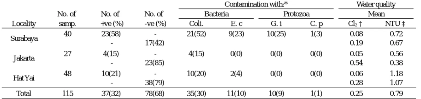
intestinalis and C. parvum were found in 9% and 1% of total samples, respectively. Of those detected, coliform was the most common and was found in all three areas with a mean detection rate of 30% (15-52%). The water samples that were positive for any of the four types of microorganisms showed a tendency to have lower residual chlorine concentrations and higher turbidities compared with negative samples. It is important to supply safe water in order to maintain people’s health because most of the people surveyed (4-88%) ordinarily drank tap water with- out treating it. Continued efforts are needed to maintain and improve drinking water quality. (193 words)
I NTRODUCTION
A safe water supply is advantageous to the health of people. On the other hand, a public water supply will have a serious risk to the health of people if water contamination should occur. Such risks are seen as unexceptional, even in developed countries, where public institutions maintain water quality. Perz et al . (1998) made a risk assessment of cryptosporidiosis in New York City, and estimated that the number of tap-water-related cases per annum was 6 to 34.
Szewzyk et al . (2000) reviewed the microbiological safety of drinking water and described many outbreaks caused by various organisms. The most prominent recorded outbreak occurred in Milwaukee in 1993, involving 403,000 people, with Cryptosporidium parvum as the causative agent (Mac Kenzie et al ., 1994). In addition to this, 703 cases of giardiasis were reported in Massachusetts during 1985 to 1986 (Kent et al ., 1988), and 6,000 cases in six outbreaks of campylobacteriosis during 1992 to 1996 were reported in Sweden (Szewzyk et al ., 2000). But in general, as Dawson
and Sartory (2000) reported, significant advances in water treatment have taken place over the last century and in de- veloped countries massive improvements have been achieved in the microbiological safety of public drinking water supplies. Incidences of illness due to poor treatment or post-treatment contamination are rare in these countries.
However, in areas with low standards of hygiene and sanitation, the contamination of water by microorganism is still common (Luksamijarulkul et al ., 1994). In these coun- tries, mainly tropical, it is difficult to maintain water quality at a safe level because of technical and/or economic prob- lems. For example, more than seven species of parasite were recovered from plant-processed drinking water in Ar- gentina (Basualdo et al ., 2000). In Bangkok, Thailand, 64% of drinking water sources tested were contaminated with coliform (Luksamijarulkul et al ., 1994). In Taiwan, 39% to 77% of treated water samples were positive for Giardia sp. and Cryptosporidium sp. (Hsu et al ., 1999). It appears that the health hazard posed to people who are sup- plied such low quality water is significant. In spite of such
Jpn. J. Trop. Med. Hyg., Vol.31, No.2,2003, pp.87‐91
1. Department of Medical Technology, Faculty of Health Science, Kobe University School of Medicine, Kobe 654-0142, Japan:
2. Water Quality Laboratory of Kobe City Waterworks Bureau, Kobe 652-0004, Japan:
3. Water Quality Center, Maezawa Industry Inc., Saitama 332-8556, Japan:
4. Laboratory of Intestinal Parasites, Tropical Disease Center, and Laboratory of Parasitology, Faculty of Veterinary Medicine, Airlangga Univer- sity, Indonesia:
5. Department of Parasitology, Faculty of Medicine, University of Indonesia, Jakarta 10430, Indonesia:
6. Department of Pathology:
7. Department of Epidemiology Faculty of Medicine, Prince of Songkla University, Thailand.
87
circumstances, little is known about how frequent tap-water is contaminated with microorganisms and how frequent tap water is used in developing countries.
This paper reports the results of a field survey done in Indonesia and Thailand to clarify the relationship between tap water quality and contamination by microorganism. In- formation regarding tap water usage was obtained through questionnaire surveys and the results were analyzed.
M ATERIALS AND M ETHODS 1. Survey areas and period
Surveys were conducted from August 2000 to August 2001 in Surabaya and Jakarta, Indonesia, and in Hat Yai, Thailand. Surabaya is the second largest city in Indonesia and is located in the east of Java Island. Jakarta, located in the west of Java Island, is the capital city of Indonesia con- taining many modern buildings and has a population of 9.1 million. Hat Yai is a city in the south of Thailand near the Thai border with Malaysia. Questionnaire surveys were performed at health centers in each area with the coopera- tion of staff members.
2. Filtration of sample water
Filtration of tap water was performed for C. parvum oocysts and Giardia intestinalis cyst [oocysts of C. parvum and cysts of G. intestinalis are expressed as (oo)cysts].
Sample water was collected directly from a faucet in each house visited. Permission to collect water and confirmation of its origin were recorded. A polycarbonate membrane (142 mm in diameter, 3 µ m pore size; Nucleopore, What- man, USA) was inserted in a filter folder (Advantec KS-142, Tokyo, Japan), and 5 to 50 liters of tap water were filtered from the pipe that was connected directly to a faucet. To es- timate the total filtered volume, the filtered water was kept in a bucket. We did not use any pressure device for the fil- tration, so we were only able to filter a small amount of tap water when the water pressure was low, or when turbidity was high. A filter membrane was put into each of the 50 ml centrifuge tubes that contained 30-40 ml of 1% formalin, kept at room temperature, and transported to Japan.
3. Parasitological measurements
In Japan, each membrane was transferred to a 200 ml beaker containing 100 ml of PBST (PBS with 0.1% of Tween 80), and (oo)cysts on the membrane were eluted by agitation for 2 min by hand and sonication for 2 min using a sonicator (IUC-7321, Tocho co., Tokyo, Japan). The (oo) cyst suspension was again filtered using a polycarbonate membrane (90 mm in diameter and 3 µ m pore size) and transferred to a 10 ml test tube (Dynal, Oslo, Norway) con-
taining 10 ml PBS. The membrane was again treated by sonication and then removed. The remaining solution was further processed by an immunomagnetic separation method (Dynal). The purified and concentrated solution was dried on a slide glass and stained with anti- Cryptosporidium/Giardia monoclonal antibodies conju- gated with fluorescein (Aqua G/C Direct, Waterborne, LA, USA) for 30 min and 6-diamidino-2-phenylindole (DAPI) for 2 min, and the (oo)cysts were then observed under a fluorescent microscope (400- to 440- nm filter in the former, and 330- to 385- nm filter in the latter).
4. Bacterial measurements
Tap water samples were examined for coliform and Es- cherichia coli using a commercially available kit (Colilert, IDEXX, Maine, USA) at the sample collection site. This kit allowed the simultaneous qualitative detection of coli- form and E. coli.
5. Water quality measurements
Water quality was examined simultaneously when each sample was collected. Residual chlorine was measured with the Lovibond 2000 kit (Tintometer Limited, England), and turbidity was measured using a turbidity meter (2100P, Hach Co., Colorado, USA).
6. Questionnaire survey
A questionnaire survey was performed in three areas to investigate number of households equipped with tap water and the frequency of drinking of tap water by local people.
Following our comprehensive explanation of the study and the inhabitants’ consent, we surveyed them on seven items including whether tap water was available in the house, or what type of drinking water was usually used.
R ESULTS 1. Contamination with microorganisms
A total of 115 water samples were examined and 37 (32%) were positive for microorganisms. These microor- ganisms were two kinds of bacteria (coliform and E. coli) and two species of protozoa (G. intestinalis and C. parvum).
Of the microorganisms detected, coliform was the most common and was found in all three areas with a mean de- tection rate of 30% (15-52%). E. coli was found with a mean of 10% (0-23%). Among protozoa, G. intestinalis was found with a mean of 9% (0-25%) (Table 1). The mean number of cysts found in 10 positive samples was 2.5 (1-8).
One C. parvum oocyst was found in one sample (S-1) in
Surabaya (Tables 1 and 2). The intensity of contamination
in the three areas was compared. In Surabaya, which
88showed the highest contamination rate, 58% of tap water samples were positive for microorganisms, and all of the four types of microorganisms recovered through this study were detected (Table 1). In Jakarta 15% of water samples were positive for coliform, and in Hat Yai 20% and 4%
were positive for coliform and E. coli, respectively (Table 1).
2. Microorganism contamination and water quality
Table 2 shows the details of 37 samples that had any of four microorganisms: 23 samples from Surabaya (S), 4 from Jakarta (J), and 10 from Hat Yai (H).
Bacterial contamination was closely related to the con- centration of residual chlorine but protozoan contamination was not. Of the 35 bacteria-positive samples, only one (3%, H-5) had 0.1 mg/l of residual chlorine and the remaining 34 (97%) had a concentration of less than 0.1mg/l. Of the 115 water samples examined, 66 (57%) had less than 0.1mg/l of residual chlorine concentration; of those, 53% were positive for bacteria. In contrast to this G. intestinalis, although only a single sample, was found even in a residual chlorine concentration of 0.6 mg/l. Five positive samples from Sura- baya (S-1, S-9, S-18, S-22, S-23) were contaminated with three kinds of microorganisms (Table 2).
When the positive and the negative samples were com- pared using the parameters of residual chlorine concentra- tion and turbidity, the former had a tendency to have lower residual chlorine concentrations and had higher turbidities than the latter (Table1).
3. Questionnaire survey
One of the aims of this survey was to confirm whether the local people used tap water as a source of drinking water. A total of 352 questionnaires were completed and collected in the three areas examined, and revealed that 80% of the people used tap water. Of the people who had tap water, more than half (54%) drank it without boiling or Table 1 Contamination of tap water with microorganisms
Contamination with:* Water quality
No. of No. of No. of Bacteria Protozoa Mean
Locality samp. +ve (%) -ve (%) Coli. E. c G. i C. p Cl2† NTU‡
Surabaya 40 23(58) - 21(52) 9(23) 10(25) 1(3) 0.08 0.72
- 17(42) 0.19 0.67
Jakarta 27 4(15) - 4(15) 0(0) 0(0) 0(0) 0.05 0.56
- 23(85) 0.54 0.38
Hat Yai 48 10(21) - 10(20) 2(4) 0(0) 0(0) 0.06 1.18
- 38(79) 0.28 1.07
Total 115 37(32) 78(68) 35(30) 11(10) 10(9) 1(1) 0.25 0.79
* E. c: Escherichia coli, G. i: Giardia intestinalis, C. p: Cryptosporidium parvum.
†Less than 0.1 mg/ml is tentatively considered as 0.05 mg/ml and calculated.
‡National Turbidity Unit.
Table 2 Relationship between microorganism contamination and tap water quality
Sample* Water Contaminated with:† Quality of water nos. filtered (L) Coli. E. c G. i C. p Cl2 NTU‡
S-1 70 + - +(2) +(1) <0.1 0.39
S-2 55 + - - - <0.1 0.44
S-3 22 + - - - <0.1 0.46
S-4 25 + - - - <0.1 0.92
S-5 20 + - - - <0.1 1.14
S-6 20 + - +(1) - <0.1 0.63
S-7 14 - - +(1) - 0.6 0.83
S-8 15 - - +(4) - <0.1 1.06
S-9 30 + + +(2) - <0.1 0.36
S-10 ND + + ND ND <0.1 1.85
S-11 ND + + ND ND <0.1 0.38
S-12 ND + + ND ND <0.1 0.57
S-13 ND + - ND ND <0.1 0.44
S-14 25 + - +(3) - <0.1 0.56
S-15 40 + - - - <0.1 0.53
S-16 30 + + - - <0.1 0.65
S-17 30 + - +(1) - <0.1 0.57
S-18 40 + + +(8) - <0.1 0.48
S-19 30 + - - - <0.1 0.6
S-20 8 + - - - <0.1 0.56
S-21 ND + + ND ND <0.1 1.28
S-22 50 + + +(2) - <0.1 0.61
S-23 30 + + +(1) - <0.1 0.93
J-1 ND + - ND ND <0.1 0.4
J-2 15 + - ND ND <0.1 0.52
J-3 50 + - - - <0.1 0.92
J-4 40 + - - - <0.1 0.4
H-1 50 + - - - <0.1 3.13
H-2 50 + - - - <0.1 0.66
H-3 50 + + - - <0.1 1.36
H-4 50 + - - - <0.1 0.86
H-5 50 + - - - 0.1 1.72
H-6 50 + + - - <0.1 1.57
H-7 50 + - - - <0.1 0.85
H-8 50 + - - - <0.1 0.71
H-9 50 + - - - <0.1 0.53
H-10 50 + - - - <0.1 0.94
*Samples were collected from Surabaya (S), Jakarta (J), and Hat Yai (H).
†Coli.: coliform, E. c: Escherichia coli, G. i: Giardia intestinalis, C. p: Crypto- sporidium parvum.
‡National Turbidity Unit.
89
filtering it. This rate varied from area to area: 88% in Sura- baya but only 4% in Hat Yai. Those who did not have tap water to the house used bottled water for drinking, but at the same time, 40-55% of them also used well water (Table 3).
D ISCUSSION
A safe water supply system is a “double-edged sword”.
If this system functions well, it can maintain the health of a population. But if not, large public health problems occur (Dawson and Sartory, 2000; Szewzyk et al ., 2000). In both developed countries where urbanization or centralization of the population has progressed, and developing countries where water quality is not maintained, such apprehensions are present. Unfortunately, the water quality found in this study was poor. As Steiner et al . (1997) stated, the leading cause of infant mortality in the developing world is infec- tious diarrhea, and the prevalence of the diarrhea pathogen is largely influenced by the quality of clean water available for drinking. Recent guidelines and legislation of the Euro- pean Union Council Directive 98/83/EC and the World Health Organization stated that drinking water should be safe and not contain pathogenic microorganisms, or contain only such low numbers that the risk for acquiring water- borne infections is below an acceptable limit (Szewzyk et al ., 2000).
Four kinds of microorganisms were detected through- out the survey. Most of these microorganisms have an ani- mal reservoir from which they are transmitted to humans, directly or via the environment. These microorganisms are resistant to environmental stress and have a very low infec- tive dose. It appears that even low-level contamination of water supply systems may lead to infections and disease in the exposed population (Szewzyk et al ., 2000).
Emerging and reemerging infectious parasites in drink- ing water have become increasingly important during the last few decades. These include newly recognized protozoa
such as C. parvum and Cyclospora cayetanensis, or G. in- testinalis known as traveler’s diarrhea (Rose and Slifko, 1999). Since C. parvum and G. intestinalis have a toler- ance to chlorine, they sometimes become causative agents of waterborne infectious diseases. Among these protozoa, the detection of C. parvum oocysts from water samples is very difficult because the oocyst is small, has little morpho- logical character and, more importantly, is only seen in small numbers in water samples. In the United States and Britain, methods for detecting oocysts from water have been reported (Fricker and Crabb, 1998). The method we devel- oped for this survey, which is simple and highly efficient in recovering oocysts, is suitable for use in developing coun- tries. A notable feature of this method is that it does not in- volve a centrifugation process during isolation because we experienced that centrifugation reduced the recovery rate.
E. coli is a bacterial indicator of faecal contamination.
In recent years, enterococci, faecal bacteria of the genera Enterococcus and Streptococcus, have also been given sig- nificant recognition as faecal indicators (Dawson and Sar- tory, 2000), though we did not examine them. It is reported that 0.2 mg/l of residual chlorine can kill 90% of E. coli within two hours (Hsu et al ., 1984). We suspect that the shortage of residual chlorine concentration was one cause for the bacterial contamination of tap water. There are many factors contributing to the decline of residual chlorine concentration in tap water. In this survey, the reason of low chlorine concentrations may be the insufficient removal of organic matter at waterworks, based on relatively high tur- bidities of tap water and river water from which the tap water is derived. When flood damaged Hat Yai City in 2000, we revealed that the mean residual chlorine concentration in 30 tap water samples was maintained at 0.5 mg/l (data not shown). That is, the public works adjustment of the chlo- rine concentration according to the situation occurred.
However, during the usual operation of the water works in the same city in 2001, 0.1 mg/l or less chlorine concentra- tion was frequently observed. Therefore, we cannot deny Table 3 Utility of water by people in three different areas as revealed by questionnaire survey
With water supply system Without water supply system No. of ques-
tionnaires
Drink tap
water without Sometimes
we drink: (%) No. (%) Sometimes we drink: (%) Locality recovered No. (%) boiling/filtering (%)
Jakarta 100 100 (100) 67 (67) bottled water (20) 0 (0)
(Indonesia) well water (7)
Surabaya 102 91 (89) 80 (88) bottled water (31) 11 (11) well water (55)
(Indonesia) well water (1) bottled water (36)
Hat Yai 150 90 (60) 4 (4) bottled water (61) 60 (40) well water (40)
(Thailand) well water (18) bottled water (13)
Total 352 281 (80) 151 (54) Bottled and well
water 71 (20) Well and bottled
water 90
the possibility of mishandled operations at the water purifi- cation plant.
Of the three areas studied, Surabaya City had the low- est water quality. Despite this, 88% of people drank tap water without treating it. As a primary prevention, an im- mediate improvement of the water-purifying environment, as well as the education of the populace, are needed. The spread rate of water supply system in Hat Yai was the low- est (60%). But at the same time, the number of the people who drank tap water without treating it was the lowest. We thought that this was probably due to knowledge accumu- lated through the experience of the local people.
Poorly managed public water supplies have the poten- tial to make a large number of people ill (Dawson and Sar- tory, 2000). Therefore, we have to continue to put efforts in supplying of safe drinking water and to educate the people who use it.
R EFERENCES
1)Basualdo, J., Pezzani, B., Luca, M.D., Cordoba, A. and
Apezteguia, M. (2000): Screening of the municipal water system of La Plata, Argentina, for human intestinal para- sites. Int. J. Hyg. Environ. Health, 203, 177-182
2)Dawson, D.J. and Sartory, D.P. (2000): Microbiology
safety of water. Br. Med. Bull., 56, 74-83
3)Fricker, C.R. and Crabb, J.H. (1998): Water-born crypto-
sporidiosis: detection methods and treatment options.
Adv. Parasitol., 40, 241-278
4)Hsu, S.C., Martin, R. and Wentworth, B.B. (1984): Isola-
tion of Legionella species from drinking water. Appl. En- viron. Microbiol., 48, 830-832
5)Hsu, B.M., Huang, C. and Jiang, G.Y. (1999): The preva-
lence of Giardia and Cryptosporidium in Taiwan water supplies. J. Toxicol. Environ. Health, Part A, 56, 149-160
6)Kent, G.P., Greenspan, J.R., Herndon, J.L., Mofenson, L.M., Harris, J. S., Eng, T.R. and Waskin, A.A. (1988): Epi- demic giardiasis caused by a contaminated public water supply. Am. J. Public Health, 78, 139-143
7)Luksamijarulkul, P., Pumsuwan, V. and Pungchitton, S.
(1994): Microbiological quality of drinking water and us- ing water of a Chao Phya River community, Bangkok.
Southeast Asian J. Trop. Med. Public Health, 25, 633-637
8)Mac Kenzie, W.R., Hoxie, N.J., Progtor, M.E., Gradus,M.S., Blair, K.A., Peterson, D. E., Kazmierczak, J.J. and Davis, J.P. (1994): A massive outbreak of Cryptosporid- ium infection transmitted through the public water supply.
N. Engl. J. Med., 331, 161-167
9)Perz, J.F., Ennever, F.K. and Le Blancq, S.M. (1998):
Cryptosporidium in tap water: comparison of predicted risks with observed levels of disease. Am. J. Epidemiol., 147, 298-301
10)Rose, J.B. and Slifko, T.R. (1999): Giardia, Crypto-
sporidium, and Cyclospora and their impact on foods: a review. J. Food Prot., 62, 1059-1070
11)Steiner, T.S., Thielman, N.M. and Guerrant, R.L. (1997):
Protozoal agents: what are the dangers for the public water supply? Annu. Rev. Med., 48, 329-340
12)Szewzyk, U., Szewzyk, R., Manz, W. and Schleifer, K.H.
(2000): Microbiological safety of drinking water. Annu.
Rev. Microbiol., 54, 81-127
91
A RAPID SINGLE-STEP SCREENING METHOD FOR GLUCOSE-6-PHOSPHATE DEHYDROGENASE
DEFICIENCY IN FIELD APPLICATIONS
K
UNII
WAI1, H
IROYUKIM
ATSUOKA1, F
UMIHIKOK
AWAMOTO2, M
EIJIA
RAI1, S
HIGETOY
OSHIDA1, M
AKOTOH
IRAI1and A
KIRAI
SHII1Accepted 25,
June
, 2003Abstract. The single-step screening method (SSS) is a qualitative rapid screening test for glucose-6-phosphate de- hydrogenase (G6PD) deficiency based on blue formazan formation on anion-exchanger. The reaction mixture con- tains equal volumes of anion exchanger, substrate mixture, coloring mixture (MTT-PMS mix) and distilled water.
We assessed the stability of the reaction mixture and evaluated its reliability with two anion exchangers, DEAE- Sephadex A-50
TMand DEAE-Sephacel
TM, for applications in tests under field conditions. The reaction mixture was sufficiently stable under conditions of incubation at 70° C for 6 hours or vigorous shaking for 24 hours at room tem- perature. The reaction mixture could be kept at 30-35° C for 14 days under indoor conditions without shielding if it contained no MTT-PMS mix. The coloring was detectable even in diluted blood with hemoglobin concentration as low as 1.6 g/dl. Under laboratory conditions, the proportion of the samples with 10% of the normal level of activ- ity that were diagnosed as ‘low activity’ was higher with DEAE-Sephacel (92%) than with DEAE-Sephadex A-50 (81%) (p=0.023). The proportion of the samples with normal activity that were diagnosed as ‘normal’ was 98%
with DEAE-Sephacel and 100% with DEAE-Sephadex A-50. In field samples obtained in Myanmar and Indonesia, the sensitivity was lower (P=0.03 using DEAE-Sephadex and p< 0.001using DEAE-Sephacel) when we used the blue formazan spot test (BFST) as the standard. Twenty-three of 27 G6PD-deficient individuals subjected to ge- netic analysis were found to have mutations. All individuals who had concordant results between the SSS and the blue formazan spot test (BFST) carried molecular mutations. One case of G6PD mutation was detected among four cases diagnosed as G6PD-deficient by SSS with DEAE-Sephacel
TM, but diagnosed as ‘normal’ by BFST. The costs of one test with the DEAE-Sephadex A-50
TMand the DEAE-Sephacel
TMsystem were 0.15 US dollar and 0.30 US dollar, respectively. (297 words)
Key words: Single-step screening test, Glucose-6-phosphate dehydrogenase deficiency, Device approval, Field trial, Malaria
I NTRODUCTION
Glucose-6-phosphate dehydrogenase (G6PD) defi- ciency is a frequent and heterogeneous X-chromosome- linked enzyme abnormality. As G6PD plays a key role in maintaining erythrocytes, G6PD deficiency possibly results in acute hemolysis after exposure to various oxidative stresses, including infections, medications, and fava beans (favism). A striking correlation is observed between the prevalence of G6PD deficiency and historical malaria en- demicity, particularly in tropical and sub-tropical areas (W.
H.O. Working Group, 1989). Therefore, it is important to detect and inform G6PD-deficient individuals in and from areas in which malaria is endemic before exposing such in- dividuals to oxidative stress in order to avoid acute hemo-
lytic attack, especially hemolytic attack caused by pri- maquine.
For field screening of G6PD deficiency, the test used should be simple to perform and affordable. It is also ad- vantageous if the test reagents can be stored and the reac- tion can be carried out at around room temperature, particu- larly in areas with an insufficient supply of electricity.
Several tests, including the G6PD spot test (Fairbanks and Beutler, 1962), fluorescent spot test (Beutler, 1966), and blue formazan spot test (Fujii et al ., 1984) have been developed for field screening of G6PD deficiency; however most of them are complicated or expensive. One of the tests, the blue formazan spot test (BFST) (Fujii et al ., 1984;
Pujades et al ., 1999), has been used for field studies be- cause of its comparatively simple procedure and sufficiently
Jpn. J. Trop. Med. Hyg., Vol.31, No.2,2003, pp.93‐97
1 Department of Medical Zoology, Jichi Medical School, Tochigi 329-0498, Japan
2 Department of International Health, Nagoya University School of Medicine, Nagoya 466-0064, Japan
93
low cost. However, BFST requires 8 hours of incubation for diagnosis, which makes immediate on-site diagnosis diffi- cult.
Recently, a simple and quick screening method, a single-step screening test (SSS) was developed (Hirono et al ., 1998). Our previous trials showed the usefulness of SSS in field studies (Tantular et al ., 1999), but the stability and reliability of the test have not been fully assessed. In this study, we assessed in detail the stability of SSS and the results of the reliability of SSS use under field conditions.
M ATERIALS AND M ETHODS
SSS is a qualitative test that assesses the formation of blue formazan along with the reduced form of nicotinamide adenine dinucleotide phosphate (NADPH) by G6PD ab- sorbed on an anion exchanger. The reaction mixture con- sists of equal volumes (200 µ l each) of substrate mixture containing 5 mM glucose-6-phosphate (G6P) and 0.4 mM oxidized nicotinamide adenine dinucleotide phosphate (NADP), coloring mixture (MTT-PMS mix) containing 0.025% 3 (4,5 dimethylthiazolyl 1-2) 2,5 diphenyltetra- zolium bromide (MTT), 0.025% phenazine methosulphate (PMS), anion exchanger (originally DEAE-Sephadex A- 50
TM) and distilled water. The test can detect G6PD- deficient individuals with less than 10% residual activity within 40 minutes without any special equipment.
The procedures and chemicals used for SSS (Hirono et al ., 1998) and BFST (Fujii et al ., 1989) were as described, with slight modifications in the procedure for SSS: 0.05%
sodium azide (NaN
3) was added and saponin was omitted in the reaction mixture to avoid the growth of microorganisms.
We assessed the reliability of two different anion exchang- ers in SSS: translucent DEAE-Sephadex A-50
TMand white- colored DEAE-Sephacel
TM(Amersham Pharmacia Biotech, Buckinghamshire, UK). When using the DEAE-Sephacel
TMsystem, 4.8mM of oxidative glutathione (GSSG) was added to optimize the reactivity (Hirono et al ., 1998).
The blood samples for laboratory assessment were drawn from one of the authors who was confirmed pheno- typically and genotypically to have normal G6PD. The G6PD gene was analyzed by direct sequencing of all coding exons. The G6PD-deficient blood was artificially made from the blood of the same volunteer by heat inactivation at 56° C for 15 minutes and proved to have no residual G6PD activity. Nine volumes of heat-inactivated blood and one volume of fresh blood were mixed to make G6PD-deficient samples with 10% of the normal activity. The samples for field comparison and genetic analysis were obtained from local residents who participated in an extensive field study with the aim of quick detection of malaria and G6PD defi-
ciency (Tantular et al . 1999) in the Taninthayi Division in Myanmar and on Buru Island in Indonesia, 1998. Prior to the study, the outline and procedures of this research were discussed within the committees of the national and local governments in Indonesia and Myanmar. The participants were orally informed of and gave consent to the examina- tion twice before screening and blood drawing. We did not obtain written informed consent from each participant be- cause of the opinion of local co-organizers about the partici- pants’ literacy and differences of local culture. The field samples were used to assess the sensitivity and specificity of SSS using BFST as the standard. We also compared the
‘G6PD-deficient’ samples of SSS with BFST, using genetic analysis as the standard.
To assess the stability of the test mixtures of SSS un- der various laboratory conditions, we simulated three differ- ent conditions which may often occur during field studies in tropical areas: 30-35° C for 14 days (storage at room tem- perature), 70° C for 6 hours in an oven (being left in a car), continuous shaking using a shaker (TAITEC R-1) at maxi- mum strength (200 rotations/minute) for 24 hours at room temperature (transporting over a rough surface). We also examined the photosensitivity of the test, because the dis- solved MTT-PMS mixture is highly photosensitive (Fair- banks and Beutler, 1962). For simulating anemia, which is a common illness and may affect the results (Fairbanks and Beutler, 1962), serial two-fold dilutions of blood with nor- mal G6PD activity from the same volunteer in other labora- tory tests were made with phosphate buffered saline (PBS) to one-eighth of the original concentration (the estimated hemoglobin concentration was then 1.6 g/dl). For all of the assessments, the combination of newly prepared testing mixture and fresh blood was used for the standard of the normal reaction.
For the evaluation of reliability, the results were cate- gorized into two groups, ‘low activity’ and ‘normal activity’, based on color development of the anion-exchanger. Two investigators who were blinded to the actual activity of sam- ples then evaluated the enzyme activity (50 deficient and 50 normal per person) independently as ‘low activity’ or ‘nor- mal activity’. The results were analyzed by the chi-square test using a 2×2 table or Fisher’s exact test. P values less than 0.05 were considered as statistically significant.
BFST was used as the standard for field assessment of the sensitivity and specificity of SSS. The SSS was per- formed at each field site and samples for BFST were ob- tained on cation-exchange paper (Whatman P-81) (Fujii et al ., 1984) at the same time and dried. The BFST was per- formed within the same day, and thus the results were ob- tained the next day. The results were analyzed statistically as described above.
94
For comparison of the results of SSS and BFST with the results of genetic analysis, the participants were screened by SSS, and a blood spot was obtained in the same way as described above. The participants were diagnosed with G6PD deficiency based on the results of SSS, and their blood was drawn for genetic analysis. The procedures for genetic analysis were described previously (Hirono et al ., 1994; Hirono et al ., 1997, Iwai et al ., 2001).
R ESULTS AND D ISCUSSION
The reaction mixture of SSS retained sufficient reactiv- ity compared with the control mixture after exposure to 70° C for 6 hours or continuous shaking for 24 hours at room temperature. Reaction mixture without the MTT-PMS mix could be stored at 30-35° C for 14 days under indoor condi- tions without shielding. These features would make this test appropriate for regular use in primary care and in field studies in rural areas in developing countries. A previously reported 1-year storage trial (Hirono et al ., 1998) showed that DEAE-Sephadex
TMcan be kept at room temperature, the MTT-PMS mix can be stored at 4° C, and the substrate mixture is stable at −20° C. If a refrigerator and freezer are available, each mixture can be safely stored for at least1year.
In our simulation of anemia, the reaction mixture yielded detectable color development when the estimated hemoglobin concentration of the G6PD-normal blood was as low as 1.6 g/dl. Because elevated levels of reticulocytes lead to higher activity of peripheral blood G6PD in the ane- mic condition (Jansen et al ., 1985), our finding implies that the G6PD-normal samples of patients with severe anemia should not be frequently misdiagnosed as ‘G6PD-deficient’.
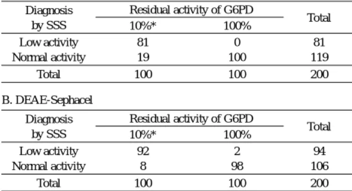
Table 1 shows the reliability of the 2 anion-exchangers.
Under laboratory conditions, a higher proportion of the samples with 10% of normal activity were diagnosed as
‘low activity’ with DEAE-Sephacel
TM(92%) than with DEAE-Sephadex A-50
TM(81%) (p=0.023). With the DEAE-Sephadex A-50
TMsystem, nearly 20% of G6PD- deficient samples were judged as normal. The red-colored hemoglobin in the reaction mixture together with the trans- lucent DEAE-Sephadex A-50
TMparticles may cause misdi- agnosis. The proportion of the samples with normal activity that were diagnosed as ‘normal’ was 98% with DEAE- Sephacel
TMand 100% with DEAE-Sephadex A-50
TM.
The sensitivity and specificity in field tests are shown in Table 2. We used BFST as the standard because a previ- ous study using the samples of genetically diagnosed G6PD-deficient patients showed that BFST had 100% sen- sitivity in hemizygotes and 75% in heterozygotes and 100%
specificity (Pujades et al ., 1999). The sensitivity was sig- nificantly higher in males than in females when we use DEAE-Sephacel as anion-exchanger (P=0.04). Neither the sensitivity nor specificity differed significantly between DEAE-Sephadex
TMand DEAE-Sephacel
TM.
Comparison of field testing with laboratory assessment revealed that the sensitivity of field testing was lower than that of laboratory assessment. The P-value was 0.03 using DEAE-Sephadex A-50 and less than 0.001 using DEAE- Sephacel. We found no difference in specificity. In field assessment, the sensitivity was significantly higher among males when we used DEAE-Sephacel (p=0.04). The actual value of the sensitivity was also high among males tested with DEAE-Sephacel, although there was no significant dif- ference (p=0.12). We suppose the discrepancy between laboratory and field assessments may be due to 3 reasons.
One is the difference of genetic status between males and females. Because G6PD deficiency is an X-chromosome- linked abnormality, the enzyme activity is particularly vari-
Table 1. Laboratory evaluation of the single-step screening method (SSS) with 2 anion exchangers
A. DEAE-Sephadex A-50 Diagnosis
by SSS
Residual activity of G6PD
Total
10%* 100%
Low activity Normal activity
81 19
0 100
81 119
Total 100 100 200
B. DEAE-Sephacel Diagnosis
by SSS
Residual activity of G6PD
Total
10%* 100%
Low activity Normal activity
92 8
2 98
94 106
Total 100 100 200
*The proportion is significantly different between the DEAE-Sephadex A-50 sys- tem and DEAE-Sephacel system
Table 2. Sensitivity and specificity of the SSS using BFST as a standard
A. DEAE-Sephadex A-50 Diagnosis
by BFST
Diagnosis by SSS (male/female) Total (male/female) G6PD deficiency G6PD- normal
G6PD deficiency G6PD normal
20(18/2) 3(2/1)
12(8/4) 187(96/91)
32(26/6) 190(98/92)
Total 23(20/3) 199(104/95) 222(124/98)
Sensitivity: male 69% (18/26), female 33% (2/6) Specificity:male 98% (96/98), female 99% (91/92) B. DEAE-Sephacel
Diagnosis by BFST
Diagnosis by SSS (male/female) Total (male/female) G6PD deficiency G6PD- normal
G6PD deficiency G6PD normal
17(13/4) 9(5/4)
9(3/6) 354(171/183)
26(16/10) 363(176/187) Total 26(18/8) 363(174/189) 389(192/197) Sensitivity: male 81% (13/16), female 40% (4/10)*
Specificity:male 97% (171/176), female 98% (183/187)
*Significantly different (P=0.04)
95
able in heterozygous females.
The crude G6PD level of blood may also be affected by anemia caused by various illnesses. Variable Hb levels together with various levels of G6PD activity in field sam- ples could partially explain the discrepancy of these 2 tests, although our laboratory assessment showed that the G6PD- deficient samples could be distinguished from G6PD- normal samples.
We also consider it possible that the discrepancy be- tween two tests may be due to inaccurate evaluation by SSS.
The definite cut-off level of coloring should be assessed by comparison of BFST and SSS using genetically confirmed samples.
Comparison of the results of SSS and BFST with the results of genetic analysis showed that 23 of 27 G6PD- deficient individuals detected by SSS, including two het- erozygous females, had mutations (Table 3). All samples for which the results of SSS coincided with those of BFST were shown to have mutations, regardless of the anion ex- changer used for the analysis. One case of class 2 G6PD mutation, 383 T→C (G6PD Vanua Lava), was detected among four cases that were diagnosed as ‘G6PD-deficient’
using SSS with DEAE-Sephacel
TMbut diagnosed as ‘G6PD normal’ with BFST. This result suggests that some cases of G6PD deficiency may be undetectable using BFST, al- though a study showed that the G6PD-deficient hemizy- gotes could be distinguished from heterozygotes and nor- mal controls using BFST (Pujades et al ., 1999).
Including all reagents and disposable supplies, the esti- mated cost for one test of SSS with DEAE-Sephadex A- 50
TMwas approximately half that of a test of SSS with the DEAE-Sephacel
TM(0.15 US dollar and 0.30 US dollar, re- spectively). The cost of both of these systems is below an acceptable limit.
SSS is a quick, simple and reliable screening test for G6PD deficiency, although a larger field study is necessary for more precise evaluation of SSS. All the procedures in this test could be completed in field conditions without any electric equipment except during some preparations prior to the study. Using either anion exchanger, it can be used for quick detection of G6PD deficiency in various situations in- cluding field surveys, mass-treatment of malaria and differ-
ential diagnosis of hemolytic anemia, particularly in areas with substandard laboratory conditions. Although the white- colored DEAE-Sephacel
TMgives better reaction visibility, the DEAE-Sephadex A-50
TMsystem is more cost-effective, and is therefore preferable financially, particularly for field studies.
A CKNOWLEDGEMENTS
The authors are indebted to Dr. Akira Hirono (Okinaka Memorial Institute for Medical Research) for valuable sug- gestions and critical reading of the manuscript. This study was supported by research grants from the Japanese Minis- try of Education, Science, Culture and Sports (Grant No. 10041205 granted to Matsuoka, and 09041179 to Kawamoto). It was also supported by Toyota Foundation (Grant No. 96B3-011, to Kawamoto), the large-scale coop- erative program (No. 2) from Japan Society for Promotion of Science (to Kawamoto), and a grant for International Health Cooperation Research from the Ministry of Health, Labour and Welfare (Grant No. 13-A-5, to Ishii).
R EFERENCES
1)Beutler E: A series of new screening procedures for pyru-
vate kinase deficiency, glucose-6-phosphate dehydroge- nase deficiency and glutathione reductase deficiency.
Blood 28: 553-562, 1966.
2)Fairbanks, V.F., and Beutler, E. (1962): A simple method
for detection of erythrocyte glucose-6-phosphate dehy- drogenase deficiency (G6PD Spot Test). Blood, 20, 591- 601
3)Fujii, H., Takahashi, K. and Miwa, S. (1984): A new sim-
ple screening method for glucose-6-phosphate dehydro- genase deficiency. Acta Haematol. Jpn., 47, 185-188
4)Hirono, A., Fujii, H. and Miwa, S. (1998): An improvedsingle-step screening method for glucose-6-phosphate dehydrogenase deficiency. Jpn. J. Trop. Med. Hyg., 26, 1-4
5)Hirono, A., Miwa, S., Fujii, H., Ishida, F., Yamada, K.
and Kubota, K. (1994): Molecular study of eight Japa- nese cases of glucose-6-phosphate dehydrogenase defi- ciency by nonradioisotopic single-strand conformation polymorphism analysis. Blood, 833, 3363-3368
6)Hirono, A., Fujii, H., Takano, T., Chiba, Y., Azuno, Y. and
Miwa, S. (1997): Molecular analysis of eight biochemi- cally unique glucose-6-phosphate dehydrogenase variants found in Japan. Blood 1997; 89: 4624-4627
7)Iwai, K., Hirono, A., Matsuoka, H., Kawamoto, F., Horie,
T., Lin, K., Tantular, I.S., Dachlan, Y.P., Notopuro, H., Hidayah, N.I., Salim, A.M.A., Fujii, H., Miwa, S. and Ishii, A. (2001): Distribution of Glucose-6-phosphate de- hydrogenase mutations in Southeast Asia. Hum. Genet.
2001; 108: 445-449 Table 3. The results of genetic analysis and the BFST of
‘G6PD-deficient’ samples diagnosed by SSS
Diagosisby BFST
DEAE-Sephadex A-50 DEAE-Sephacel Analyzed Variants Analyzed Variants Deficient
Normal
17 1
17(1) 0
5 4
5(1) 1
Total 18 17(1) 9 6(1)
Heterozygotes in parentheses 96
8)Jansen G, Koenderman L, Rijksen G, Cats BP, Staal GEJ:
Characteristics of hexokinase, pyruvate kinase, and glucose-6-phosphate dehydrogenase during adult and neonatal reticulocyte maturation. Am. J. Hematol. 20:
203-215, 1985.
9)Pujades, A., Lewis, M., Salvati, A.M., Miwa, S., Fujii, H.,
Zarza, R., Alvarez, R., Rull, E., and Corrons, J.L.V.
(1999): Evaluation of the blue formazan spot test for screening glucose 6 phosphate dehydrogenase deficiency.
Int. J. Hematol., 69, 234-236
10)Tantular, I.S., Iwai, K., Lin, K., Basuki, S., Horie, T.,
Htay, H.H., Marwoto, H., Wongsrichanalai, C., Dachlan, Y.P., Kojima, S., Ishii, A. and Kawamoto, F. (1999): Field trials of a rapid test for G6PD deficiency in combination with a rapid diagnosis of malaria. Trop. Med. Int. Health, 4, 245-250
11)W.H.O. Working Group (1989): Glucose-6-phosphate de-
hydrogenase deficiency. Bull. W.H.O., 67, 601-611
97
Short communication
NATURAL INFECTIONS WITH FILARIAL LARVAE IN TWO SPECIES OF BLACK FLIES (DIPTERA:
SIMULIIDAE) IN NORTHERN THAILAND
M
ASAKOF
UKUDA1,2, W
EJC
HOOCHOTE3, O
DILEB
AIN4, C
HIHARUA
OKI2and H
IROYUKIT
AKAOKA2Accepted 18,
July
, 2003Abstract: To find out the natural infection with filarial larvae, female adult black flies were collected on a human attractant in December, 2001 at Tambol Ban Laung (altitude 750 m), Doi Inthanon National Park, in northern Thai- land. The total number of females collected was 823: of which 557 (67.7%) were identified as Simulium asakoae Takaoka et Davies, 144 (17.5%) as S. nigrogilvum Summers, 97 (11.8%) as S. nakhonense Takaoka et Suzuki and 25 (3%) as other six simuliid species. By dissections, eight third- and one second-stage larvae of unidentified filar- ial species were found in one of 138 S. nigrogilvum and one of 484 S. asakoae, respectively. Non-filarial nema- todes were found in 1.03% (5/484) of S. asakoae. This is the first report of natural infections of two black-fly spe- cies, S. nigrogilvum and S. asakoae, with a filarial larva.
Key words: black fly, filaria, natural infection, Onchocerca, Simuliidae, Thailand
I NTRODUCTION
Simuliidae or black flies have been well known to be a pest of humans and animals and also vectors of some para- sites and pathogens (Crosskey, 1990). The main medical significance is the transmission of Onchocerca volvulus to humans by the bites of the flies in Africa and Central and South America; certain simuliid species are suspected to be a probable vector of zoonotic onchocerciasis which occurs sporadically in Japan, North America and Europe (e.g., Hashimoto et al., 1990; Takaoka et al., 1996).
In the Oriental Region, several man-biting simuliid species have been reported, e.g., Simulium asishi Datta, S.
himalayense Puri, S. indicum Becher, S. japonicum Matsu- mura, S. nodosum Puri and S. tenuistylum Datta (Datta, 1992; Lewis, 1974; Takaoka, 1977). Several other species have been known as a pest of animals (Datta, 1992; Datta and Dasgupta, 1975; Friederichs, 1925). Recently, we ex- amined adult female black flies captured on humans and water buffalos at Ban Pang Fan (250 m in altitude), Chiang Mai Province, in northern Thailand, and reported for the
first time the natural infections of S. nodosum with Oncho- cerca larvae (Takaoka et al ., 2003).
To get further information on natural filarial infections of black flies in northern Thailand, we collected adult black flies at Tambol Ban Laung (750 m in altitude) in Chiang Mai Province. Here we report two more black-fly species naturally infected with a filarial larva.
M ATERIALS AND M ETHODS
Study area
Adult black flies were collected at a site exposed to the sun in the village of Tambol Ban Laung (18° 25’-18° 37’ N and 98° 27’-98° 42’ E: ca. 750 m in altitude), Doi Inthanon National Park, Chiang Mai Province, northern Thailand.
Collection of adult black flies
The collection was made on 16 December of 2001, for 12 hr from 06.00 to 18.00 hours using a human attractant (WC, one of the authors) with his legs below the knees ex- posed. Female black flies landing on or flighting around the
Jpn. J. Trop. Med. Hyg., Vol.31, No.2,2003, pp.99‐102
1 Institute of Scientific Research, Oita Medical University, Oita, Japan
2 Department of Infectious Diseases, Oita Medical University, Oita, Japan
3 Department of Parasitology, Faculty of Medicine, Chiang Mai University, Chiang Mai, Thailand
4 Parasitologie comparée et Modèles expérimentaux, associé à l’INSERM (U567), et École pratique des Hautes Études, Muséum National d’His- toire Naturelle, Paris, France
Correspondence: M. Fukuda, Department of Infectious Diseases, Oita Medical University, Hasama, Oita 879-5593, Japan. E-mail: mfukuda@oita -med.ac.jp
99