Glossanodon microcephalus, a New Argentine Fish from Japan and the South China Sea (Protacanthopterygii: Argentinidae)
Hiromitsu Endo
1and Kazuya Nashida
21
Laboratory of Marine Biology, Faculty of Science, Kochi University, 2–5–1 Akebono-cho, Kochi 780–8520, Japan
E-mail: endoh@kochi-u.ac.jp
2
Kochi Kuroshio Research Laboratory, National Research Institute of Fisheries Science, Fisheries Research Agency (FRA), 6–1–21 Sanbashi-dori, Kochi 780–8010, Japan
E-mail: knashi@affrc.go.jp
Abstract A new argentinid species, Glossanodon microcephalus, is described based on 30 speci- mens (53–97 mm SL) from Tosa Bay, off Kochi Prefecture, Shikoku Island, southern Japan, and from the southwest South China Sea. The new species clearly differs from its congeners in having the following combination of characters: dorsal-¿n rays 11–12; anal-¿n rays 10–12; pectoral-¿n rays 17–19; pelvic-¿n rays 10–12; branchiostegal rays 5; gill-rakers on ¿rst arch 27–30; vertebrae 44–46; small conical teeth in 1–2 and 1–3 irregular rows on vomer and palatines respectively;
10–21 small conical teeth on lower jaws, and 3–7 on tip of tongue; head length 25–27% SL; snout length 30–33% HL; anus immediately anterior to anal-¿n origin; a longitudinal black stripe above lateral line interrupted, alternately composed of short bars and faint blotches; dense pigmentation just behind chin; no melanophore on isthmus to abdomen; maturation at small size (the minimum sizes of mature males and females are 65 mm SL and 72 mm SL respectively). The new species is a benthopelagic dweller on muddy and sandy bottoms in depths of about 100–200 m.
Key words: Argentinidae, Glossanodon, new species, Japan, South China Sea, precocious.
Argentines of the genus Glossanodon Guichenot, 1867 are small benthopelagic ¿shes (attaining ca. 7–20 cm SL), occurring on offshore bottoms and sea mounts of tropical to temperate waters in world oceans (Kobilyansky, 1998). The genus is composed of 14 known species, classi-
¿ed into two subgenera: Glossanodon Guichenot, 1867 and the monotypic Prosoarchus Cohen, 1958 containing Glossanodon pygmaeus Cohen, 1958 (Endo and Nashida, 2010). Further, the 13 species of the subgenus Glossanodon are tenta- tively divided into three species groups by Kobi- lyansky (1998), and one species recently described by Endo and Nashida (2010): “polli”
group (8 species), “leioglossus” group (2 spe- cies), and “lineatus” group (3 species). Of these, three Japanese species, Glossanodon kotakamaru Endo and Nashida, 2010, Glossanodon semifasciatus (Kishinouye, 1904), and Glossanodon lineatus
(Matsubara, 1943) belong to the “polli” group,
“leioglossus” group, and “lineatus” group respec- tively.
During bottom trawl surveys by the R/V
Kotaka-maru in central Tosa Bay from 2007 to
2010, and from sampling at the Mimase ¿sh mar-
ket of Kochi City in March of 2010, we obtained
about 100 small specimens of a Glossanodon
species (46–97 mm SL). While somewhat similar
to the young of G. semifasciatus, they differ from
their congeners in having a smaller head, a
shorter snout, an interrupted longitudinal dark
stripe above the lateral line, and lower counts of
all ¿n-rays, gill-rakers, and vertebrae. In addi-
tion, most specimens about 70 mm SL and larger
were mature, suggesting that this is a small Glos-
sanodon species. Subsequent to collecting the
specimens, we found three specimens of Glos-
sanodon (72–78 mm SL) deposited in the ¿sh
collection of Kochi University (BSKU) that were collected by the R/V Hakuho-maru from the South China Sea in 1972 (Fig. 1). These subse- quent specimens were considered conspeci¿c with the specimens recently collected from Tosa Bay and from the Mimase ¿sh market, and we herein describe this new species.
Materials and Methods
Specimens examined are deposited in the fol- lowing institutions: Australian Museum, Sydney (AMS); Laboratory of Marine Biology, Faculty of Sciences, Kochi University (BSKU); National Museum of Nature and Science (NSMT, for- merly National Science Museum, Tokyo). Otter trawls by the R/V Kotaka-maru (NRIFS:
National Research Institute of Fisheries Science, Japan) in central Tosa Bay were planned and operated by K. Nashida (NRIFS).
Counts and measurements follow Cohen (1958) and Kobilyansky (1998), which were dis- cussed by Endo and Nashida (2010). Proportions in the diagnosis and description are based on 24 adult specimens ranging from 72–97 mm SL. The longest ray of each ¿n was not measured because of damaged (lacking) distal sections except in a few specimens. Observation of dentition and bony elements were made by Alizarin Red S
stained specimens except in the holotype. Total length, standard length, and head length are abbreviated as TL, SL, and HL respectively. Fin rays and vertebrae were counted from radio- graphs.
Glossanodon microcephalus sp. nov.
(New English name: Small-head argentine) (New Japanese name: Tsumari-nigisu)
(Figs. 2–7, Tables 1–3)
Glossanodon semifasciatus (not Kishinouye, 1904): Taka- gi et al., 2010: 179 (photograph of fresh specimen trawled off southwest of Shikoku, landed at Fukaura
¿shing port, Ainan-cho, Ehime, Shikoku Island,
Japan).
Holotype. NSMT-P 106647 (formerly BSKU 103750), 97 mm SL, female, Tosa Bay, 33°18.3ƍN, 133°36.9ƍE–33°19.7ƍN, 133°38.1ƍE, 120–116 m, R/V Kotaka-maru, bottom trawl, St.
Fig. 1. Map showing two localities of Glossanodon microcephalus sp. nov.
Fig. 2. Glossanodon microcephalus sp. nov., NSMT-P
106647, holotype, 97 mm SL in fresh (above and
middle) and in preserved (below) condition. Photo-
graphed by N. Nakayama (above and middle) and
H. Endo (below).
T1ƍ-1, coll. by K. Nashida and K. Kenmotsu, 6 July 2010.
Paratypes. 29 specimens. Tosa Bay (27 speci- mens): AMS I. 45674-001 (formerly BSKU 104074, BSKU 104075), 88 mm SL, female, 89 mm SL, male, 33°18.39ƍN, 133°36.25ƍE–
33°16.85ƍN, 133°34.08ƍE, 120–122 m, R/V Kotaka-maru, bottom trawl, St. T1ƍ-1, coll. by K.
Nashida and K. Kenmotsu, 8 Mar. 2010; BSKU 90672 (stained), 64 mm SL, female, 33°16.45ƍN, 133°32.77ƍE–33°15.27ƍN, 133°31.60ƍE, 119–122 m, R/V Kotaka-maru, bottom trawl, St. T1ƍ-2, coll. by K. Nashida and N. Nakayama, 13 June 2007; BSKU 90673 (stained), 66 mm SL, female, 33°13.86ƍN, 133°34.35ƍE–33°12.60ƍN, 133°32.56ƍE, 149–151 m, R/V Kotaka-maru, bottom trawl, St.
T3-2, coll. by K. Nashida and N. Nakayama, 13 June 2007; BSKU 92320, 91 mm SL, male, BSKU 92321, 73 mm SL, female, BSKU 92323, 93 mm SL, female, 33°13.85ƍN, 133°34.30ƍE–
33°12.63ƍN, 133°32.25ƍE, 148–156–143 m, R/V Kotaka-maru, bottom trawl, St. T3-2, coll. by K.
Nashida and N. Nakayama, 23 Aug. 2007; BSKU 101347, 77 mm SL, male, BSKU 101348, 59 mm SL sex unknown, BSKU 101349, 81 mm SL, female, 33°18.15ƍN, 133°35.86ƍE–33°16.98ƍN, 133°33.54ƍE, 120–119 m, R/V Kotaka-maru, bot- tom trawl, St. T1ƍ-1, coll. by K. Nashida and S.
Yamamoto, 16 Nov. 2009; BSKU 102586, 88 mm SL, male, 33°18.42ƍN, 133°36.28ƍE–
33°17.19ƍN, 133°34.17ƍE, 120–121 m, R/V Kotaka-maru, bottom trawl, St. T1ƍ-1, coll. by K. Nashida and S. Yamamoto, 15 Jan. 2010;
BSKU 102896, 82 mm SL, male, 33°17.78ƍN, 133°35.49ƍE–33°16.34ƍN, 133°34.40ƍE, 121–
123 m, R/V Kotaka-maru, bottom trawl, St. T 1ƍ-1, coll. by K. Nashida and K. Kenmotsu, 15 Apr. 2010; BSKU 104068, 87 mm SL, female, BSKU 104069, 85 mm SL, female, BSKU 104070, 89 mm SL, female, BSKU 104071, 92 mm SL, female, BSKU 104072, 96 mm SL, female, BSKU 104073, 85 mm SL, male, same data as AMS I. 45674-001; BSKU 104076, 83 mm SL, male, BSKU 104077, 85 mm SL, male, BSKU 104078, 81 mm SL, male, BSKU 104079, 88 mm SL, male, 33°18.39ƍN,
133°36.25ƍE–33°16.85ƍN, 133°34.08ƍE, 120–122 m, R/V Kotaka-maru, bottom trawl, St. T1ƍ-1, coll. by K. Nashida and K. Kenmotsu, 8 Mar.
2010; BSKU 104080, 89 mm SL, female, BSKU 104081, 91 mm SL, female, BSKU 104082, 76 mm SL, male, BSKU 104083, 72 mm SL, female, 33°18.0 ƍN, 133°35.2ƍE–33°17.0ƍN, 133°33.3ƍE, 118–118 m, R/V Kotaka-maru, bottom trawl, St.
T1ƍ-1, coll. by K. Nashida and K. Kenmotsu, 1 June 2010; NSMT-P 106648 (formerly BSKU 101346), 85 mm SL, male, same data as BSKU 102586; NSMT-P 106649 (formerly BSKU 102588), 53 mm SL, sex unknown, same data as BSKU 103286. South China Sea (2 specimens):
BSKU 17162, 78 mm SL, female, BSKU 17163, 75 mm SL, male, 06°51.6ƍN, 108°47.2ƍE–06°51.6ƍN, 108°48.9ƍE, 137–135 m (shelf edge), R/V Hakuho-maru (KH-72-1), St. 50, beam trawl, coll. by O. Okamura, 10–11 July 1972.
Non-types. 72 specimens. Tosa Bay: BSKU 101349, 82 mm SL, female, same data as NSMT- P 106648; BSKU 102672, 88 mm SL, male, central Tosa Bay, 120 m, R/V Kotaka-maru, bottom trawl, St. T1ƍ-1, coll. by K. Nashida, 12 Feb. 2010; BSKU 104187, 87 mm SL, male, 33°18.7ƍN, 133°36.7ƍE–33°19.6ƍN, 133°38.2ƍE, 120–116 m, R/V Kotaka-maru, bottom trawl, St.
T1ƍ-1, coll. by K. Nashida and K. Kenmotsu, 1 Sept. 2010; BSKU 104223 (stained), 65 mm SL, male, BSKU 104224 (stained and dissected), 93 mm SL, female, BSKU 104225 (stained), 84 mm SL, male, BSKU 104226 (stained), 86 mm SL, male, BSKU 104227 (stained), 90 mm SL, male, BSKU 104228 (10 stained specimens), 81–95 mm SL, BSKU 106339 (21 specimens), 74–95 mm SL, Mimase ¿sh market,
Fig. 3. Glossanodon microcephalus sp. nov., BSKU
17163, paratype, 75 mm SL, male, in preserved
condition. Photographed by H. Endo.
Kochi City, Kochi Prefecture, Japan, offshore trawl by Kousei-maru, coll. by N. Nakayama and M. Doi, 19 March 2010; BSKU 106340 (18 specimens), 46–88 mm SL, same data as BSKU 104068; BSKU 106341 (11 specimens), 78–93 mm SL, males, BSKU 106342 (3 speci- mens), 90–93 mm SL, females, same data as holotype. South China Sea: BSKU 17164, 72 mm SL (damaged), male, same data as BSKU 17162 and BSKU 17163.
Diagnosis. A new species of Glossanodon with the following combination of characters:
dorsal-¿n rays 11–12; anal-¿n rays 10–12; pecto- ral-¿n rays 17–19; pelvic-¿n rays 10–12; bran- chiostegal rays 5; gill-rakers on ¿rst arch 27–30;
vertebrae 44–46; small conical teeth in 1–2 and 1–3 irregular rows on vomer and palatines respectively; 10–21 small conical teeth on lower jaw, and 3–7 on tip of tongue; head length 25–27% SL; snout length 30–33% HL; anus located immediately anterior to anal-¿n origin; a longitudinal black stripe above lateral line, inter- rupted, alternately composed of short bars and faint blotches; anterior part of gular just behind chin densely pigmented; isthmus to thorax, and abdominal region unpigmented externally.
Description. Proportions and counts are shown in Tables 1–3. Data for the holotype are given in brackets. Body slender, moderately deep, depth at dorsal-¿n base about 13–14% SL, nearly
square in cross-section at pectoral-¿n origin.
Dorsal-¿n origin above joint between 15th and 16th vertebrae to middle of 16th vertebra [between 15th and 16th], predorsal length shorter than postdorsal. First two dorsal ¿n-rays unbranched, others bifurcated: 1st ray short (5.2% SL in NSMT-P 106648), slender, unseg- mented; 2nd elongated (10.9% SL in BSKU 102586). Adipose ¿n slender, moderate in size, origin located above middle of anal-¿n base.
Anal-¿n origin below 35th vertebrae. Dorsal-¿n base somewhat longer than anal-¿n base. Caudal peduncle moderately long, depth 57–68% of length [57]. Pectoral ¿n positioned ventrolater- ally, its base at an angle of about 35 degrees. Pre- pectoral length almost equal to HL. Pelvic-¿n origin below 6th dorsal-¿n ray and 18th vertebra;
anterior tip of pelvic girdle below 15th vertebra;
length from pectoral- to pelvic-¿n origins shorter than that from pelvic to anal ¿n. Caudal ¿n forked.
Head small, length 25–27% SL. Nape Àat- tened, nearly square in cross-section. Cephalic lateral-line system on dorsal side of occipital region well developed with numerous branched tubes and pores; skin naked, fragile, damaged in most specimens (Fig. 4). Snout short, lateral pro-
¿le nearly an equilateral triangle, its length almost equal to eye diameter. Eye moderately large, about one-fourth of HL. Interorbital width
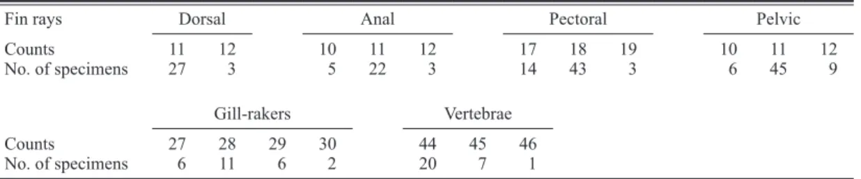
Table 1. Counts, dentition and depth ranges of 4 Glossanodon species. Data from the followings: 1: —Cohen (1958), 2—Kobilyansky (1998), and 3—this study. Numbers of teeth on lower jaw in parentheses.
G. microcephalus G. leioglossus G. semifasciatus G. pseudolineatus
No. of specimens 30 4 32 5
SL (mm) 53–97 51–111 94–206 72–80
Dorsal-¿n rays 11–12 13–14 11–13 10–11
Anal-¿n rays 10–12 11–13 11–13 10
Pectoral-¿n rays 17–19 20–22 18–22 18–19
Pelvic-¿n rays 10–12 12 10–12 11
Gill-rakers on 1st arch 27–30 36 35–40 24–26
Branchiostegal rays 5 5 5 4
Vertebrae 44–46 49 46–49 43–44
Lateral-line scales 44–45 — 50–53 —
Teeth on lower jaw entirely (10–17) partly (2–8) partly (0–10) entirely
Teeth on tongue 4–6 2–3 2–6 2–3
Depth ranges (m) 116–156 80–360 70–240 150–156
References 3 1 1, 3 2
slightly less than eye diameter. Mouth moder- ately small, most of maxillary covered dorso-lat- erally by lachrymal. Lower jaw projecting slightly beyond upper, extending posteriorly to vertical line at anterior rim of eye. No teeth on upper jaws. 10–21 small conical teeth entirely on lower jaw [left 12, right 14]. 1–2 rows and 1–3 irregular rows of small conical teeth arrayed on vomer (about 5–30 teeth, generally 10–20) and palatines (about 15–30 teeth) respectively [5 on vomer and 2 irregular rows of about 25 teeth on each palatine]. 3–7 (generally 4–5) small canine- like conical teeth present on tip of tongue [4].
Gill rakers elongate, lath-like, closely arranged.
Branchiostegal rays thin, Àat: 1st slender, reduced, 2nd to 5th expanded; ¿rst two attached medially on ceratohyal, others laterally on epi- hyal (Fig. 5). Anus immediately anterior to anal-
¿n origin. Pyloric caeca 10–11 (10 in BSKU 104224 and BSKU 104225, and 11 in BSKU 104226, BSKU 104227, BSKU 104228-9, and BSKU 104228-10).
Scales large, deciduous; lateral-line scales elongate transversely, posterior margins with a medial dent; 44–45 lateral-line scales (in 11 specimens; scale pockets were dif¿cult to count in most specimens) [44].
Color when fresh (Fig. 2). Head and body whitish and silvery. Lateral head from lachrymal to opercular region, iris, and a longitudinal band on side of body bright silver: the band narrow
near each end, widest at the middle of dorsal- and anal-¿n origins. Anterior of snout, upper jaws, and pupil, and dorsal of eye, side of nape, and supracleithrum blackish. A longitudinal bro- ken stripe above lateral line dark, sparsely pig- mented: alternately composed of short bars and blotches, rather obscure near head. All ¿n bases except adipose ¿ns blackish: densely black near base of ¿rst two dorsal-¿n rays; a blotch on upper lobe of caudal-¿n base yellowish; lower lobe of caudal ¿n near its base with a blackish triangular blotch.
Color in alcohol (Figs. 2–4, 6). Body light yel- low to ocher (all bright silver coloration faded).
Iris darkish with white crescent above whitish pupil. Anterior snout, anterior rim of lachrymal, premaxillary, anteroventral of maxillary, anteri- ormost portion of gular region just behind chin densely pigmented. Anterior of dentary and man- dibular rami sparsely pigmented. Posteroventral corner of orbit (anterior surface of 4th suborbital within the orbital cavity) densely pigmented.
Buccal cavity partly pigmented on posterior of lower jaw, palatines and ectopterygoids. No melanophores on gular region except just behind chin, branchiostegal membrane, and ventral side of body from isthmus to anus. Ventral margins of interopercle and subopercle, and posterior rim of opercle with some melanophores. Dorsal-most part of opercle, upper part of supracleithrum, posttemporal, and supratemporals densely pig- mented. Opercle mostly translucent except the
Fig. 4. Cephalic sensory canals and pores on nape of Glossanodon microcephalus sp. nov., holotype, NSMT-P 106647, 97 mm SL (stained by cyanine).
Photographed by H. Endo.
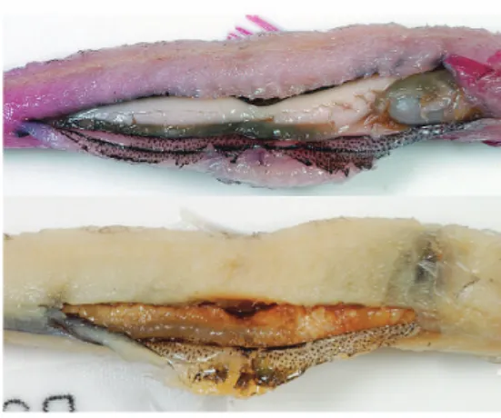
Fig. 5. Hyoid arch of Glossanodon microcephalus sp.
nov., BSKU 104224, 93 mm SL, left lateral
(above) and medial (below) views. Bar indicates
2 mm.
upper part and rim. A longitudinal broken stripe above lateral line, composed of densely pig- mented short bars and blotches, on a continuous pale stripe ¿nely pigmented: ¿rst two bars and blotches pale. Dorsal midline of body pigmented internally except behind adipose ¿n. Ventral mid- line behind anal ¿n moderately to heavily pig- mented internally. All ¿n bases pigmented:
densely on ¿rst two rays of dorsal, pectoral, pel-
vic, and anal ¿n; internally on adipose ¿n; 2 blackish blotches present externally on upper and lower caudal ¿n base, lower heavily pigmented internally. Dorsal-¿n membrane unpigmented;
posterior margin of dorsal-¿n rays sparsely pig- mented except distal half of middle rays and all of last 3 rays. Pectoral-¿n membrane partly pig- mented near base [pigmentation absent on one- third of lower rays]. Dorsal and ventral sides of
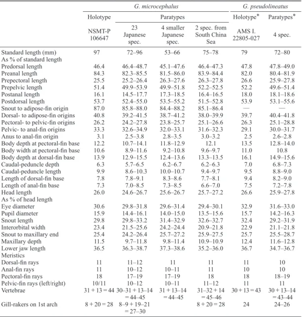
Table 2. Proportional measurements and counts of Glossanodon microcephalus and G. pseudolineatus. Asterisk indicates data from the original description.
G. microcephalus G. pseudolineatus
Holotype Paratypes Holotype* Paratypes*
NSMT-P 106647
23 Japanese
spec.
4 smaller Japanese
spec.
2 spec. from South China
Sea
AMS I.
22805-027 4 spec.
Standard length (mm) 97 72–96 53–66 75–78 79 72–80
As % of standard length
Predorsal length 46.4 46.4–48.7 45.1–47.6 46.4–47.3 47.8 47.8–49.0
Preanal length 84.3 82.3–85.5 81.5–86.0 83.9–84.4 82.0 80.4–81.9
Prepectoral length 25.5 25.2–26.4 26.3–27.6 26.3–27.8 26.6 25.9–27.8
Prepelvic length 51.4 49.9–53.9 49.9–51.8 52.2–52.5 52.2 49.6–51.4
Postanal length 16.1 14.5–17.7 17.3–18.5 16.4–16.5 18.0 18.1–18.6
Postdorsal length 53.7 52.4–55.0 53.5–55.2 51.5–52.8 53.9 53.1–55.6 Snout to adipose-¿n origin 87.0 85.8–88.0 84.4–88.2 85.1–86.4 — — Dorsal- to adipose-¿n origins 40.8 39.2–41.5 38.7–41.2 38.0–39.9 39.7 40.4–41.8 Pectoral- to pelvic-¿n origins 26.2 24.2–27.8 23.8–25.7 25.1–26.6 26.3 25.1–28.8 Pelvic- to anal-¿n origins 33.3 32.6–34.9 32.0–33.1 31.6–32.3 29.1 30.0–31.7
Anus to anal-¿n origin 3.1 2.5–3.8 2.8–3.5 3.0–3.2 2.5 2.6–2.8
Body depth at pectoral-¿n base 12.2 10.7–14.1 11.8–12.9 12.1 13.5 12.8–14.0 Body width at pectoral-¿n base 10.6 8.9–11.6 9.2–10.8 9.6–9.7 11.0 10.8 Body depth at dorsal-¿n base 13.9 12.9–15.5 12.4–13.6 13.3–13.5 16.1 14.9–15.6
Caudal-peduncle depth 6.3 5.7–6.5 6.2–6.7 6.2–6.3 7.0 6.8–7.3
Caudal-peduncle length 9.9 8.6–10.3 10.0–10.7 9.4–9.7 9.5 8.8–9.0
Length of dorsal-¿n base 7.8 7.8–9.1 8.3–8.6 7.7–8.1 9.4 8.2–9.0
Length of anal-¿n base 7.3 7.0–8.5 7.3–8.5 6.6–7.0 7.5 7.2–7.8
Head length 26.0 24.6–26.7 25.6–26.7 25.7–27.2 26.6 25.9–27.8
As % of head length
Eye diameter 30.6 29.8–31.8 29.6–31.4 29.4–30.1 32.9 31.6–33.0
Pupil diameter 15.9 14.4–16.1 14.0–15.0 13.5–15.6 15.7 14.2–16.3
Snout length 29.8 29.8–33.2 31.4–32.9 32.6–32.7 32.4 29.2–31.9
Interorbital width 23.4 21.5–25.6 24.2–24.4 20.9–21.8 22.9 21.1–21.8 Snout to maxillary end 25.4 24.2–26.4 25.7–27.2 25.9–27.5 25.7 25.5–28.7
Maxillary depth 11.5 9.7–11.8 9.8–11.4 10.9–10.9 12.4 11.6–12.8
Lower jaw length 36.5 36.3–38.7 37.3–38.6 35.2–36.0 36.7 34.7–36.7
Meristics
Dorsal-¿n rays 11 11–12 11 11 11 10
Anal-¿n rays 11 10–12 10–11 11 10 10
Pectoral-¿n rays 18 17–19 17–19 18 18 18–19
Pelvic-¿n rays (left/right) 10/11 10–12 10–11 11–12 11 11
Vertebrae 31+13=44 30–31+13–14
=44–45
31+13–14
=44–45
31–32+14
=45–46
30+13=43 30+13–14
=43–44
Gill-rakers on 1st arch 8+20=28 8–9+19–21
=27–30